
Types of Hernia Abdominal hernia Diaphragmatic hernia Perineal

Types of Hernia Abdominal hernia, Diaphragmatic hernia, Perineal hernia, Inguinal hernia, Scrotal hernia, Umbilical hernia Surgical approaches to thoracic wall
TYPES OF HERNIA q Inguinal Hernia q Scrotal Hernia q Diaphragmatic Hernia q Umbilical Hernia (Omphalocele) q Perineal Hernia q Ventral Abdominal Hernia
INGUINAL HERNIA
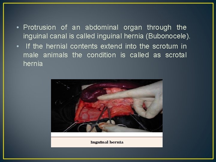
• Protrusion of an abdominal organ through the inguinal canal is called inguinal hernia (Bubonocele). • If the hernial contents extend into the scrotum in male animals the condition is called as scrotal hernia
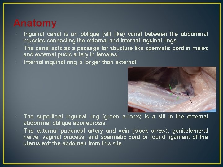
Anatomy Inguinal canal is an oblique (slit like) canal between the abdominal muscles connecting the external and internal inguinal rings. The canal acts as a passage for structure like spermatic cord in males and external pudic artery in females. Internal inguinal ring is longer than external. The superficial inguinal ring (green arrows) is a slit in the external abdominal oblique aponeurosis. The external pudendal artery and vein (black arrow), genitofemoral nerve, vaginal process, and spermatic cord or round ligament of the uterus exit the abdomen from this site.
• Inguinal hernia can be classified as indirect & direct. • Indirect inguinal hernia more common. In this condition abdominal viscera pass through an enlarge vaginal ring into the cavity of the vaginal tunic or process which forms the hernial sac. • In case of direct inguinal hernia, a separate out pouching of the peritoneum, cranial to vaginal tunic or process, act as the sac.
Causes May be congenital or acquired – Accidental slipping causes streching of the hind limbs outwards which may dilate inguinal canal.
Symptoms In bitches appreciable swelling is noticed in the inguinal region. Difficulty in defecation. Refuse to move due to pain Abduction of hind limbs Systemic signs are evident only when the hernia gets strangulated. Hernial contents includes intestine , urinary bladder , uterus in female and omentum.
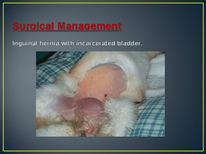
Surgical Management Inguinal hernia with incarcerated bladder.
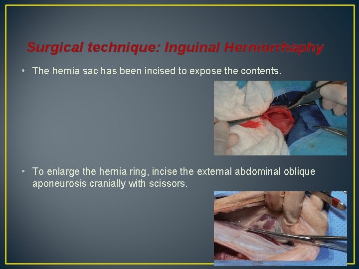
Surgical technique: Inguinal Herniorrhaphy • The hernia sac has been incised to expose the contents. • To enlarge the hernia ring, incise the external abdominal oblique aponeurosis cranially with scissors.
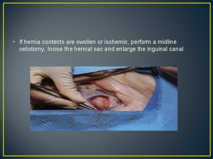
• If hernia contents are swollen or ischemic, perform a midline celiotomy. Incise the hernial sac and enlarge the inguinal canal
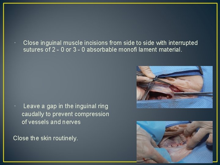
Close inguinal muscle incisions from side to side with interrupted sutures of 2 - 0 or 3 - 0 absorbable monofi lament material. Leave a gap in the inguinal ring caudally to prevent compression of vessels and nerves Close the skin routinely.
Postoperative Considerations • Exercise should be limited during recovery, • Analgesics are usually administered for several days. • Postoperative complications ; • • swelling incisional infection dehiscence peritonitis sepsis Vomiting recurrence
SCROTAL HERNIA
Etiology: Scrotal hernia are indirect hernias that result from a defect in the inguinal ring, allowing abdominal contents to protrude into vaginal process beside contents of the spermatic cord. These hernia are rare occurance. Most case reports are in young male dog.
Clinical Signs • Pain, swelling, and frequently organ dysfunction. • This hernia is predominantly unilateral. • The external appearance of this hernia depends the sac contents and amount of vascular obstruction at hernial ring.
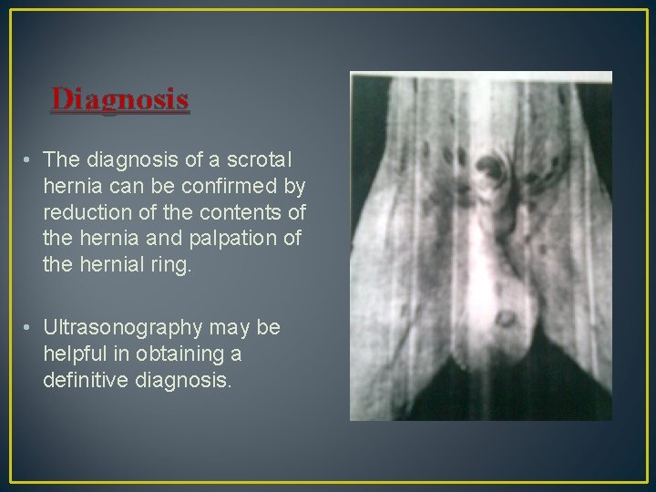
Diagnosis • The diagnosis of a scrotal hernia can be confirmed by reduction of the contents of the hernia and palpation of the hernial ring. • Ultrasonography may be helpful in obtaining a definitive diagnosis.
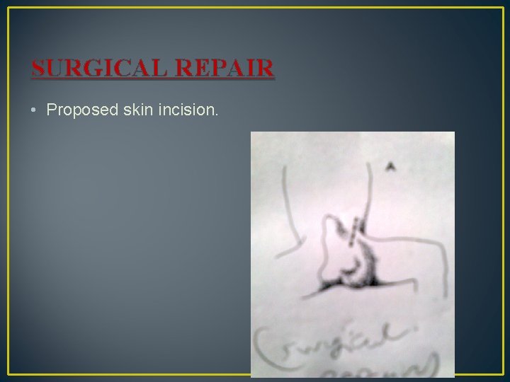
SURGICAL REPAIR • Proposed skin incision.
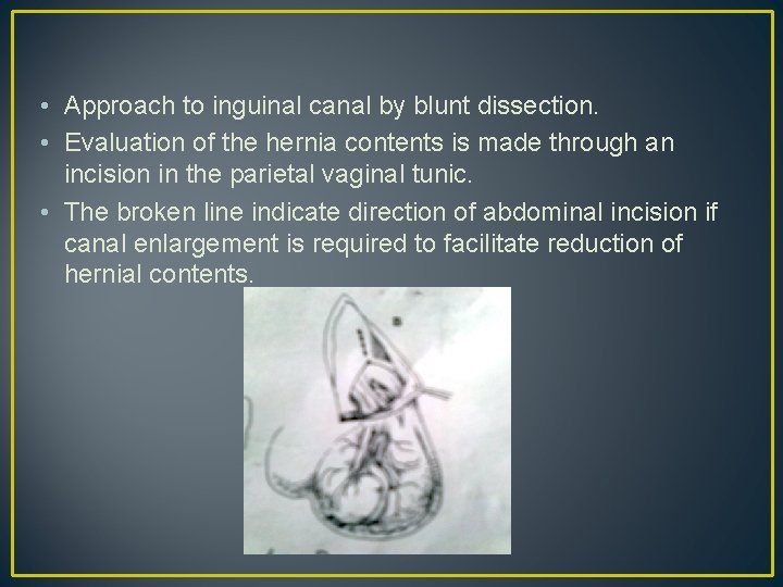
• Approach to inguinal canal by blunt dissection. • Evaluation of the hernia contents is made through an incision in the parietal vaginal tunic. • The broken line indicate direction of abdominal incision if canal enlargement is required to facilitate reduction of hernial contents.
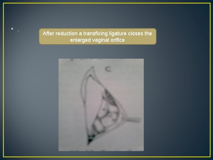
• . After reduction a transfixing ligature closes the enlarged vaginal orifice
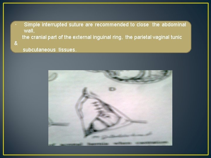
Simple interrupted suture are recommended to close the abdominal wall, the cranial part of the external inguinal ring, the parietal vaginal tunic & subcutaneous tissues.
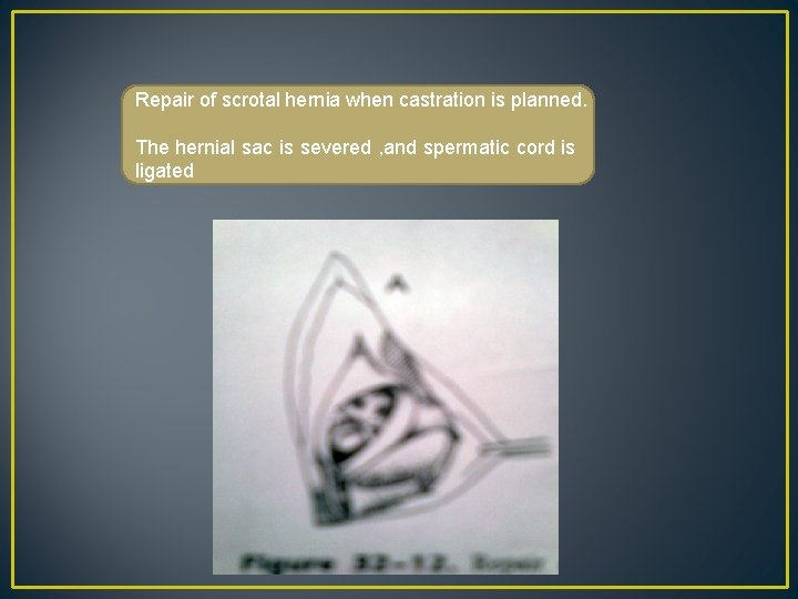
Repair of scrotal hernia when castration is planned. The hernial sac is severed , and spermatic cord is ligated
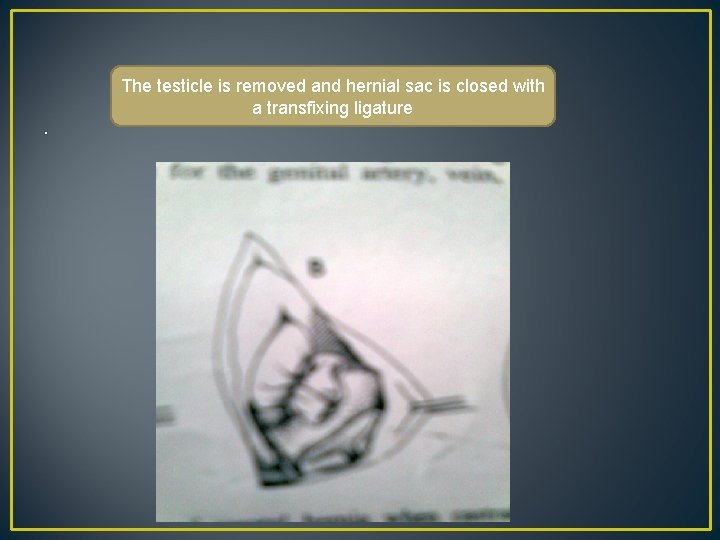
. The testicle is removed and hernial sac is closed with a transfixing ligature
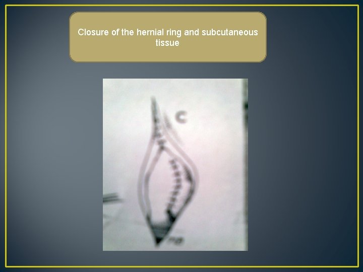
Closure of the hernial ring and subcutaneous tissue
DIAPHRAGMATIC HERNIA
A diaphragmatic hernia is caused by the passage of abdominal viscera into thoracic cavity, through tear or a congenital defect in diaphragm. Etiology: congenital defect in the diaphragm. trauma e. g. , getting run over by an automobile, falling from a height etc.
Symptoms: Abdominal breathing. Peculiar cough. Tendency to vomit after feeding. Difficulty and pain while walking down from height. Respiratory distress if the stomach is full and causing pressure on lungs. Respiratory sound may be absent on the affected side.
• Differential Diagnosis : • Conditions like hydrothorax and aspiration pneumonia • Diagnosis: • A lateral radiograph of thorax & • A ventro-dorsal radiograph of thorax.
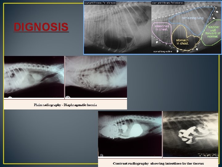
DIGNOSIS
TREATMENT Surgical treatment: • Thoracic approach: • The sixth or seventh intercostal site may be chosen • On entering the thoracic cavity care is taken to prevent injury to lungs. The wound edges retracted by proper retractor. • The respiratory pump is so adjusted that lung will be inflated to only about half their normal capicity.
• The hernia reduced and tear in diaphragm is sutured with No. 1 chromic catgut. • Closure of intercostal incision is made by placing sutures around adjacent ribs. • When the intercostal wound sutured in this manner , the nagative pressure in plural cavity should be established. • The skin edges are united by apposition sutures.
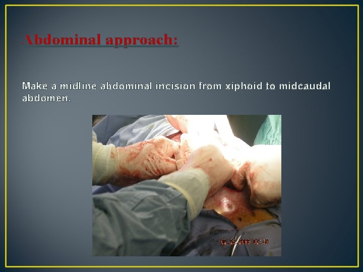
Abdominal approach: Make a midline abdominal incision from xiphoid to midcaudal abdomen.
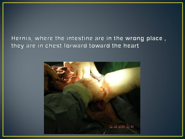
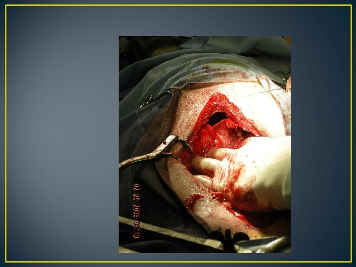
Hernia, where the intestine are in the wrong place , they are in chest forward toward the heart
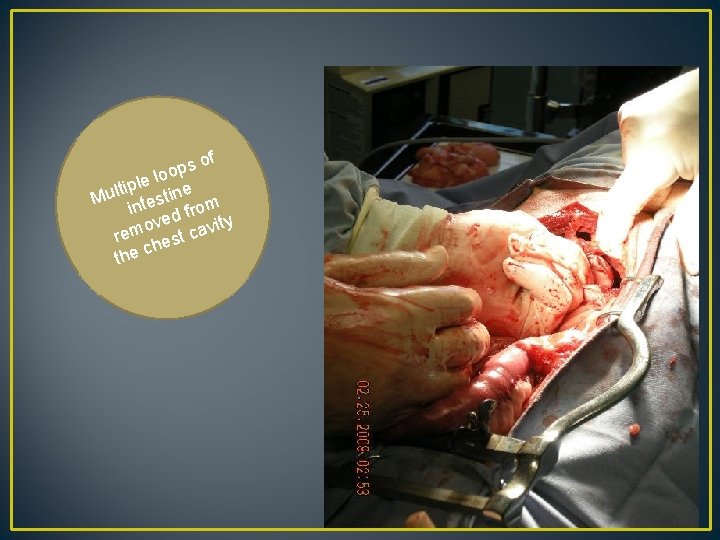
of s p loo e l tip Mul ntestine m i fro y d e ov vit rem est ca ch the
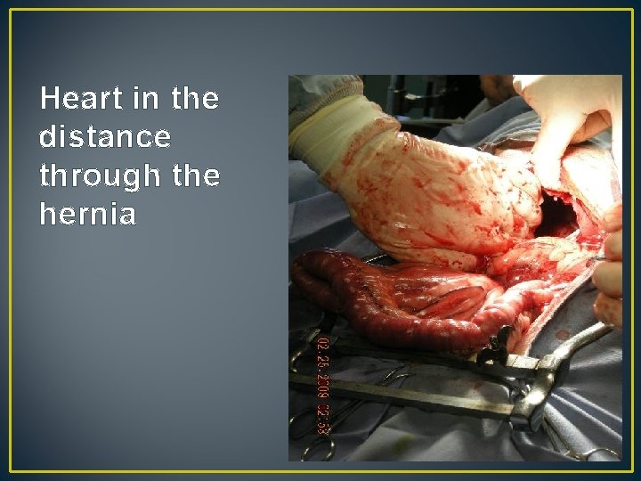
Heart in the distance through the hernia
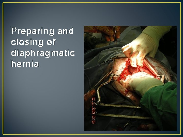
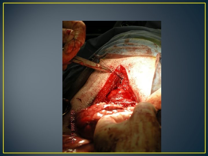
Preparing and closing of diaphragmatic hernia
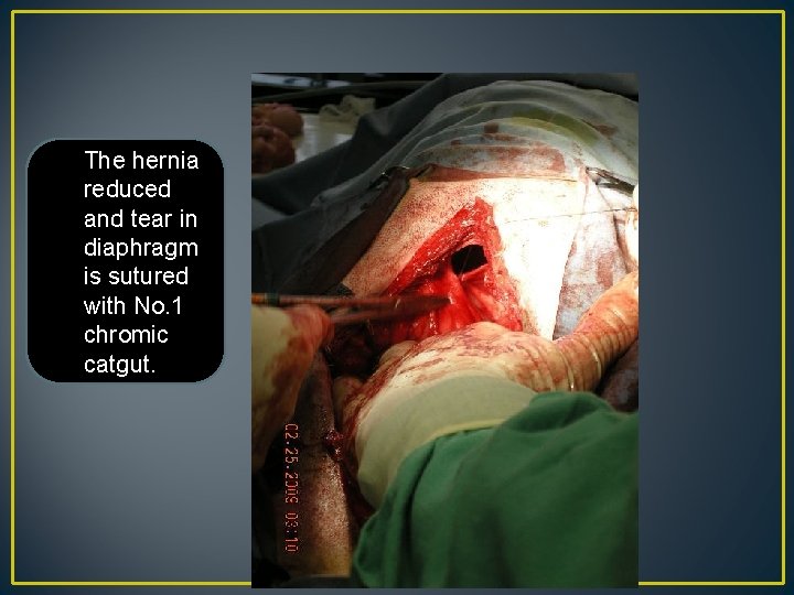
The hernia reduced and tear in diaphragm is sutured with No. 1 chromic catgut.
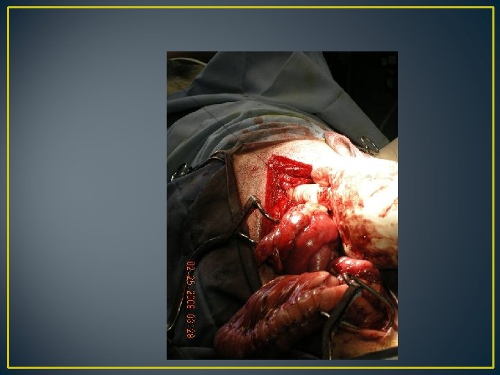
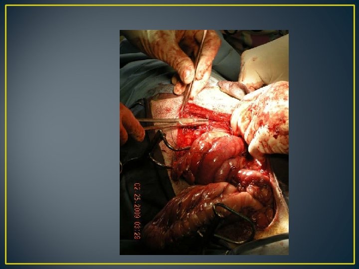
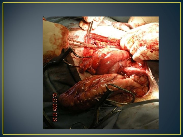
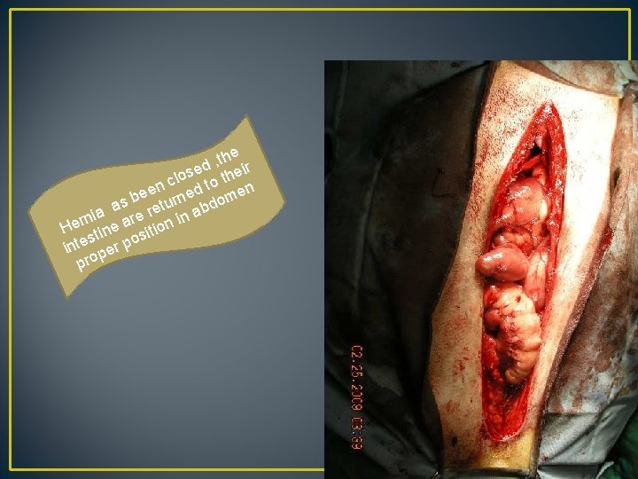
e h t , d eir e s clo to th n n bee turned dome s a e re n ab a i r rn i He tine a sition s o inte per p pro
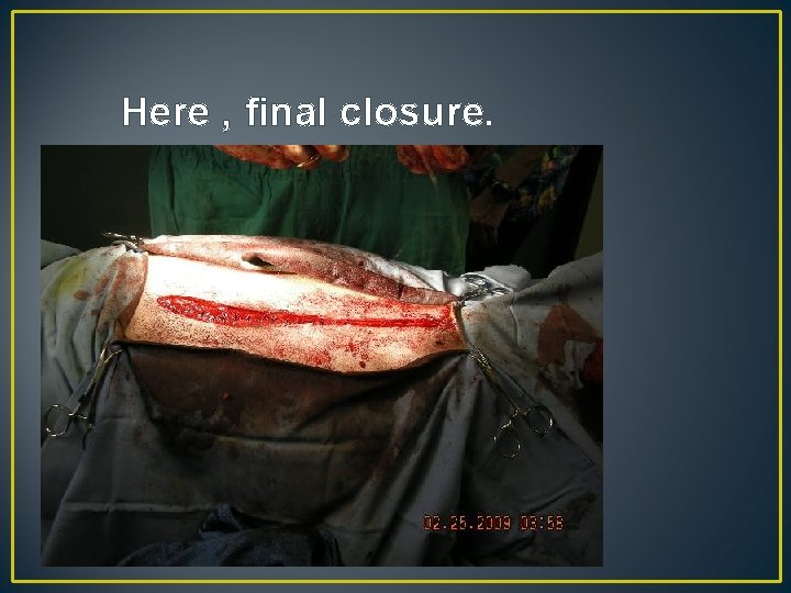
Here , final closure.

• Potential complications include cardiac or respiratory arrest, pulmonary edema, persistent pneumothorax, and organ failure. • Mortality rates after surgical repair of traumatic diaphragmatic hernias are 10% to 18%. • Prognosis is worse for older cats, dogs with gastric entrapment and dilatation, and animals with concurrent injuries.

UMBILICAL HERNIA
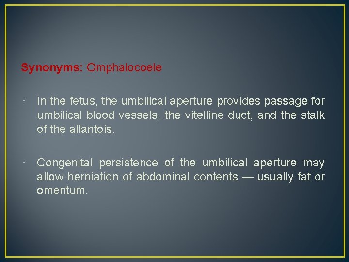
Synonyms: Omphalocoele In the fetus, the umbilical aperture provides passage for umbilical blood vessels, the vitelline duct, and the stalk of the allantois. Congenital persistence of the umbilical aperture may allow herniation of abdominal contents — usually fat or omentum.
Causes • The condition can be congenital or acquired. • Acquired hernial ring may be primarily due to • Trauma • Resection of cord too close to abdominal wall • Excessive straining due to diarrhoea/ constipation • Infection of the cord may also prevent natural closure of umbilicus.

Clinical signs A discrete spherical swelling at umbilicus Hernial contents are usually fat and omentum Larger hernial sac contains loops of small intestine Sac is formed by skin, fibrous tissue and peritoneum Presence of adhesions/ umbilical abscess may prevent reduction. Rarely, the contents get strangulated with symptoms of pain and intestinal obstruction
Treatment • Conservative methods (only for a small reducible hernia) • Surgical method
conservative methods (only for a small reducible hernia) • Apply abdominal pressure bandages or clamps • Reduce the hernia and the hernial ring to close by cicatrisation.
surgical method Operation is done at the age of three months. If the swelling is too big , treatment is attempted immediately. Anaesthesia : General anaethesia or sedation combined with local anaethesia.
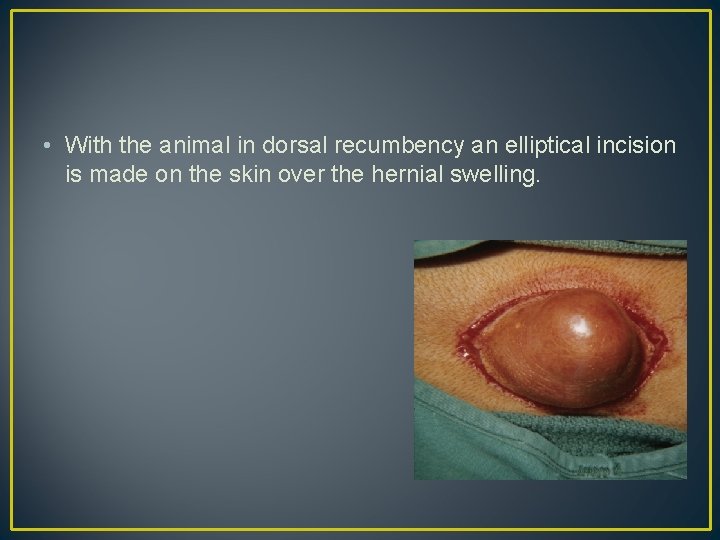
• With the animal in dorsal recumbency an elliptical incision is made on the skin over the hernial swelling.
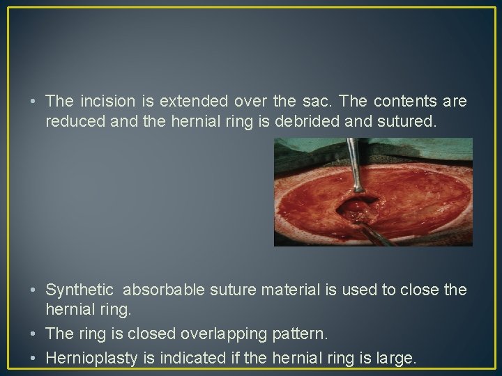
• The incision is extended over the sac. The contents are reduced and the hernial ring is debrided and sutured. • Synthetic absorbable suture material is used to close the hernial ring. • The ring is closed overlapping pattern. • Hernioplasty is indicated if the hernial ring is large.
PERINEAL HERNIA

most predominantly seen in old uncastrated male dogs. Etiology : Weakening of the pelvic diaphragm and hernia can occur due to the following factors. • Hormonal disorders • Prostatic diseases • Rectal diseases and • Anatomical factors – rectal deviations, diverticula etc.
The levator ani and coccygeal muscles form the pelvic diaphragm, which supports the rectal wall and helps to form a natural partition between the abdominal cavity and ischiorectal fossa. Muscular atrophy or stress from chronic straining can weaken the pelvic diaphragm, resulting in herniation of the rectal wall or abdominal organs into the perineal region. Hernial Content : - Deviated rectal wall - Serous or serosanguinous fluid - Pelvic or retroperitoneal fat - Prostate, prostatic cysts, - Bladder - Intestines
Clinical Signs & Symptoms Tenesmus Dyschezia Perineal swelling. Animals with partial or complete urethral obstruction may have stranguria, anuria, or incontinence.
• Fluctuating /hard swelling ventral and lateral to the anus (in the ischiorectal fossa) • Swelling may be unilateral or bilateral • The hernial contents are usually rectum, enlarged prostate and perineal fat. • Retroflexion of the bladder is common. Incarceration of bladder in the perineal hernia should be considered as an emergency. • Hard swelling should be differentiated from perianal neoplasm.
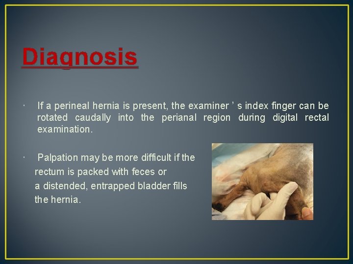
Diagnosis If a perineal hernia is present, the examiner ’ s index finger can be rotated caudally into the perianal region during digital rectal examination. Palpation may be more difficult if the rectum is packed with feces or a distended, entrapped bladder fills the hernia.
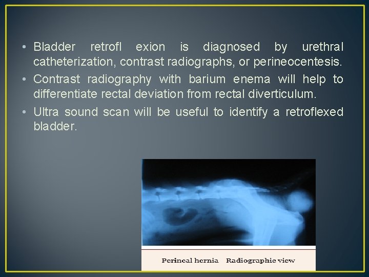
• Bladder retrofl exion is diagnosed by urethral catheterization, contrast radiographs, or perineocentesis. • Contrast radiography with barium enema will help to differentiate rectal deviation from rectal diverticulum. • Ultra sound scan will be useful to identify a retroflexed bladder.
Preoperative management • Serum chemistries, complete blood count, and urinalysis are evaluated for metabolic abnormalities and evidence of sepsis or infection. • If the urinary tract is obstructed, a urethral catheter should be passed into the bladder to keep it decompressed until surgery. • Enemas can be given to constipated animals but should be avoided within 12 hours of surgery, since liquefied feces are more likely to leak into the sterile field.
• Epidural nerve block is performed for intraoperative and postoperative analgesia • The perineum and base of the tail are clipped and prepped. • Intact male dogs should also be prepped for caudal or prescrotal castration. • The rectum is evacuated digitally, and a purse - string suture is placed in the anus. • For herniorraphy, patients are placed in a perineal position with the tail pulled forward.
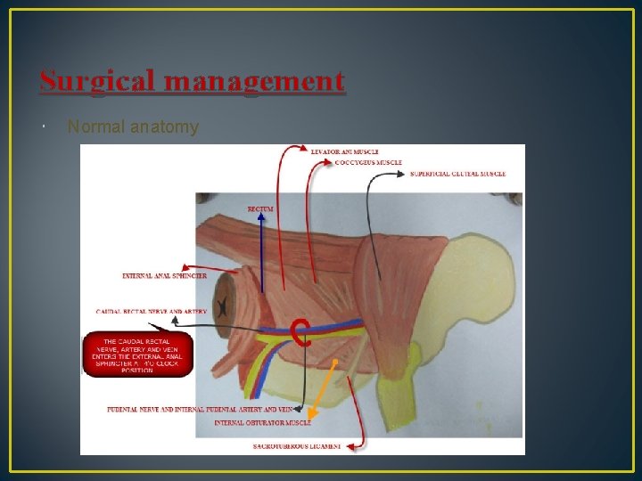
Surgical management Normal anatomy
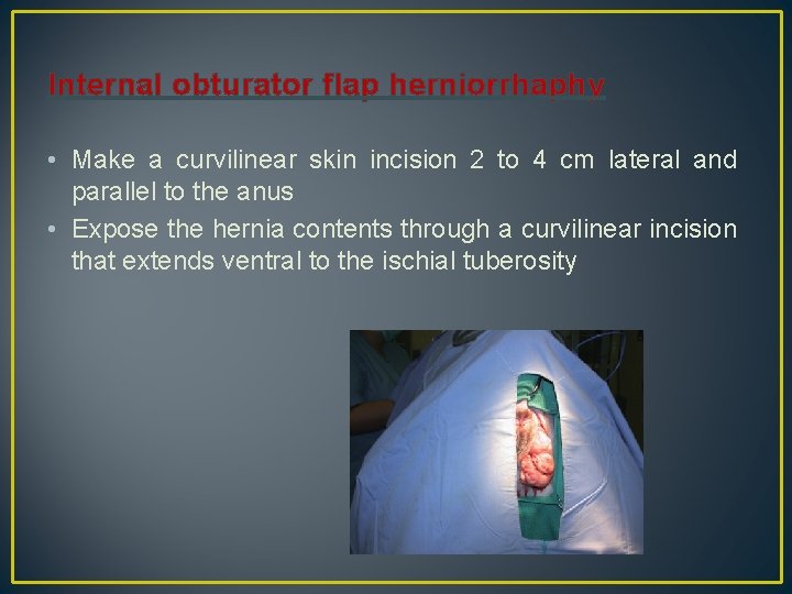
Internal obturator flap herniorrhaphy • Make a curvilinear skin incision 2 to 4 cm lateral and parallel to the anus • Expose the hernia contents through a curvilinear incision that extends ventral to the ischial tuberosity
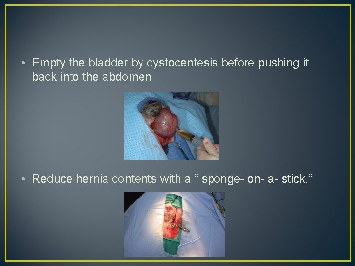
• Empty the bladder by cystocentesis before pushing it back into the abdomen • Reduce hernia contents with a “ sponge- on- a- stick. ”
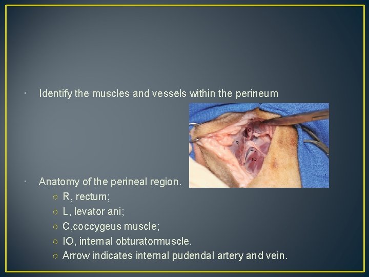
Identify the muscles and vessels within the perineum Anatomy of the perineal region. ○ R, rectum; ○ L, levator ani; ○ C, coccygeus muscle; ○ IO, internal obturatormuscle. ○ Arrow indicates internal pudendal artery and vein.
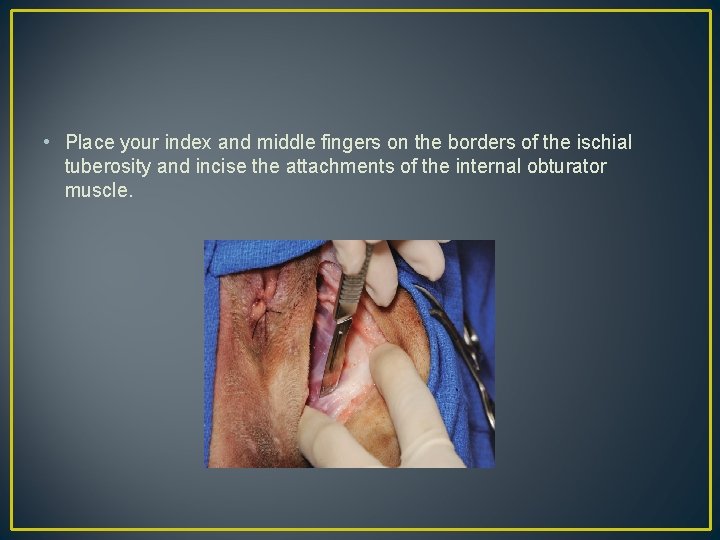
• Place your index and middle fingers on the borders of the ischial tuberosity and incise the attachments of the internal obturator muscle.
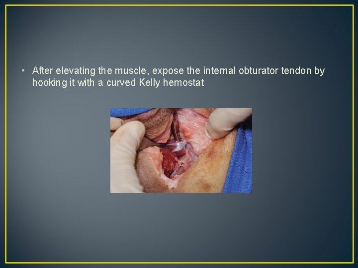
• After elevating the muscle, expose the internal obturator tendon by hooking it with a curved Kelly hemostat
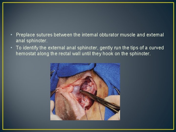
• Preplace sutures between the internal obturator muscle and external anal sphincter. • To identify the external anal sphincter, gently run the tips of a curved hemostat along the rectal wall until they hook on the sphincter.
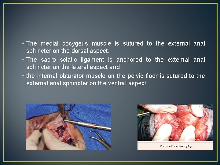
• The medial cocygeus muscle is sutured to the external anal sphincter on the dorsal aspect. • The sacro sciatic ligament is anchored to the external anal sphincter on the lateral aspect and • the internal obturator muscle on the pelvic floor is sutured to the external anal sphincter on the ventral aspect.
Postoperative considerations • • Antibiotic Analgesics Stool softener such as lactulose Elizabethan collars are recommended for 1 week to prevent self - trauma.
Common complications : • Swelling • Wound infection • Tenesmus • rectal prolapse • Fecal or urinary incontinence & • Hernia recurrence.

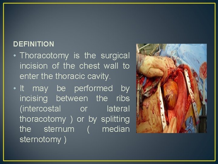
DEFINITION • Thoracotomy is the surgical incision of the chest wall to enter the thoracic cavity. • It may be performed by incising between the ribs (intercostal or lateral thoracotomy ) or by splitting the sternum ( median sternotomy )
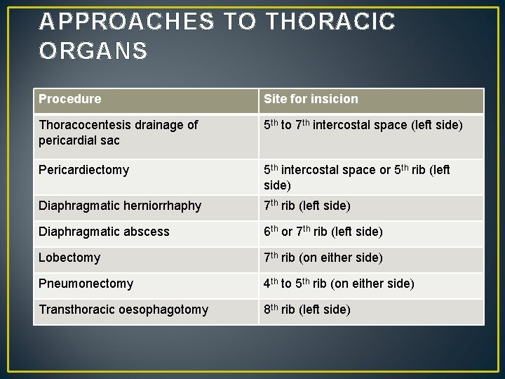
APPROACHES TO THORACIC ORGANS Procedure Site for insicion Thoracocentesis drainage of pericardial sac 5 th to 7 th intercostal space (left side) Pericardiectomy 5 th intercostal space or 5 th rib (left side) Diaphragmatic herniorrhaphy 7 th rib (left side) Diaphragmatic abscess 6 th or 7 th rib (left side) Lobectomy 7 th rib (on either side) Pneumonectomy 4 th to 5 th rib (on either side) Transthoracic oesophagotomy 8 th rib (left side)
• SURGICAL ANATOMY • Thoracic cavities of dogs and cats are compressed laterally so that the greater dimension is dorso-ventral. • The first and last sternebrae are known as the manubrium and xiphoid, respectively. • The 10 th , 11 th, 12 th ribs do not articulate with sternum, but instead form the costal area bilaterally. • The space between the ribs is generally three times as wide as the adjacent rib, in conjunction with a satellite vein and nerve.
ANAESTHETIC CONSIDERATIONS • Endotrachial Intubation must be accomplished rapidly in animals with respiratory dysfunction and anaesthesia should be maintained with an inhalation anaesthetic (i. e) isoflurane (or) halothane) • All animals with open chest cavities requires intermittent positive pressure ventilation • High ventilation pressure should be avoided in patients with chronically collapsed lung • Thoracotomy procedure often produces substantial pain and hence post operative analgesic therapy is indicated.
GENERAL PRINCIPLES • Animals with traumatic lesions that impair respiration eg. those with acute respiratory impairment (ie) ruptured bullae, ruptured pulmonary abscess (often require emergency stabilization eg. stabilization of rib segments, thoracocentesis, oxygen therapy) prior to surgery. • Equipments for thoracocentesis and should be readily available • clinician should be familiar with technique. • Positioning the animal • Blood gas analysis (or) evaluation of pulse oxymetry
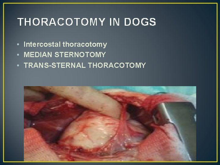
THORACOTOMY IN DOGS • Intercostal thoracotomy • MEDIAN STERNOTOMY • TRANS-STERNAL THORACOTOMY
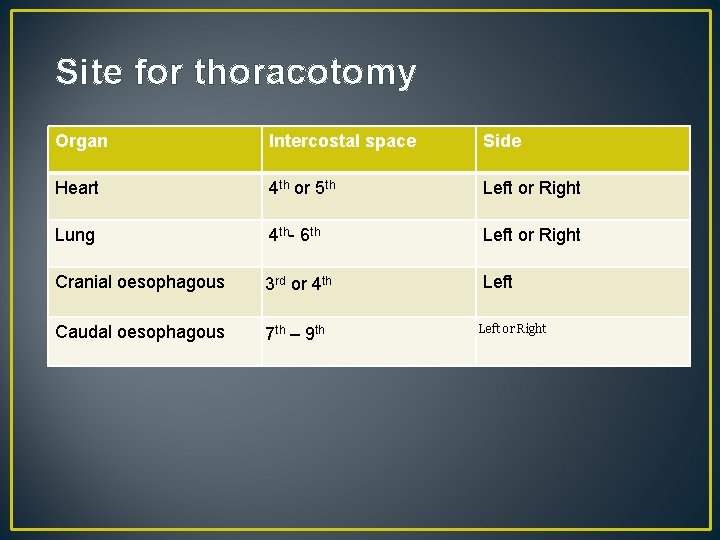
Site for thoracotomy Organ Intercostal space Side Heart 4 th or 5 th Left or Right Lung 4 th- 6 th Left or Right Cranial oesophagous 3 rd or 4 th Left Caudal oesophagous 7 th – 9 th Left or Right
• Median sternotomy is the only thoracic approach that allows access to the entire thoracic cavity, and therefore is indicated when exploration of the thoracic cavity is necessary. • Trans-sternal thoracotomy is similar to intercostal thoracotomy • performed with the animal in dorsal recumbency. • Intercostal thoracotomies in the same intercostal space on both sides are connected by ligation of internal thoracic vessels and a transverse osteotomy of the sternum.
THORACOTOMY IN CATTLE • Intercostal incision • Rib resection • Split rib resection
• INTERCOSTAL THORACOTOMY It provides access to the thoracic cavity. • It is employed when exposure to a specific region of the thoracic cavity is needed, but access to structures in the area of thoracotomy is limited. • In general Intercostal thoracotomy allows access to about one third of ipsilateral thoracic cavity and mediastinal structures. • The site of an intercostal thoracotomy may be third through the tenth intercostal space depending on the structures being exposed
• Rib resection thoracotomy provides more exposure than a simple intercostal thoracotomy and about the same exposure as an intercostal thoracotomy with osteotomies of a rib a potential • Advantage of this approach is that it results in fewer adhesions between the lung and incision • A disadvantage of rib resection is that secure closure is more difficult to obtain, especially in large dog that has undergone wide retraction of the rib during surgery. • Rib resection thoracotomy is used infrequently in small animals and is reserved for specific indications.

k n • a h T u o Y
- Slides: 86