Pleura v v Closed serous sac lined by

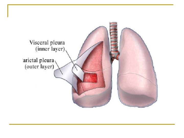
Pleura v v Closed serous sac, lined by mesothelium Invaginated by lungs Parietal pleura Visceral pleura

Pleural cavity n Potential space between the two layers n n Contains pleural fluid Allows the lungs to glide freely

Visceral Pleura n n Adherent to the surfaces and fissures of the lungs Continuous with the parietal pleura at the hilum & pulmonary ligament Parietal Pleura n n Lines the q Thoracic wall q Mediastinum q Diaphragm Can be separated from the surface

Parietal pleura n Costal pleura n Diaphragmatic pleura n Mediastinal pleura n Cervical pleura

Cervical pleura n n Covers the apex of the lung Exetrnally - Suprapleural membrane

Costal pleura v lines the inner surface of thoracic wall v Endothoracic fascia

Mediastinal pleura Lines the corresponding surfaces of mediastinum

Pulmonary ligament n Mediastinal pleura forms fold beyond the root of the lung n Contains loose areolar tissue, lymphatics n Provide dead space for pulmonary veins during increased venous return

Diaphragmatic pleura q Lines the superior aspect of diaphragm

Pleural recesses n Potential spaces between two parts of parietal pleura n Acts as a reserve spaces for the lungs n Two recesses on each side q Costomediastinal recess q Costodiaphragmatic recess

Costomediastinal recess n n n Present between the costal and mediastinal pleurae Lies behind the sternum and costal cartilages Obvious in the region of cardiac notch of the left lung

Costodiaphragmatic recess n Lies inferiorly between the costal and diaphragmatic pleurae n Vertically it measures 5 cm n Most dependent part

Surface marking of the pleura n Anterior margin Costo-mediastinal line of reflection

n Posterior margin

n Inferior margin Costo–diaphragmatic line of reflection

Nerve supply n n Parietal pleura Somatic nerves Visceral pleura Autonomic nerves Intercostal nerves

Blood supply Arterial supply n Intercostal arteries n Internal thoracic artery n Musculo phrenic artery Venous drainage v Azygos vein v Internal thoracic vein

Development

Applied aspects

Pleurisy Inflammation of the pleura

Pleural effusion

Hemo thorax Collection of blood

Chylothorax and Empyema Presence of lymph and pus in the pleural cavity respectively

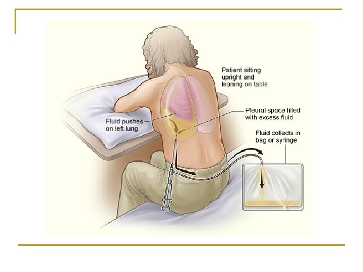
Paracentesis thoracis
- Slides: 26