CNS pathology Third year medical students lecture 9


 • CNS")

 ,")


 astrocytoma • 2.")


 • or vascular proliferation")


- Slides: 54
CNS pathology Third year medical students lecture 9 CNS tumors/1 Dr Heyam Awad FRCPath 2017
CNS TUMORS/ epidemiology • Tumors in the CNS can arise in the brain or the spinal cord. • Brain tumors : around 10 - 17/ 100 000 population • Spinal cord tumors : around 1 -2 / 100 000
• CNS tumors can be primary or secondary • Secondary account for 1/4 th to ½ of all CNS tumors • CNS tumors account for 20% of childhood tumors. • 70% of childhood tumors arise in the posterior fossa • 70% of adulthood tumors arise within the cerebral hemispheres above the tentorium.
CNS tumors in Jordan according to 2013 cancer registry ( latest published) • CNS tumors are the 10 th most common tumor among Jordanians • Second most common among Jordanian children
CNS tumors in Jordan/ 2013 stats according to Jordan Cancer registry CNS tumors in 2013 Males Females Nubers in percentag No 2013 e of total cancers in 2013 Females % Total No Total % 95 2% 153 2. 8% 3. 7% 58
Frequency of incidence of cases among Jordanians by site and agegroup (Males ) , 2013 CNS tumors , age distribution, males 30 25 20 15 10 5 0 0 to 9 10 to 19 20 -29 30 -39 40 -49 50 -59 60 -69 70 -79
CNS tumours/ characteristics • No premalignant or in situ stage • Low grade lesions can widely infiltrate with serious clinical deficit • Anatomical site important in outcome regardless of type, grade • Rarely spread outside CNS
Classification of CNS tumors • According to the cell of origin: 1. gliomas 2. Neuronal tumors 3. Embryonal (primitive) neoplasms: 4. Others: lymphoma and germ cell tumors 5. Meningioma 6. Metastatic tumors
WHO classification of CNS tumors • The international classification of human tumors was published by the World Health Organization (WHO) to establish a classification and grading of human tumors that is accepted and used worldwide. • The first edition on the histological typing of tumors of the nervous system was published in 1979 • 4 th edition 2007 • Newest edition 2016
• Throughout the years, the classification was based on the consensus of an international Working Group. • in 2007 the consensus group contained 25 pathologists and geneticists , and the results of their deliberations and those of an additional 50 contributors were contained in the 2007 WHO classification of tumors of the central nervous system
2016 classification/ a shakeup of the traditional views on CNS tumors • For the first time, the WHO classification of CNS tumors uses molecular parameters in addition to histology to define tumor entities, thus formulating a concept for how CNS tumor diagnoses should be structured in the molecular era. • As such, the 2016 CNS WHO presents a major restructuring of several tumors including diffuse gliomas. • The 2016 edition has added newly recognized neoplasms, and has deleted some entities, variants and patterns that no longer have diagnostic and/or biological relevance.
SO: • The 20016 classification has changed our understanding of CNS tumors. • It relies on genetic changes plus morphology (integrated layered diagnosis) • The changes in classification are important because they have better prognostic and therapeutic implications.
NOTE • In most centers in Jordan we are still using the 2007 WHO classification, because the new system needs expensive immunohistochemical and molecular tests to decide on genetic mutations. • King Hussain medical center is better funded and they started working on the new system! • So, in these lectures we will mainly discuss the classical classification but will give some examples of the new changes.
TOO MANY ENTITIES!!
Classification in general
note • Classification deals with types of tumors and entities • We also need a grading system, That’s why there is a WHO grading system for CNS tumors • Regarding stage: note that CNS tumors rarely metastasize so we don’t usually use the TNM staging for these tumors. • Grading in CNS tumors determines outcome, although site is also important.
WHO grading
Gliomas/ tumors of glial cells
gliomas • Are the most common primary brain tumors. • Gliomas are classified to: astrocytomas, oligodendrogliomas and ependymomas. • It is now thought that these three types originate from a progenitor cell that can differentiate to these three morphologic types.
Astrocytomas • Two major types • 1. diffuse ( infiltrating ) astrocytoma • 2. localized astrocytomas, the most important one is the PILOCYTIC ASTROCYTOMA
WHO classification of astrocytomas WHO designation WHO grade • pilocytic astrocytoma I • Astrocytoma, well diff II • anaplastic astrocytoma III • glioblastoma IV
Diffuse astrocytoma • • Account for 80% of adult gliomas. Present at 40 - 60 years of age Location: cerebral hemispheres Present with: seizures, headache, focal neurologic deficit
Diffuse astrocytoma • • Spectrum of histological differentiation: Well differentiated. . WHO grade 2 Anaplastic astrocytoma… grade 3 Glioblastoma…. Grade 4 • Prognosis is affected by grade • Note: there are no grade 1 diffuse astrocytomas
Well differentiated astrocytoma • • Can be static for several years But progress Mean survival is more than five years When progress: rapid deterioration + anaplastic histological features develop.
Well differentiated astrocytoma gross features • Poorly defined grey, infiltrative tumors that invade the brain without forming a discrete mass:
Well diff astro
Mowell diff astrorphology
Microscopic features • Well diff: mild to moderate increase in glial cells. • Some nuclear pleomorphism • Background: fibrillary due to fine astrocytic processes. . These are positive with glial fibrillary acidic protein (GFAP)
Well diff astro
GFAP staining in astro
Anaplastic astro • More cellular than well diff astro. • More pleomorphism • Mitotic figures
Anaplastic astrocytoma. . note the high cellularity
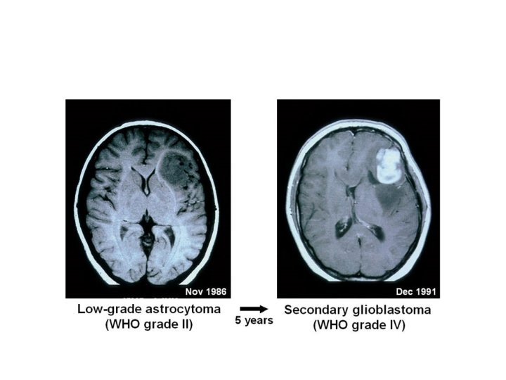
glioblastoma -Poor prognosis -15 months survival - Survival rate improved ; 25% live up 2 years or more with resection followed by chemo and radiotherapy -Can result due to progression from a previous astrocytoma ( secondary glioblastoma) or the tumor can start as glioblastoma from the beginning ( Primary astrocytoma)
morphology • Glioblastoma: charectarized histologically by variation of the tumor appearance (that’s why it was called glioblastoma multiforme); • So there will be soft , necrotic and hemorrhagic areas.
glioblastoma
glioblastoma
Glioblastoma multiforme Looks like anaplastic plus • Necrosis (usually pesudopalisading) • or vascular proliferation
palisade -high fence made of pointed stakes that was used in the past to protect a building or area -palisades : a line of steep cliffs especially along a river or ocean
Glioblastoma/ palisaded nuclei around necrotic area
glioblastoma
Vascular proliferation in glioblastoma • Manifests as tufts of cells that pile up and bulge into the lumen. • Minimal criteria to diagnose vascular proliferation: is the presence of double endothelial layer. • If it is marked and severe it forms: glumeruloid body.
Glomeruloid body
Radiological appearance • High grade astro contains abnormal leaky vessels • This gives contrast enhancement on imaging studies • Contrast given before MRI scanning has limited capacity to reach the brain tissue due to blood brain barrier (BBB). • If there is defect in BBB ( like in the leaky vessels ), the contrast material reaches the brain and forms obvious lesions.
Contrast enhancement / This slide is for your information. Not for the test!! • In general, the terms 'enhancing' or 'non-enhancing' lesion refer to the uptake of Gadolinium-based contrast agent in the lesion. • The difference between enhancing an non-enhancing is very pronounced in brain tissue, where the blood-brain barrier effectively hinders the contrast agent from accumulating in the tissue in normal circumstances. When the blood-brain barrier is leaking, e. g. due to an inflammatory process in a lesion or due to cancerous angiogenesis, the contrast can extravasate and accumulate in the tissue. • Specifically in Multiple Sclerosis, 'active' and 'chronic' MS lesions are often differentiated based on their contrast enhancement, based on the fact that an active lesion exhibits acute inflammation and breakdown of the BBB, whereas chronic MS lesions usually don't enhance.
Enhancing lesion
genetics • 80% of astrocytomas have a mutation in IDH 1 and IDH 2 ( isocitrate dehydrogenase). • This mutation is important in diagnosis and prognosis. • This can be detected by immunohistochemistry and molecular studies. • The mutations drive increased methylation in gliomas. so affect the epigenetics • Gliomas with mutated IDH 1 and IDH 2 have better prognosis compared to gliomas with wild-type IDH. • No drugs currently target mutated IDH, although this remains an area of active research.
2016 classification of glioblastoma Glioblastomas are divided in the 2016 CNS WHO into • (1) glioblastoma, IDH-wildtype (about 90 % of cases), which corresponds most frequently with the clinically defined primary or de novo glioblastoma and predominates in patients over 55 years of age • (2) glioblastoma, IDH-mutant (about 10 % of cases), which corresponds closely to so-called secondary glioblastoma with a history of prior lower grade diffuse glioma and preferentially arises in younger patients • (3) glioblastoma, NOS, a diagnosis that is reserved for those tumors for which full IDH evaluation cannot be performed.
note • Primary and secondary or IDH mutated or wild type glioblastomas are histopathologically similar
IDH 1 staining in anaplastic astrocytoma
IDH staining
FYI
FYI