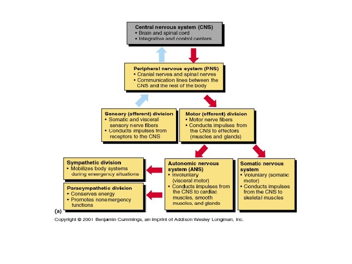
CNS and PNS Central Nervous System CNS CNS

 ¡CNS consists of brain and spinal cord")
 Cerebellum")


 Thalamus Posterior commissure Pineal gland")

 Optic chiasma Optic nerve")
, and temperature regulation. ¡Substantia")
 Medial lemniscus Red nucleus Substantia nigra Fibers")


 ¡Caudate nucleus ¡Putamen ¡Globus pallidus")


 Thalamus Posterior commissure Pineal gland")


 superficial layer of gray matter ¡ 40% mass")


 Location of the")


 cortex ¡ Extreme posterior tip of occipital lobe ¡")


 cortex ¡ Medial aspect of temporal lobes (in piriform")


 ¡ Sends impulses to")

 ¡Membranes (meninges) ¡Watery cushion (cerebrospinal fluid) ¡Blood brain")


- Slides: 70
CNS and PNS
Central Nervous System (CNS) ¡CNS consists of brain and spinal cord
Adult Brain Regions 1. 2. 3. 4. Brain stem (midbrain, pons, and medulla) Cerebellum Diencephalon Cerebral hemispheres
Cerebral hemisphere Diencephalon Cerebellum Brain stem • Midbrain • Pons • Medulla oblongata
Brain Stem ¡ Three regions ¡ Midbrain ¡ Pons ¡ Medulla oblongata
Corpus callosum Fornix Lateral ventricle (covered by septum pellucidum) Thalamus Posterior commissure Pineal gland Third ventricle Epithalamus Corpora quadrigemina Cerebral aqueduct Anterior commissure Hypothalamus Arbor vitae Fourth ventricle Optic chiasma Cerebellum Mammillary body Pons Medulla oblongata Midbrain
Brain Stem ¡Controls automatic behaviors necessary for survival ¡ Breathing ¡ Circulation ¡ Digestion ¡ Swallowing
Frontal lobe Olfactory bulb (synapse point of cranial nerve I) Optic chiasma Optic nerve (II) Optic tract Mammillary body Midbrain Pons Temporal lobe Medulla oblongata Cerebellum Spinal cord
Midbrain ¡Associated with vision, hearing, motor control, sleep/wake, arousal (alertness), and temperature regulation. ¡Substantia nigra—functionally linked to basal nuclei
Tectum Periaqueductal gray matter Oculomotor nucleus (III) Medial lemniscus Red nucleus Substantia nigra Fibers of pyramidal tract Dorsal Cerebral aqueduct Reticular formation Ventral Midbrain Superior colliculus Crus cerebri of cerebral peduncle
Pons ¡Fibers of pons ¡Connect higher brain centers and spinal cord ¡Nuclei that relay signals from the forebrain to the cerebellum, ¡along with nuclei that deal primarily with sleep, respiration, swallowing, bladder control, hearing, equilibrium, taste, eye movement, facial expressions, facial sensation, and posture.
Superior cerebellar peduncle Trigeminal main sensory nucleus Trigeminal motor nucleus Middle cerebellar peduncle Trigeminal nerve (V) Medial lemniscus Pons Fourth ventricle Reticular formation Pontine nuclei Fibers of pyramidal tract
Medulla Oblongata: Functions ¡Cardiovascular center ¡ Cardiac center adjusts force and rate of heart contraction ¡Respiratory centers ¡ Generate respiratory rhythm ¡ Control rate and depth of breathing (with pontine centers)
Medulla Oblongata ¡Additional centers regulate ¡ Vomiting ¡ Hiccuping ¡ Swallowing ¡ Coughing ¡ Sneezing
Cerebellum ¡Input from cortex, brain stem and sensory receptors ¡Allows smooth, coordinated movements ¡ Learn to ride a bicycle
Anterior lobe Arbor vitae Cerebellar cortex Pons Fourth ventricle Medulla oblongata Posterior lobe Flocculonodular lobe Choroid plexus
Anterior lobe Primary fissure Posterior lobe Horizontal fissure Vermis
Cerebellar Processing of Motor Activity ¡Cerebellum receives impulses from cerebral cortex of intent to initiate voluntary muscle contraction ¡Continuously "inform" cerebellum of body's position and momentum ¡Proprioception ¡Smoothly coordinate muscle contraction
Basal Nuclei (Ganglia) ¡Caudate nucleus ¡Putamen ¡Globus pallidus
Striatum Caudate nucleus Putamen Thalamus Tail of caudate nucleus
Anterior Cerebral cortex Cerebral white matter Corpus callosum Anterior horn of lateral ventricle Head of caudate nucleus Putamen Globus pallidus Thalamus Tail of caudate nucleus Third ventricle Inferior horn of lateral ventricle Posterior
Functions of Basal Nuclei ¡Functions thought to be ¡ Helps in balance
Diencephalon ¡ Thalamus ¡ Hypothalamus ¡ Epithalamus
Cerebral hemisphere Corpus callosum Fornix Choroid plexus Septum pellucidum Interthalamic adhesion (intermediate mass of thalamus) Thalamus (encloses third ventricle) Posterior commissure Pineal gland Interventricular foramen Anterior commissure Hypothalamus Optic chiasma Pituitary gland Epithalamus Corpora quadrigemina Midbrain Cerebral aqueduct Mammillary body Pons Medulla oblongata Spinal cord Arbor vitae (of cerebellum) Fourth ventricle Choroid plexus Cerebellum
Thalamic Function ¡“Router” to cerebral cortex ¡Sorts, edits, and relays ascending input
Hypothalamus ¡Underneath the Thalamus ¡Contains many nuclei
Paraventricular nucleus Anterior commissure Preoptic nucleus Anterior hypothalamic nucleus Supraoptic nucleus Suprachiasmatic nucleus Optic chiasma Infundibulum (stalk of the pituitary gland) Fornix Arcuate nucleus Pituitary gland The main hypothalamic nuclei. Dorsomedial nucleus Posterior hypothalamic nucleus Lateral hypothalamic area Ventromedial nucleus Mammillary body
Hypothalamic Function ¡Controls autonomic nervous system (e. g. , blood pressure, rate and force of heartbeat, digestive tract motility, pupil size) ¡Physical responses to emotions (limbic system) ¡ Perception of pleasure, fear, and rage, and in biological rhythms and drives
Hypothalamic Function ¡Regulates sleep-wake cycles ¡Controls endocrine system ¡ Controls secretions of anterior pituitary gland ¡ Produces posterior pituitary hormones
Hypothalamic Function ¡Regulates body temperature – sweating/shivering ¡Regulates hunger and satiety in response to nutrient blood levels or hormones ¡Regulates water balance and thirst
Cerebral hemisphere Corpus callosum Fornix Choroid plexus Septum pellucidum Interthalamic adhesion (intermediate mass of thalamus) Thalamus (encloses third ventricle) Posterior commissure Pineal gland Interventricular foramen Anterior commissure Hypothalamus Optic chiasma Pituitary gland Epithalamus Corpora quadrigemina Midbrain Cerebral aqueduct Mammillary body Pons Medulla oblongata Spinal cord Arbor vitae (of cerebellum) Fourth ventricle Choroid plexus Cerebellum
Epithalamus ¡Connect the limbic system to other parts of the brain ¡ Regulation of emotions ¡Pineal gland secretes melatonin ¡ Involved in circadian rhythms © 2013 Pearson Education, Inc.
Corpus callosum Fornix Lateral ventricle (covered by septum pellucidum) Thalamus Posterior commissure Pineal gland Third ventricle Epithalamus Corpora quadrigemina Cerebral aqueduct Anterior commissure Hypothalamus Arbor vitae Fourth ventricle Optic chiasma Cerebellum Mammillary body Pons Medulla oblongata Midbrain
© 2013 Pearson Education, Inc.
© 2013 Pearson Education, Inc.
Lateralization of Cortical Function ¡Left hemisphere ¡ Controls language, math, and logic ¡Right hemisphere ¡ Visual-spatial skills, intuition, emotion, and artistic and musical skills ¡Hemispheres communicate almost instantaneously via fiber tracts and functional integration
© 2013 Pearson Education, Inc.
Cerebral Hemispheres ¡Five lobes ¡ Frontal ¡ Parietal ¡ Temporal ¡ Occipital ¡ Insula
Cerebral Cortex ¡Thin (2– 4 mm) superficial layer of gray matter ¡ 40% mass of brain ¡Site of conscious mind: awareness, sensory perception, voluntary motor initiation, communication, memory storage, understanding
4 General Considerations of Cerebral Cortex 1. Three types of functional areas ¡ Motor areas—control voluntary movement ¡ Sensory areas—conscious awareness of sensation ¡ Association areas—integrate diverse information 2. Each hemisphere concerned with contralateral side of body
Cerebral Hemispheres ¡Central sulcus ¡ Separates precentral gyrus of frontal lobe and postcentral gyrus of parietal lobe
Precentral gyrus Frontal lobe Central sulcus Postcentral gyrus Parietal lobe Parieto-occipital sulcus (on medial surface of hemisphere) Lateral sulcus Fissure (a deep sulcus) Occipital lobe Temporal lobe Transverse cerebral fissure Cerebellum Pons Medulla oblongata Spinal cord Gyrus Cortex (gray matter) Sulcus White matter Lobes and sulci of the cerebrum
Primary Motor Cortex ¡Motor homunculi - upside-down caricatures represent contralateral motor innervation of body regions
Frontal lobe Central sulcus Gyri of insula Temporal lobe (pulled down) Location of the insula lobe
Posterior Eye b Toes Genitals Face Jaw Swallowing e Ey se No e c Fa s Lip h Teet s Gum Jaw Tongue Lips Tongue rs m b Sensory map in postcentral gyrus Th u um Nec k Bro w Knee Leg Hip Trunk Neck Head Ar Elb m o Fo w rea rm Ha nd Foot Knee s er Th Anterior Hip Trunk d n Ha ng Fi der Shoul Arm ow Elb t s Wri Motor map in precentral gyrus Sensory Fi ng e Motor Primary motor cortex (precentral gyrus) Primary somatosensory cortex (postcentral gyrus) Pharynx Intraabdominal
Primary Somatosensory Cortex ¡In postcentral gyri of parietal lobe ¡Receives general sensory information Somatosensory homunculus upside-down caricatures represent contralateral sensory input from body regions
Posterior rs Sensory map in postcentral gyrus Th Foot um b Fi ng e Neck Head Ar Elb m o Fo w rea rm Ha nd Knee Leg Hip Trunk Anterior Sensory Genitals e Ey se No e c Fa s Lip h Teet s m Gu Jaw Tongue Primary somatosensory cortex (postcentral gyrus) Pharynx Intraabdominal
Visual Areas ¡Primary visual (striate) cortex ¡ Extreme posterior tip of occipital lobe ¡ Receives visual information from retinas
Motor areas Central sulcus Primary motor cortex Premotor cortex Frontal eye field Broca's area (outlined by dashes) Sensory areas and related association areas Primary somatosensory cortex Somatic Somatosensory sensation association cortex Gustatory cortex (in insula) Prefrontal cortex Working memory for spatial tasks Executive area for task management Working memory for object-recall tasks Solving complex, multitask problems Wernicke's area (outlined by dashes) Primary visual cortex Visual association area Auditory association area Primary auditory cortex Lateral view, left cerebral hemisphere Primary motor cortex Taste Motor association cortex Primary sensory cortex Sensory association cortex Vision Hearing Multimodal association cortex
Auditory Areas ¡Primary auditory cortex ¡ Superior margin of temporal lobes ¡ Interprets information from inner ear as pitch, loudness, and location
Motor areas Central sulcus Primary motor cortex Premotor cortex Frontal eye field Broca's area (outlined by dashes) Sensory areas and related association areas Primary somatosensory cortex Somatic Somatosensory sensation association cortex Gustatory cortex (in insula) Prefrontal cortex Working memory for spatial tasks Executive area for task management Working memory for object-recall tasks Solving complex, multitask problems Wernicke's area (outlined by dashes) Primary visual cortex Visual association area Auditory association area Primary auditory cortex Lateral view, left cerebral hemisphere Primary motor cortex Taste Motor association cortex Primary sensory cortex Sensory association cortex Vision Hearing Multimodal association cortex
OIfactory Cortex ¡Primary olfactory (smell) cortex ¡ Medial aspect of temporal lobes (in piriform lobes)
Motor areas Central sulcus Primary motor cortex Premotor cortex Frontal eye field Broca's area (outlined by dashes) Sensory areas and related association areas Primary somatosensory cortex Somatic Somatosensory sensation association cortex Gustatory cortex (in insula) Prefrontal cortex Working memory for spatial tasks Executive area for task management Working memory for object-recall tasks Solving complex, multitask problems Wernicke's area (outlined by dashes) Primary visual cortex Visual association area Auditory association area Primary auditory cortex Lateral view, left cerebral hemisphere Primary motor cortex Taste Motor association cortex Primary sensory cortex Sensory association cortex Vision Hearing Multimodal association cortex
Gustatory Cortex ¡In insula just deep to temporal lobe ¡Involved in perception of taste
Premotor cortex Cingulate Primary gyrus motor cortex Corpus callosum Central sulcus Primary somatosensory cortex Frontal eye field Parietal lobe Somatosensory association cortex Parieto-occipital sulcus Prefrontal cortex Occipital lobe Processes emotions related to personal and social interactions Orbitofrontal cortex Visual association area Olfactory bulb Olfactory tract Fornix Temporal Primary lobe olfactory cortex Parasagittal view, right cerebral hemisphere Primary motor cortex Motor association cortex Primary sensory cortex Uncus Calcarine sulcus Parahippocampal gyrus Sensory association cortex Primary visual cortex Multimodal association cortex
Limbic System: Emotion and Cognition ¡React emotionally to things we consciously understand to be happening ¡Consciously aware of emotional richness in our lives ¡Hippocampus and amygdaloid body—play a role in memory
Septum pellucidum Diencephalic structures of the limbic system Anterior thalamic nuclei (flanking 3 rd ventricle) Hypothalamus Corpus callosum Fiber tracts connecting limbic system structures Fornix Anterior commissure Cerebral structures of the limbic system Cingulate gyrus Septal nuclei Amygdaloid body Mammillary body Olfactory bulb Hippocampus • Dentate gyrus • Parahippocampal gyrus
Reticular Formation: RAS and Motor Function ¡Reticular activating system (RAS) ¡ Sends impulses to cerebral cortex to keep it conscious and alert ¡ Filters out repetitive, familiar, or weak stimuli (~99% of all stimuli!) ¡ ADHD ¡ Inhibited by sleep centers, alcohol, drugs ¡ Severe injury results in permanent unconsciousness (coma)
Radiations to cerebral cortex Visual impulses Auditory impulses Reticular formation Ascending general sensory tracts (touch, pain, temperature) Descending motor projections to spinal cord
Protection of the Brain ¡Bone (skull) ¡Membranes (meninges) ¡Watery cushion (cerebrospinal fluid) ¡Blood brain barrier
Meninges ¡Cover and protect CNS ¡Protect blood vessels and enclose venous sinuses ¡Contains cerebrospinal fluid (CSF) ¡Forms partitions in skull
Meninges ¡Three layers ¡ Dura mater ¡ Arachnoid mater ¡ Pia mater ¡Meningitis ¡ Inflammation of meninges
Skin of scalp Periosteum Superior sagittal sinus Subdural space Subarachnoid space Bone of skull Dura mater • Periosteal layer • Meningeal layer Arachnoid mater Pia mater Arachnoid villus Blood vessel Falx cerebri (in longitudinal fissure only)
Dura Mater ¡ Strongest layer
Arachnoid Mater ¡Middle layer with weblike extensions ¡Separated from dura mater by subdural space ¡Subarachnoid space contains CSF and largest blood vessels of brain ¡Arachnoid villi protrude into superior sagittal sinus and permit CSF reabsorption
© 2013 Pearson Education, Inc.
Skin of scalp Periosteum Superior sagittal sinus Subdural space Subarachnoid space Bone of skull Dura mater • Periosteal layer • Meningeal layer Arachnoid mater Pia mater Arachnoid villus Blood vessel Falx cerebri (in longitudinal fissure only)
Pia Mater ¡Delicate vascularized connective tissue that clings tightly to brain
© 2013 Pearson Education, Inc.