CHAPTER 4 Microscopy Staining and Classification MICROSCOPY AND


 Electron gun Magnetic lenses Primary electrons Secondary")


- Slides: 43
CHAPTER 4 Microscopy, Staining, and Classification
MICROSCOPY AND STAINING Familiarize yourself with the microscope again in lab ANIMATION Microscopy and Staining: Overview
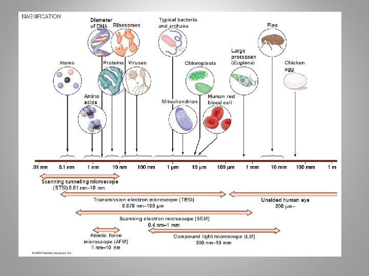
TABLE 4. 1 METRIC UNITS OF LENGTH
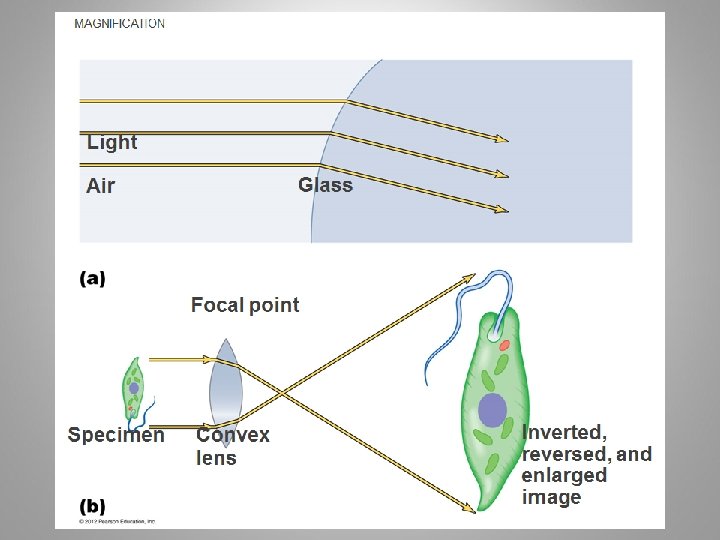
• General Principles of Microscopy – Wavelength of radiation • How well 2 wavelengths can be discriminated against is how enhanced the image will be – Magnification • Results when a beam of radiation is refracts (bent) • Degree of magnification – thickness of lens, curvature, the speed of light through substance – Resolution • Ability to distinguish objects that are close together • Blue filter allows shorter wavelengths to pass which increases resolution – Contrast • Intensity between an object and its background © 2012 Pearson Education Inc.
• General Principles of Microscopy – Contrast • Differences in intensity between two objects, or between an object and background • Important in determining resolution • Staining increases contrast • Use of light that is in phase increases contrast © 2012 Pearson Education Inc.
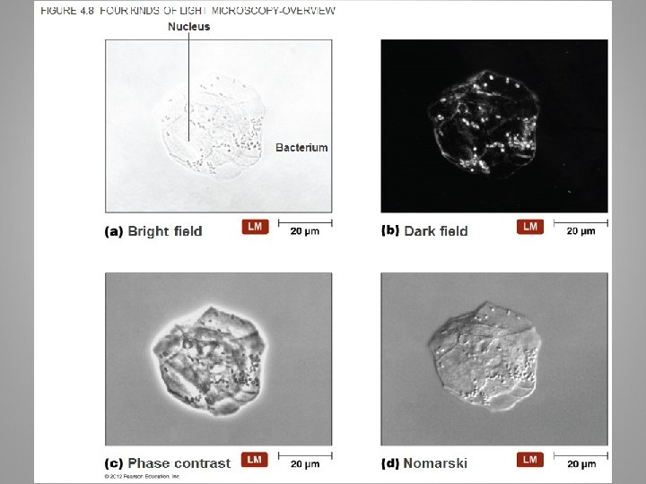
• Light Microscopy – Bright-field microscopes • Simple – Contain a single magnifying lens – Similar to magnifying glass – Leeuwenhoek used simple microscope to observe microorganisms © 2012 Pearson Education Inc.
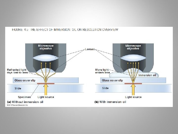
• Light Microscopy – Bright-field microscopes • Compound – Series of lenses for magnification – Light passes through specimen into objective lens – Oil immersion lens increases resolution – Have one or two ocular lenses – Total magnification (objective lens X ocular lens) – Most have condenser lens (direct light through specimen) © 2012 Pearson Education Inc.
• Light Microscopy – Dark-field microscopes • Best for observing pale objects • Only light rays scattered by specimen enter objective lens • Specimen appears light against dark background • Increases contrast and enables observation of more details © 2012 Pearson Education Inc.
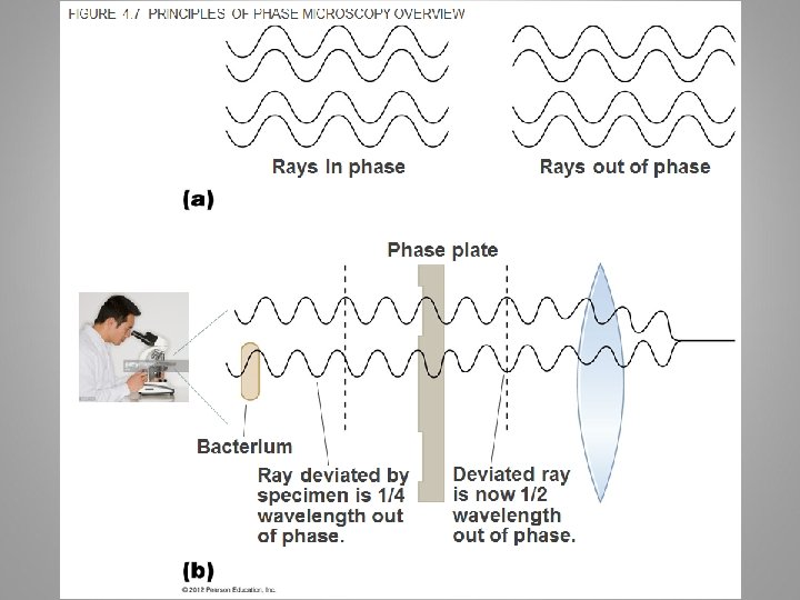
• Light Microscopy – Phase microscopes • Examine living organisms or specimens that would be damaged/altered by attaching them to slides or staining • Contrast is created because light waves are out of phase and produce darker image of specimen • Two types – Phase-contrast microscope – Differential interference contrast microscope © 2012 Pearson Education Inc.
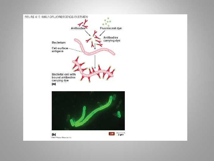
• Light Microscopy – Fluorescent microscopes • Direct UV light source at specimen • Specimen radiates energy back as a visible wavelength • UV light increases resolution and contrast • Some cells are naturally fluorescent; others must be stained • Used in immunofluorescence to identify pathogens and to make visible a variety of proteins © 2012 Pearson Education Inc.
• Light Microscopy – Confocal microscopes • Use fluorescent dyes • Use UV lasers to illuminate fluorescent chemicals in a single plane • Resolution increased because light passes through pinhole aperture • Computer constructs 3 -D image from digitized images © 2012 Pearson Education Inc.
• Electron Microscopy – Light microscopes cannot resolve structures closer than 200 nm – Greater resolving power and magnification – Magnifies objects 10, 000 X to 100, 000 X – Detailed view of bacteria, viruses, ultrastructure, and large atoms – Two types • Transmission electron microscopes • Scanning electron microscopes © 2012 Pearson Education Inc.
FIGURE 4. 12 SCANNING ELECTRON MICROSCOPE (SEM) Electron gun Magnetic lenses Primary electrons Secondary electrons Specimen holder Vacuum system Beam deflector coil Scanning circuit Photomultiplier Detector Monitor
FIGURE 4. 13 SEM IMAGES-OVERVIEW
• Probe Microscopy – recent advance – Magnifies more than 100, 000 X – Two types • Scanning tunneling microscopes – Passes metallic probe (that is sharpened to end in a single atom) back and forth ABOVE surface of specimen • Atomic force microscopes – Goes back and forth lightly on surface – Magnify surfaces of bacteria, viruses, proteins, etc © 2012 Pearson Education Inc.
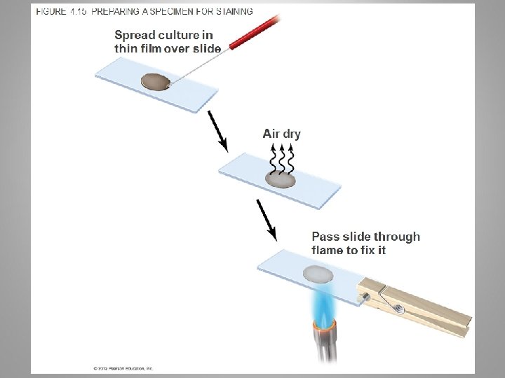
• Principles of Staining • Staining increases contrast and resolution by coloring specimens with stains/dyes • Smear of microorganisms (thin film) made prior to staining • Microbiological stains • Usually salts, positive cation and negative anion • One of these will be colored – chromophore • Anionic dyes - acidic dyes that stain alkaline structures • Cationic dyes - Basic dyes that stain acidic structures • Most ofen used – positively charged so they will attach to negatively charged cells © 2012 Pearson Education Inc.
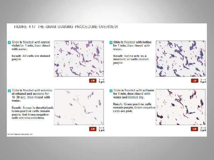
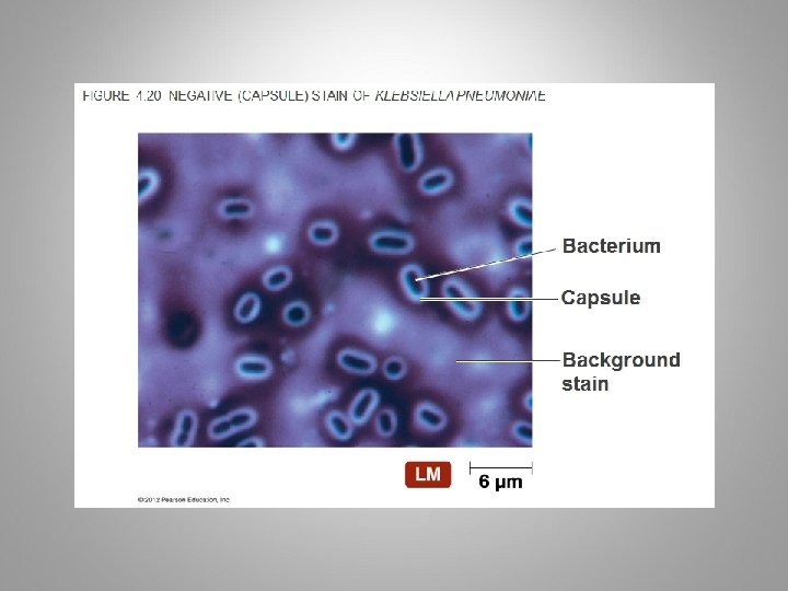
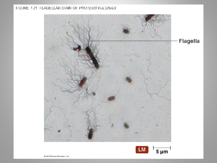
• • • Simple Stains – Involve soaking the specimen in one stain Differential Stains – Gram stain – Gram positive and Gram negative cells – Acid-fast stain – Stains genera Mycobacterium and Nocardia (waxy lipid in cell walls) – Endospore stain – Endospore wall are impermeable to many chemicals, stain uses heat to drive the staining agent into endospore – Histological stain – Used to visualize things like fungi and cancer cells in tissue samples Special Stains – Negative (capsule) stain • Acidic dye – repelled by negative charges on surface of cell – Flagellar stain – Increase diameter of flagella to be seen © 2012 Pearson Education Inc.
FIGURE 4. 16 SIMPLE STAINS-OVERVIEW
FIGURE 4. 18 THE ZIEHL-NEELSEN ACID-FAST STAIN
FIGURE 4. 19 SCHAEFFER-FULTON ENDOSPORE STAIN OF BACILLUS ANTHRACIS
• Differential Stains – Histological stain • Two popular stains for histological specimens – Gomori methenamine silver (GMS) » Presence of fungi and carbs in tissues – Hematoxylin and eosin (HE) » Presence of cancer cells © 2012 Pearson Education Inc.
• Staining for Electron Microscopy – Transmission electron microscopy uses chemicals containing heavy metals • Absorb electrons – Stains may bind molecules in specimens or the background © 2012 Pearson Education Inc.
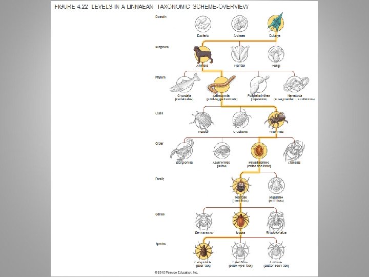
CLASSIFICATION AND IDENTIFICATION OF MICROORGANISMS – Taxonomy consists of classification, nomenclature, and identification – Organize large amounts of information about organisms – Make predictions based on knowledge of similar organisms • Linnaeus and Taxonomic Categories – Linnaeus • Classified organisms based on characteristics in common • Organisms that can successfully interbreed called species • Used binomial nomenclature in his system – Linnaeus proposed only two kingdoms – Later taxonomic approach based on five kingdoms » Animalia, Plantae, Fungi, Protista, and Prokaryotae © 2012 Pearson Education Inc.
CLASSIFICATION AND IDENTIFICATION OF MICROORGANISMS • Linnaeus and Taxonomic Categories – Linnaeus’s goal was to classify organisms to catalogue them – Modern goal is to understand relationships among groups of organisms • Reflect phylogenetic hierarchy • Emphasis on comparison of organisms’ genetic material – Led to proposal to add domain © 2012 Pearson Education Inc.
CLASSIFICATION AND IDENTIFICATION OF MICROORGANISMS • Domains – Carl Woese compared nucleotide sequences of r. RNA subunits – Proposal of three domains as determined by ribosomal nucleotide sequences • Eukarya, Bacteria, and Archaea – Cells in the three domains differ by other characteristics © 2012 Pearson Education Inc.
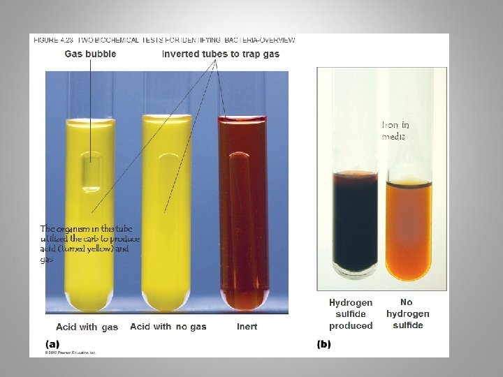
CLASSIFICATION AND IDENTIFICATION OF MICROORGANISMS • Taxonomic and Identifying Characteristics – Physical characteristics – Biochemical tests – Serological tests – Phage typing – Analysis of nucleic acids © 2012 Pearson Education Inc.
FIGURE 4. 24 ONE TOOL FOR THE RAPID IDENTIFICATION OF BACTERIA, THE AUTOMATED MICROSCAN SYSTEM EACH WELL CONTAINS DIFFERENT BIOCHEMICAL TEST Wells
FIGURE 4. 25 AN AGGLUTINATION TEST, ONE TYPE OF SEROLOGICAL TESTOVERVIEW Negative result Positive result
FIGURE 4. 26 PHAGE TYPING SPECIFIC BACTERIOPHAGES WILL ONLY INFECT CERTAIN BACTERIA – SO IF YOU KNOW THE PHAGE YOU INNOCULATE WITH, THEN YOU CAN DETERMINE SPECIES OF BACTERIA Bacterial lawn Plaques
• Taxonomic Keys – Dichotomous keys • Series of paired statements where only one of two “either/or” choices applies to any particular organism – Key directs user to another pair of statements, or provides name of organism © 2012 Pearson Education Inc.