microscopy There are three wellknown branches of microscopy

microscopy There are three well-known branches of microscopy: optical, electron, and scanning probe microscopy.
 Diffraction limits resolution to approximately 0.")
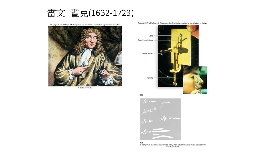
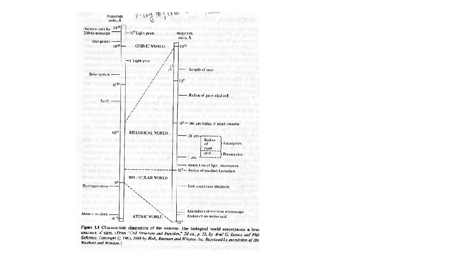
Optical microscopy standard optical microscopy (bright field microscopy) Diffraction limits resolution to approximately 0. 2 micrometres. This limits the practical magnification limit to ~1500 x.

Optical microscopy • Phase contrast is a widely used technique that shows differences in refractive index as difference in contrast. It was developed by the Dutch physicist Frits Zernike in the 1930 s (for which he was awarded the Nobel Prize in 1953).

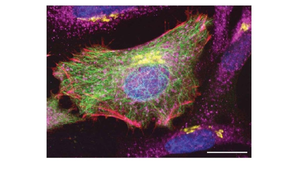
Optical microscopy • When certain compounds are illuminated with high energy light, they emit light of a lower frequency. This effect is known as fluorescence.

Electron microscopy • An electron microscope is a microscope that uses a beam of accelerated electrons as a source of illumination. Because the wavelength of an electron can be up to 100, 000 times shorter than that of visible light photons, the electron microscope has a higher resolving power than a light microscope and can reveal the structure of smaller objects.

Transmission electron microscope • The original form of electron microscope, the transmission electron microscope (TEM) uses a high voltage electron beam to create an image.
 has allowed the production of images")
• the high-resolution transmission electron microscopy (HRTEM) has allowed the production of images with resolution below 50 picometres.
 an image of an ant • the electron beam of")
Scanning electron microscope (SEM) an image of an ant • the electron beam of the SEM does not at any time carry a complete image of the specimen. When the electron beam interacts with the specimen, it loses energy by a variety of mechanisms which provide signals carrying information about the properties of the specimen surface. • The image resolution: an order of magnitude poorer than that of a TEM. • to image bulk samples • a great depth of field images the three-dimensional shape of the sample.
- Slides: 12