
FUNDAMENTAL MICROBIOLOGY By Debashish Gardia M Pharm Pharmacology
FUNDAMENTAL MICROBIOLOGY By Debashish Gardia M. Pharm (Pharmacology)
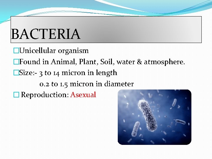
BACTERIA �Unicellular organism �Found in Animal, Plant, Soil, water & atmosphere. �Size: - 3 to 14 micron in length 0. 2 to 1. 5 micron in diameter � Reproduction: Asexual
CLASSIFICATION [Based on shape of bacteria] �COCCI: Spherical bodies with a smooth outline �Size: 0. 75 mm-3. 0 mm Based on the characteristic arrangement �Coccus: Cells are arranged singly �Diplococci: Cells are arranged in pairs �Tetracocci: The cells are arranged in group of four �Streptococci: Cells are arranged in Chains of diff. Length �Staphylococci: cells are arranged in clusters like a bunch of grapes

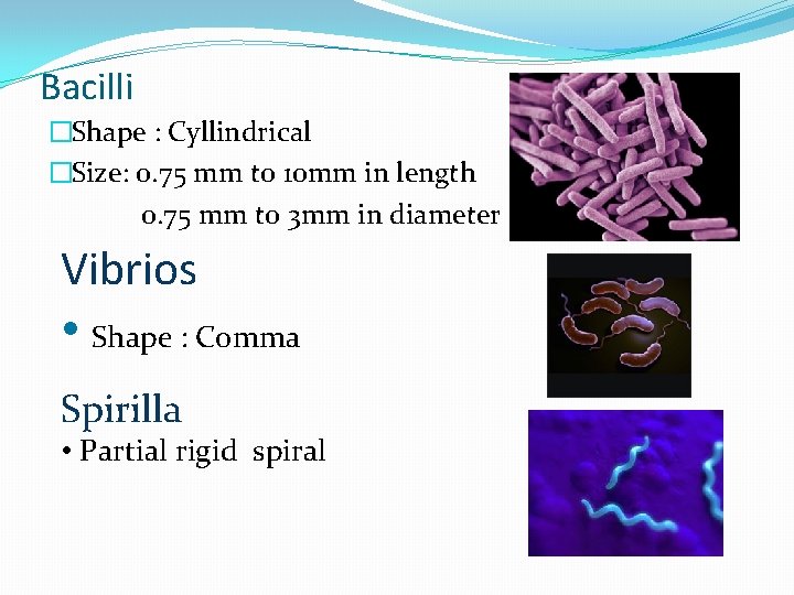
Bacilli �Shape : Cyllindrical �Size: 0. 75 mm to 10 mm in length 0. 75 mm to 3 mm in diameter Vibrios • Shape : Comma Spirilla • Partial rigid spiral
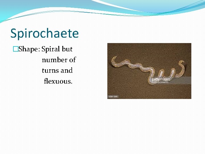
Spirochaete �Shape: Spiral but number of turns and flexuous.

Bacterial Cell
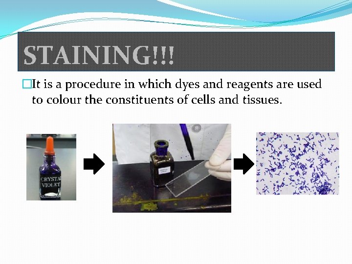
STAINING!!! �It is a procedure in which dyes and reagents are used to colour the constituents of cells and tissues.
STAINING METHOD 1) Simple Staining (Crystal Violet, Methylene blue, fuschin, safranin) 2) Negative staining (Nigrosin, Indian Ink) 3) Differential staining (Crystal Violet, Methyl Violet, Gentian Violet) I. II. Gram Staining Acid Fast Staining 4) Fluorescence staining (Auramine O, Acridine orange )
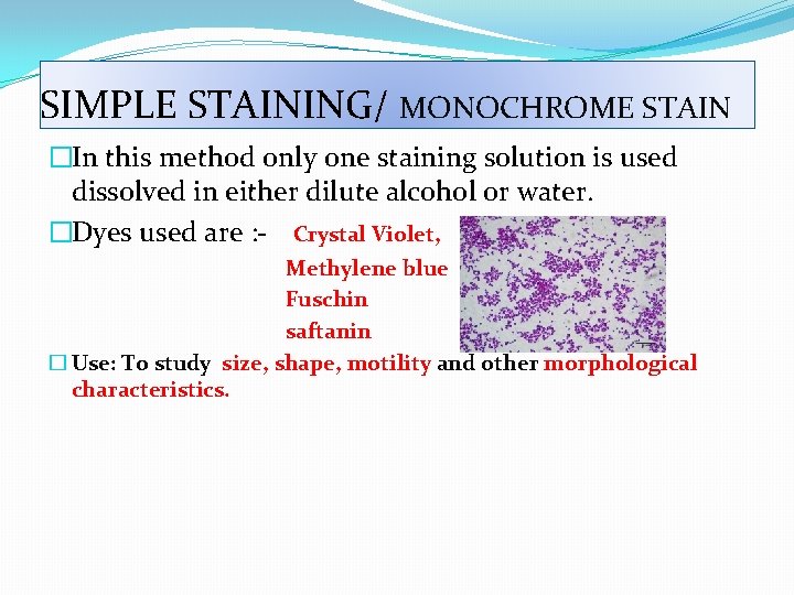
SIMPLE STAINING/ MONOCHROME STAIN �In this method only one staining solution is used dissolved in either dilute alcohol or water. �Dyes used are : - Crystal Violet, Methylene blue Fuschin saftanin � Use: To study size, shape, motility and other morphological characteristics.
Differential Staining �More than one dye is used �Mostly used against all types of bacteria. �Use: To study Bacterial Cells, Spores and capsules �Types: �Gram Staining � Gram + � Gram – �Acid Fast Staining
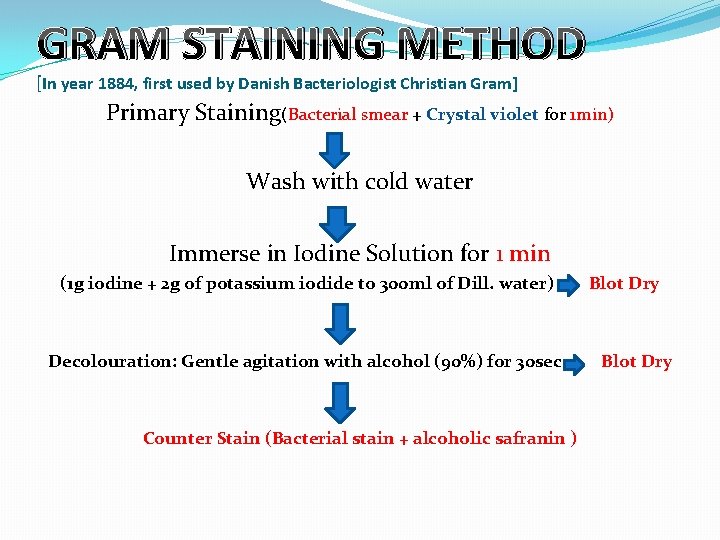
GRAM STAINING METHOD [In year 1884, first used by Danish Bacteriologist Christian Gram] Primary Staining(Bacterial smear + Crystal violet for 1 min) Wash with cold water Immerse in Iodine Solution for 1 min (1 g iodine + 2 g of potassium iodide to 300 ml of Dill. water) Decolouration: Gentle agitation with alcohol (90%) for 30 sec Counter Stain (Bacterial stain + alcoholic safranin ) Blot Dry
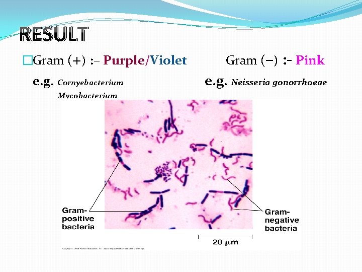
RESULT �Gram (+) : – Purple/Violet e. g. Cornyebacterium Mycobacterium Gram (–) : - Pink e. g. Neisseria gonorrhoeae
FAQs �What exactly happens to the gram +ve bacterium that they turn blue but gram –ve bacterium remain pink? �How staining technique is useful in maintenance of health of human being?
- Slides: 14