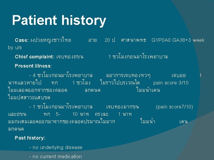
Case study 21 Facilitator Pawin Puapornpong Patient history







- Slides: 49
Case study 21 Facilitator: Pawin Puapornpong
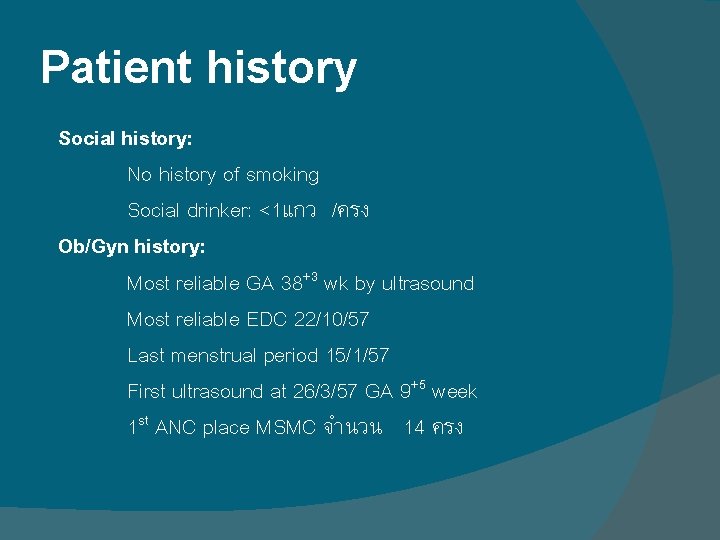
Patient history Social history: No history of smoking Social drinker: <1แกว /ครง Ob/Gyn history: Most reliable GA 38+3 wk by ultrasound Most reliable EDC 22/10/57 Last menstrual period 15/1/57 First ultrasound at 26/3/57 GA 9+5 week 1 st ANC place MSMC จำนวน 14 ครง
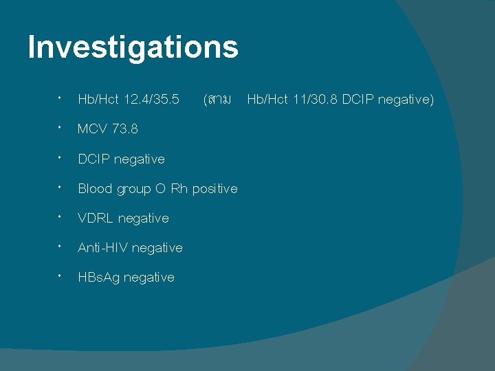
Investigations Hb/Hct 12. 4/35. 5 (สาม Hb/Hct 11/30. 8 DCIP negative) MCV 73. 8 DCIP negative Blood group O Rh positive VDRL negative Anti-HIV negative HBs. Ag negative
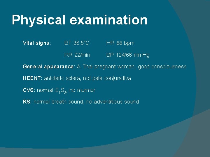
Physical examination Vital signs: BT 36. 5˚C HR 88 bpm RR 22/min BP 124/66 mm. Hg General appearance: A Thai pregnant woman, good consciousness HEENT: anicteric sclera, not pale conjunctiva CVS: normal S 1 S 2, no murmur RS: normal breath sound, no adventitious sound
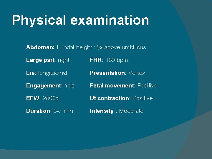
Physical examination Abdomen: Fundal height : ¾ above umbilicus Large part: right FHR: 150 bpm Lie: longitudinal Presentation: Vertex Engagement: Yes Fetal movement: Positive EFW: 2800 g Ut contraction: Positive Duration: 5 -7 min Intensity : Moderate
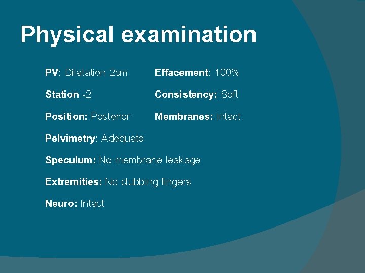
Physical examination PV: Dilatation 2 cm Effacement: 100% Station -2 Consistency: Soft Position: Posterior Membranes: Intact Pelvimetry: Adequate Speculum: No membrane leakage Extremities: No clubbing fingers Neuro: Intact
ANTENATAL CARE SCREENING FOR HIGH RISK PREGNANCY
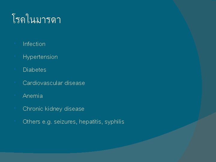
โรคในมารดา Infection Hypertension Diabetes Cardiovascular disease Anemia Chronic kidney disease Others e. g. seizures, hepatitis, syphilis
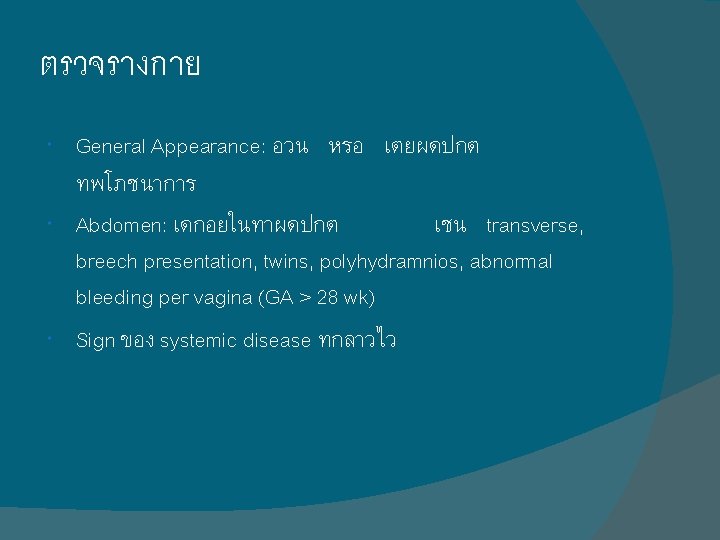
ตรวจรางกาย General Appearance: อวน หรอ เตยผดปกต ทพโภชนาการ Abdomen: เดกอยในทาผดปกต เชน transverse, breech presentation, twins, polyhydramnios, abnormal bleeding per vagina (GA > 28 wk) Sign ของ systemic disease ทกลาวไว
การตรวจทางหองปฏบตการ HIV (voluntary screening) VDRL Persistent proteinuria Persistent glucosuria Hb < 10 g/d. L, Hct < 30% Group B Streptococci
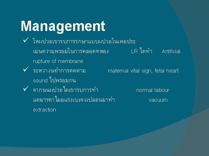
Patient history Impression: G 1 P 0 A 0 GA 38+3 by U/S with labour pain Management: - Artificial rupture of membranes - vaginal delivery
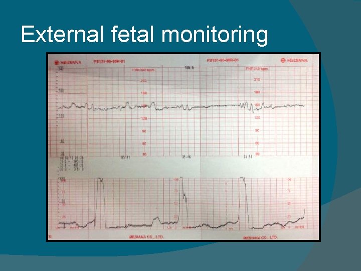
External fetal monitoring
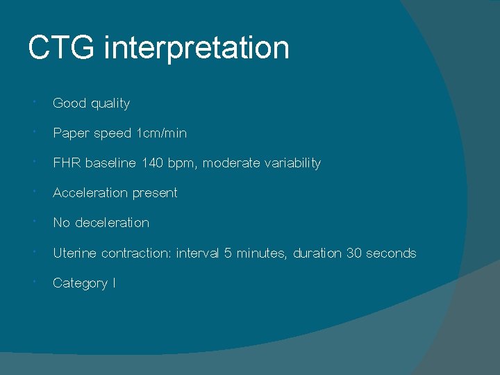
CTG interpretation Good quality Paper speed 1 cm/min FHR baseline 140 bpm, moderate variability Acceleration present No deceleration Uterine contraction: interval 5 minutes, duration 30 seconds Category I
ARTIFICIAL RUPTURE OF MEMBRANES
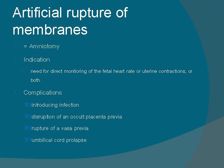
Artificial rupture of membranes = Amniotomy Indication need for direct monitoring of the fetal heart rate or uterine contractions, or both Complications introducing infection disruption of an occult placenta previa rupture of a vasa previa umbilical cord prolapse
Artificial rupture of membranes Elective Amniotomy Membrane rupture with the intention of accelerating labor amniotomy at about 5 cm dilation accelerated spontaneous labor by 1 to 2 hours Amniotomy Induction The main disadvantage of amniotomy used alone for labor induction is the unpredictable and occasionally long interval to labor onset early amniotomy at 1 to 2 cm associated with significant 4 -hour shorter labor Amniotomy Augmentation amniotomy with oxytocin augmentation for arrested active-phase labor shortened the time to delivery by 44 minutes compared with that of oxytocin alone.
VACUUM EXTRACTION
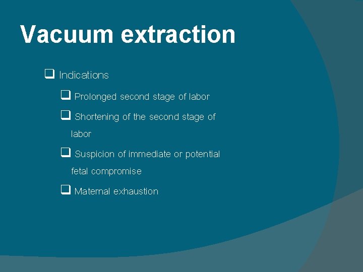
Vacuum extraction q Indications q Prolonged second stage of labor q Shortening of the second stage of labor q Suspicion of immediate or potential fetal compromise q Maternal exhaustion
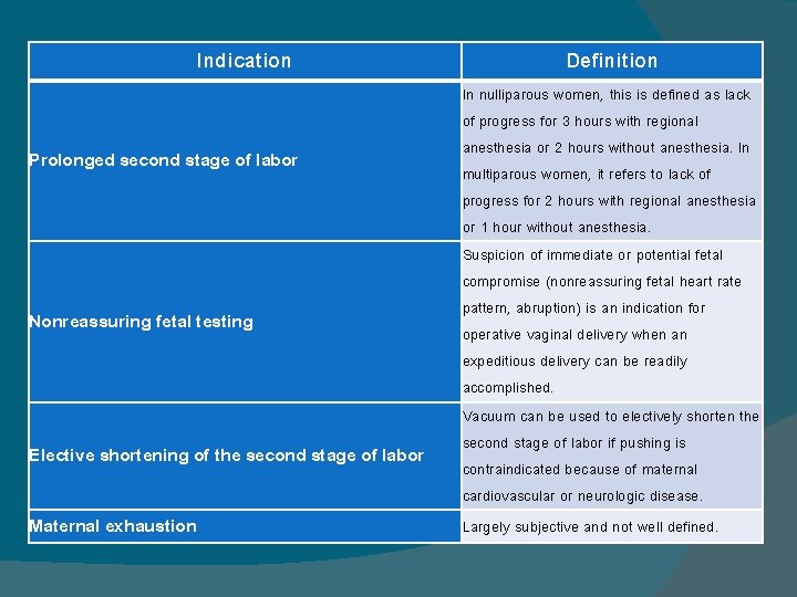
Indication Prolonged second stage of labor Nonreassuring fetal testing Elective shortening of the second stage of labor Maternal exhaustion Definition In nulliparous women, this is defined as lack of progress for 3 hours with regional anesthesia or 2 hours without anesthesia. In multiparous women, it refers to lack of progress for 2 hours with regional anesthesia or 1 hour without anesthesia. Suspicion of immediate or potential fetal compromise (nonreassuring fetal heart rate pattern, abruption) is an indication for operative vaginal delivery when an expeditious delivery can be readily accomplished. Vacuum can be used to electively shorten the second stage of labor if pushing is contraindicated because of maternal cardiovascular or neurologic disease. Largely subjective and not well defined.
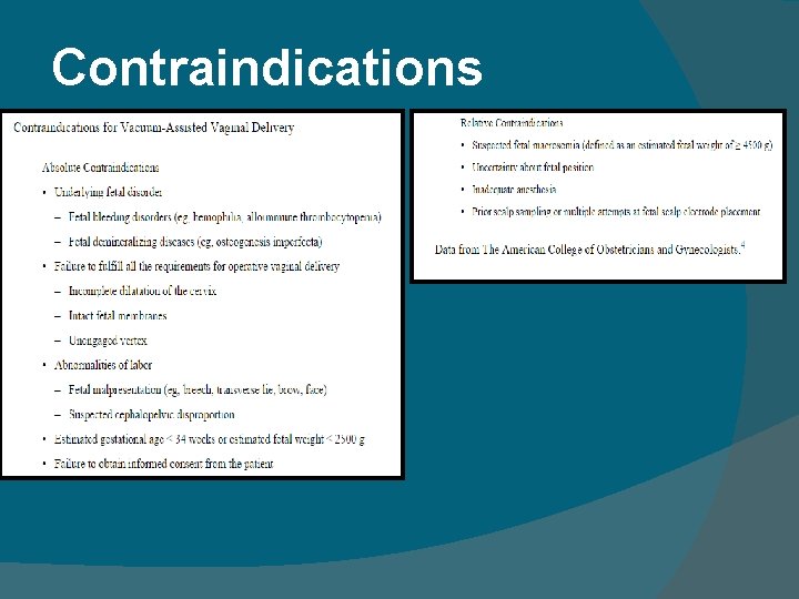
Contraindications
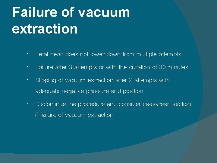
Failure of vacuum extraction Fetal head does not lower down from multiple attempts Failure after 3 attempts or with the duration of 30 minutes Slipping of vacuum extraction after 2 attempts with adequate negative pressure and position Discontinue the procedure and consider caesarean section if failure of vacuum extraction
Maternal complications Perineal pain at delivery and post partum Perineal lacerations Hematomas Blood loss and anemia Urinary retention Long-term urinary and fecal incontinence
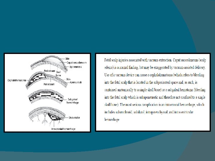
Neonatal complications Scalp lacerations Cephalhematoma Subgaleal hematoma Intracranial hemorrhage Facial nerve palsies Retinal hemorrhage
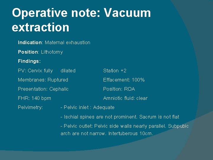
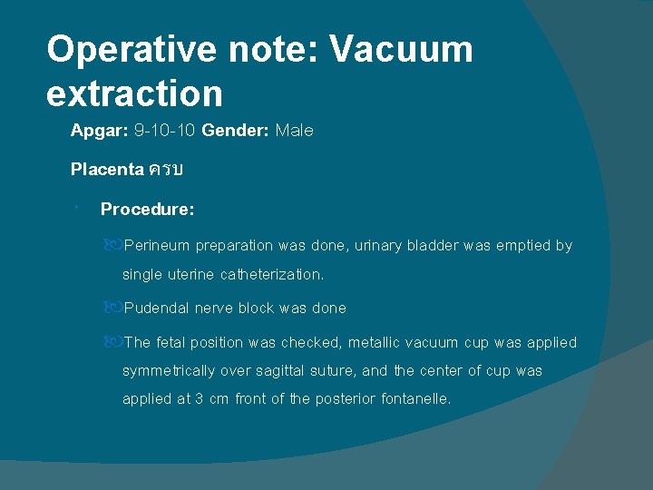
Operative note: Vacuum extraction Indication: Maternal exhaustion Position: Lithotomy Findings: PV: Cervix fully dilated Station +2 Membranes: Ruptured Effacement: 100% Presentation: Cephalic Position: ROA FHR: 140 bpm Amniotic fluid: clear Pelvimetry: - Pelvic inlet : Adequate - Ischial spines are not prominent. Sacrum is not flat - Pelvic outlet: Pelvic side walls nearly parallel. Subpubic arch are not narrow. Intertuberous 10 cm.
Operative note: Vacuum extraction Apgar: 9 -10 -10 Gender: Male Placenta ครบ Procedure: Perineum preparation was done, urinary bladder was emptied by single uterine catheterization. Pudendal nerve block was done The fetal position was checked, metallic vacuum cup was applied symmetrically over sagittal suture, and the center of cup was applied at 3 cm front of the posterior fontanelle.
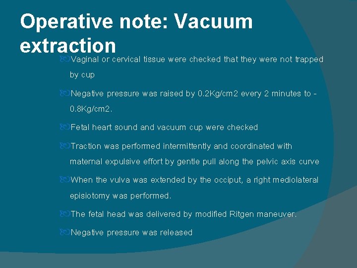
Operative note: Vacuum extraction Vaginal or cervical tissue were checked that they were not trapped by cup Negative pressure was raised by 0. 2 Kg/cm 2 every 2 minutes to 0. 8 Kg/cm 2. Fetal heart sound and vacuum cup were checked Traction was performed intermittently and coordinated with maternal expulsive effort by gentle pull along the pelvic axis curve When the vulva was extended by the occiput, a right mediolateral episiotomy was performed. The fetal head was delivered by modified Ritgen maneuver. Negative pressure was released
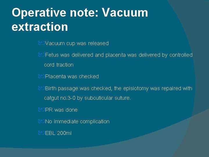
Operative note: Vacuum extraction Vacuum cup was released Fetus was delivered and placenta was delivered by controlled cord traction Placenta was checked Birth passage was checked, the episiotomy was repaired with catgut no. 3 -0 by subcuticular suture. PR was done No immediate complication EBL 200 ml
Management - Record vital signs, uterine contraction q 15 minutes until stable for 2 hours - 5%DN/2 (1, 000) IV + synto 20 unit IV 120 ml/hr - Observe bleeding per vagina - Observe voiding - Methergin 0. 2 mg IV - Regular diet - Paracetamol 500 mg for pain control - Ferrous fumerate 200 mg to prevent anemia - Amoxycillin 500 mg
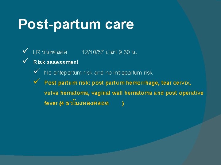
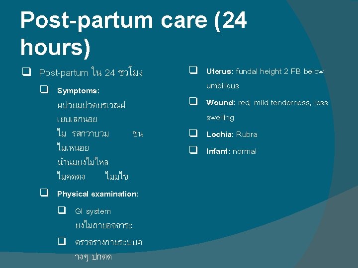
Post-partum care ü LR วนทคลอด 12/10/57 เวลา 9. 30 น. ü Risk assessment ü No antepartum risk and no intrapartum risk ü Post partum risk: post partum hemorrhage, tear cervix, vulva hematoma, vaginal wall hematoma and post operative fever (4 ชวโมงหลงคลอด )
Management ü observe ท LR ประเมนแลวพบม cervix tear และ hematoma ดงกลาว จงไดทำ explore vagina (12/10/57 18. 00น. )
EXPLORE VAGINA
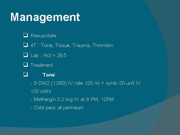
Management Resuscitate q 4 T : Tone, Tissue, Trauma, Thrombin q Lab : Hct = 26. 5 q Treatment q ‘Tone’ - 5 DN/2 (1, 000) IV rate 120 ml + synto 20 unit IV 120 ml/hr - Methergin 0. 2 mg IV at 8 PM, 12 PM - Cold pack at perineum q
If…medical failure Tamponade Uterine artery/ Ovarian vessels ligation B-Lynch sutures Abdominal packing
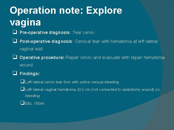
Operation note: Explore vagina Pre-operative diagnosis: Tear cervix q Post-operative diagnosis: Cervical tear with hematoma at left lateral vaginal wall q Operative procedure: Repair cervix and evacuate with repair hematoma wound q Findings: q q Left lateral cervix tear 6 cm with active venous bleeding q Left lateral vaginal hematoma 2 X 2 cm (not connected to episiotomy wound) no bleeding q EBL 150 ml
Operation note: Explore vagina Procedure: Insert AP retractor Sponge forcep was grabbed around cervix to identify site of cervical tear Vicryl 2 -0 was sutured by continuous lock at cervical tear Incision at hematoma at left lateral vaginal wall Blood clot from hematoma was evacuated Suture stopped bleeding at hematoma wound Bleeding was detected and stop Off AP retractor
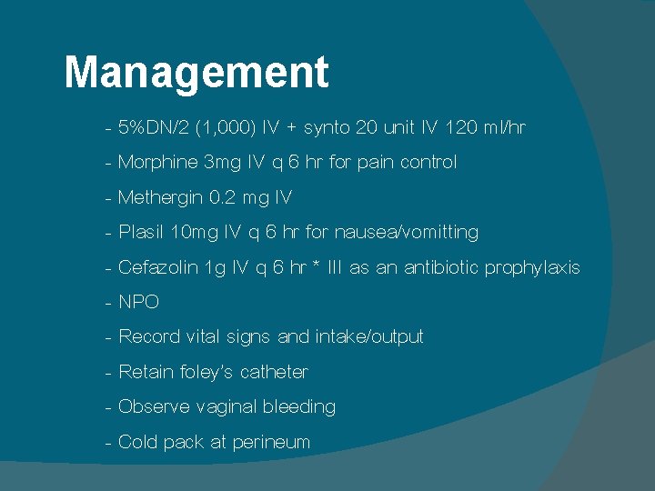
Management - 5%DN/2 (1, 000) IV + synto 20 unit IV 120 ml/hr - Morphine 3 mg IV q 6 hr for pain control - Methergin 0. 2 mg IV - Plasil 10 mg IV q 6 hr for nausea/vomitting - Cefazolin 1 g IV q 6 hr * III as an antibiotic prophylaxis - NPO - Record vital signs and intake/output - Retain foley’s catheter - Observe vaginal bleeding - Cold pack at perineum
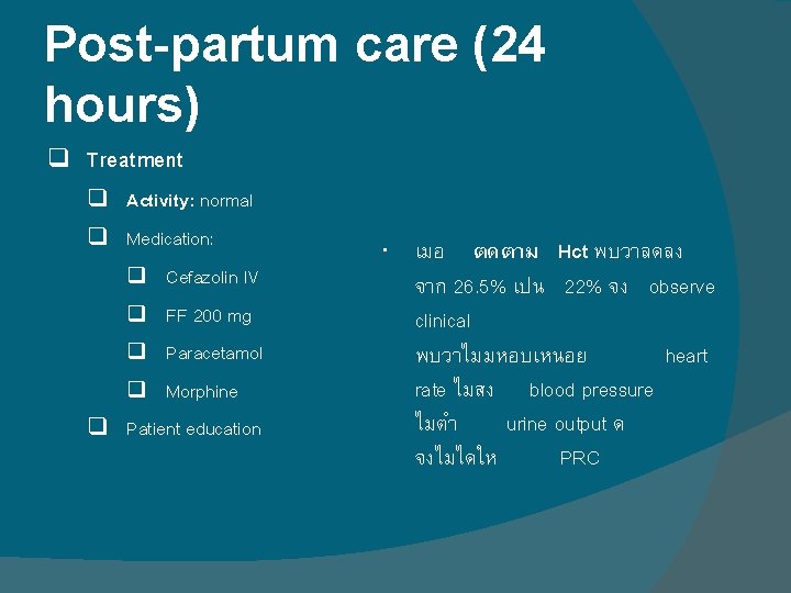
Post-partum care (24 hours) q Treatment q Activity: normal q Medication: q Cefazolin IV q FF 200 mg q Paracetamol q Morphine q Patient education เมอ ตดตาม Hct พบวาลดลง จาก 26. 5% เปน 22% จง observe clinical พบวาไมมหอบเหนอย heart rate ไมสง blood pressure ไมตำ urine output ด จงไมไดให PRC
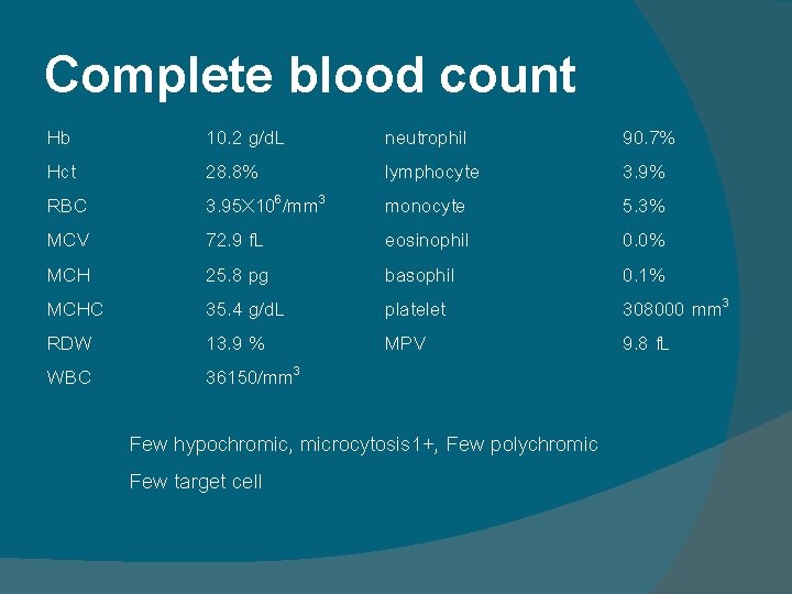
Complete blood count Hb Hct RBC MCV MCHC RDW WBC 10. 2 g/d. L 28. 8% 3. 95 X 106/mm 3 72. 9 f. L 25. 8 pg 35. 4 g/d. L 13. 9 % 36150/mm 3 neutrophil lymphocyte monocyte eosinophil basophil platelet MPV Few hypochromic, microcytosis 1+, Few polychromic Few target cell 90. 7% 3. 9% 5. 3% 0. 0% 0. 1% 308000 mm 3 9. 8 f. L
Thank you