Cardiovascular System Heart General Physiology Tony Serino Ph


 Semilunar valves closed Semilunar valves opened AV valves closed Systole:")


 node and")


")
")


")
 = End Diastolic Volume – End Systolic")

")


- Slides: 36
Cardiovascular System: Heart General Physiology Tony Serino, Ph. D. Biology Department Misericordia University
Heart as a Dual Pump • Cardiac muscle arranged as whorls that squeeze the blood • Twin pumps: systemic and pulmonary • Four chambers: 2 atria and 2 ventricles
Cardiac Muscle Cells
Cardiac Muscle Depolarization
Depolarization and Tension
Fig. 12. 17 Allows for Ca++ plateau
Heart: Internal Anatomy
Cardiac Cycle (mechanical action) Semilunar valves closed Semilunar valves opened AV valves closed Systole: Period of isovolumic contraction. Systole: Period of ejection. Systole: Period of isovolumic contraction. Ventricular contraction causes the AV valves to close, which is the beginning of ventricular systole. The semilunar valves were closed in the previous diastole and remain closed during this period.
Semilunar valves opened AV valves closed Systole: Period of ejection. Continued ventricular contraction pushes blood out of the ventricles, causing the semilunar valves to open. Semilunar valves closed AV valves closed Diastole: Period of isovolumic relaxation.
Semilunar valves closed AV valves closed Semilunar valves closed AV valves opened Diastole: Passive ventricular filling. Diastole: Period of isovolumic relaxation. Blood flowing back toward the relaxed ventricles causes the semilunar valves to close, which is the beginning of ventricular diastole. Note that the AV valves closed, also.
Diastole: Passive ventricular filling. The AV Semilunar valves closed valves open and blood flows into the relaxed ventricles, accounting for most of the ventricular filling. AV valves opened Diastole: Active ventricular filling. Semilunar valves closed AV valves opened Diastole: Passive ventricular filling.
Semilunar valves closed AV valves closed Semilunar valves closed AV valves opened Diastole: Active ventricular filling. Systole: Period of isovolumic contraction. Diastole: Active ventricular filling. The atria contract and complete ventricular filling.
Conduction System of Heart 1. Action potentials originate in the sinoatrial (SA) node and Sinoatrial travel across the wall of the (SA) node atrium (arrows) from the SA node to the atrioventricular Atrioventricular (AV) node 2. Action potentials pass through the AV node and along the atrioventricular (AV) bundle, which extends from the AV node, through the fibrous skeleton, into the interventricular septum. Atrioventricular (AV) bundle 3. The AV bundle divides into right and left bundle branches, and action potentials descend to the apex of each ventricle along the bundle branches. Left and Right bundle 4. Action potentials are carried branches by the Purkinje fibers from Purkinje the bundle branches to the fibers ventricular walls. Left atrium 1 2 3 Left ventricle 4 Apex
Pacemaker Potential
Control of Pacemaker
ECG Correlates Electrical Events
Normal ECG
ECG Leads Einthoven’s Triangle
ECG Normal Sinus Rhythm Junctional Rhythm (AV node rhythm)
Second Degree Heart Block Ventricular Fibrillation (V-fib)
Fig. 12. 16
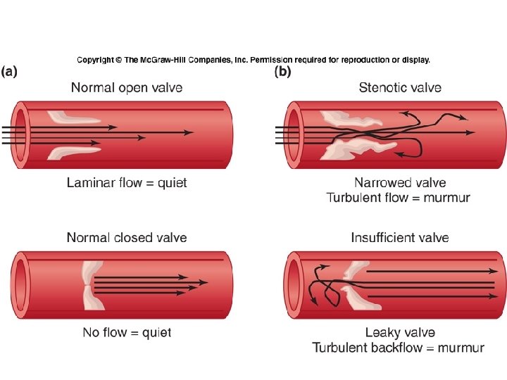
Heart Sounds • “Lub-dub” • Sound associated with valve closing producing turbulent blood flow
Cardiac Cycle
(ml/min)
Factors Affecting SV • Stroke Volume (SV) = End Diastolic Volume – End Systolic Volume • SV = EDV – ESV (ml/beat) • EDV affected by: – Venous return which is dependent on venous tone, skeletal muscle pumps, etc. • ESV – As the heart fills it is stretched which allows for better overlap of the contractile proteins which will affect the force of contraction and the ESV (Starling’s Law of the Heart) – Increasing the force of contraction at any EDV will decrease the ESV and increase the SV (sympathetic stimulation and epinephrine)
EDV is affected by Venous Return
At any EDV, increased force of contraction will increase SV (↓ESV)
As the heart fills, the muscle is stretched allowing for better overlap of contractile proteins which increases force of contraction (↓ESV)
Sympathetic Stimulation • Leads to increase HR • Increases in Ca++ release from SR, increase Ca++ through membrane and increase myosin crossbridge cycling • Increases force of contraction
Heart Rate Control • Sinus Rhythm = normal SA node control • Autonomic Activity – Sympathetic = accelerator (tachycardia) – Parasympathetic = brake (bradycardia) • Hormones – epinephrine • Drugs -caffeine, nicotine, atropine, etc.
Summary ↑ stretch ↑ Contraction Strength
CV Effects during Exercise
Exercise Benefits • Heart’s maximums are increased • Effort is decreased
Fig. 12. 07