Cardiovascular Physiology 12022022 1 Cardiovascular system CVS CVS

 CVS consists of the heart and a series of blood vessels")


 & 2 ventricles (lower)")

; blood goes")

 The right heart pump: Ø receives deoxygenated blood from all")
 The left heart pump: Ø receives oxygenated blood from the")


- Slides: 30
Cardiovascular Physiology 12/02/2022 1
Cardiovascular system (CVS) CVS consists of the heart and a series of blood vessels (arteries, veins and capillaries). 12/02/2022 2
Heart Anatomy • The heart is a strong muscular pump that contracts and relaxes all life. • It is the size of the fist of the hand. • It is formed of 4 chambers: Two thin walled low pressure reservoirs, the atria and two thick walled pumping chambers, the ventricles. 12/02/2022 3
12/02/2022 4
Introduction Ø A functional cardiovascular system is vital for supplying oxygen and nutrients to tissues and removing wastes from them. ØThe heart is the strongest muscle in the body ØThe heart must pump blood throughout the body day & night ØThe heart is 2 pumps working side by side. On your right side is the heart that pumps blood to your lungs where it picks up O 2. On your left side is the heart that pumps this O 2 -soaked blood out to your body. ØHeart pumps 200, 000000 L blood in a lifetime ØBoth pumps are divided into 2 spaces called chambers so your heart is actually a 4 -chambered pumper ØThe 2 sides do not work independently; they are precisely timed.
What Does C-V System do? • Circulate blood throughout entire body for – Transport of oxygen to cells – Transport of CO 2 away from cells – Transport of nutrients (glucose) to cells – Movement of immune system components (cells, antibodies) – Transport of endocrine gland secretions – Generating blood pressure
How does it do it? • Heart is pump • Arteries and veins are main tubes (plumbing) – Arteries Away from Heart – Veins to Heart • Diffusion happens in capillaries (oxygen, CO 2, glucose diffuse in or out of blood)
MAIN FUNCTIONS OF THE CIRCULATORY SYSTEM • Transport and distribute essential substances to the tissues. • Remove metabolic byproducts. • Adjustment of oxygen and nutrient supply in different physiologic states. • Regulation of body temperature. • Humoral communication.
Cardiovascular system
The Blood Vessels • The cardiovascular system has three types of blood vessels: • Arteries (and arterioles), Capillaries, Veins (and venules) • The Arteries • Arteries and arterioles take blood away from the heart. • The largest artery is the aorta. • The middle layer of an artery wall consists of smooth muscle that can constrict to regulate blood flow and blood pressure. • Arterioles can constrict or dilate, changing blood pressure
• Capillaries have walls only one cell thick to allow exchange of gases and nutrients with tissue fluid. • Capillary beds are present in all regions of the body but not all capillary beds are open at the same time. • Contraction of a sphincter muscle closes off a bed and blood can flow through an arteriovenous shunt that bypasses the capillary bed. • Veins • Venules drain blood from capillaries, then join to form veins that take blood to the heart. • Veins have much less smooth muscle and connective tissue than arteries. • Veins often have valves that prevent the backward flow of blood when closed. • Veins carry about 70% of the body’s blood and act as a reservoir during hemorrhage.
Figure 15 -3: Metarterioles
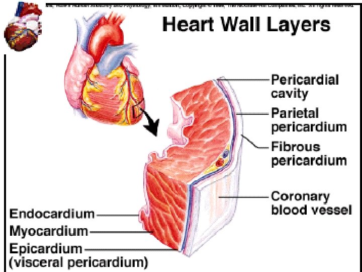
Heart Wall Epicardium: thin serous membrane of the outer surface of the heart. Also called the visceral pericardium Myocardium: thick middle layer composed of cardiac muscle. Endocardium: simple squamous epithelium over a layer of connective tissue, continuous with all blood vessels of the body. 12/02/2022 13
The heart is located near the center of the thoracic cavity between the lungs and is contained in the pericardial sac. The pericardial sac supports the heart and contains some fluid for lubrication.
Location of Heart in Thorax pg 501
Heart Valves • Atrioventricular – Tricuspid – Bicuspid or mitral • Semilunar – Aortic – Pulmonary • Prevent blood from flowing back 20 -17
The. heart is divided into 4 hollow chambers-2 atria (upper) & 2 ventricles (lower) Right chambers & valves: 1) right atrium Receives blood from 2 large veins called the superior vena cava & the inferior vena cava; coronary sinus also drains blood into the right atrium from the myocardium 1) Tricuspid valve (3 cusps) guards the atrioventricular orifice between the right atrium & the right ventricle; it permits blood to move from the right atrium into the right ventricle & doesn’t allow it to move in the opposite direction;
Chambers of the heart q The atria are smaller with thin walls, while the ventricles are larger with thick walls (much stronger) • The left ventricle has thicker wall than the right because it needs to pump blood to the whole body. 19
Right ventricle pumps blood a short distance to the pulmonary trunk (lungs); blood goes to pulmonary trunk which divides to form the left & right pulmonary arteries (deoxygenated blood) 1) Pulmonary valve (3 cusps) – guards the base of the pulmonary trunk; opens as the right ventricle contracts Left chambers & valves: 1) left atrium receives blood from the lungs through 4 pulmonary veins – 2 from right & 2 from left lungs 2) The blood passes from the left atrium into the left ventricle through the atrioventricular orifice; bicuspid or mitral valve guards the left atrioventricular orifice; it prevents blood from flowing back into the left atrium from the ventricle when the ventricle contracts 3) The left ventricle pumps blood by way of the aorta (large artery) into systemic circulation an aortic valve guards the base of the aorta
The Heart: Pumps 1) The right heart pump: Ø receives deoxygenated blood from all parts of the body except the lungs (through superior & inferior vena cava) Ø pumps deoxygenated blood to the lungs Pulmonary circulation 22
The Heart: Pumps 2) The left heart pump: Ø receives oxygenated blood from the lungs (through pulmonary veins) Ø pumps oxygenated blood to the rest of the body Systemic circulation 23
Blood Flow Through Heart 20 -25
The double pump 26
Passage of Blood Through the Heart • Blood follows this sequence through the heart: superior and inferior vena cava → right atrium → tricuspid valve → right ventricle → pulmonary semilunar valve → pulmonary trunk and arteries to the lungs → pulmonary veins leaving the lungs → left atrium → bicuspid valve → left ventricle → aortic semilunar valve → aorta → to the body.
The Heartbeat ØEach heartbeat is called a cardiac cycle. ØWhen the heart beats, the two atria contract together, then the two ventricles contract; then the whole heart relaxes. ØSystole: - is the contraction of heart chambers; ØDiastole: - is the relaxation of heart chambers ØThe heart sounds: - Lub-dup, Ø Lub: - is due to the closing of the atrioventricular valves, ØDup: - is due to closing of the semilunar valves.
The Heart: Systole/Diastole Systole It is the contraction of the ventricles. • When the ventricles contract they force the blood into the great arteries. Ø From the left ventricle into the aorta Ø From the right ventricle into the pulmonary artery. • The increased pressure that result due to the contraction of the ventricles is called systolic pressure. 29
The Heart: Systole/Diastole It is the relaxation of the ventricles. q When the ventricles relax they receive the blood from the atria. q The decreased pressure due to the relaxation of the ventricles is called diastolic pressure. 30

 Anatomy and physiology unit 7 cardiovascular system
 Capillary bed labeled
 What makes up the cardiovascular system
 Pithed rat model
 Totally tubular dude
 Circulatory system crash course
 Cengage chapter 5
 Figure 11-9 is a diagram of the hepatic portal circulation
 Figure 11-8 arteries
 Lesson 11 cardiovascular system
 Lesson 11 cardiovascular system
 Cardiorespiratory system includes
 Chapter 11 the cardiovascular system
 Introduction of heart
 Stent placement
 Cardiovascular system diseases and disorders chapter 8
 Chapter 13 cardiovascular system
 Chapter 11 the cardiovascular system figure 11-2
 The cardiovascular system includes the
 Blood vesel
 Chapter 19 the cardiovascular system blood vessels
 Concurrent version system
 Trachea
 Human.respiratory.system
 Appendicular skeleton pectoral girdle
 Riesgo cardiovascular por perimetro abdominal
 Azoulay maniobra
 Rias cardiovascular
 National cardiovascular partners
 Cardiovascular drift
 Chapter 46 the child with a cardiovascular alteration


