Avascular Necrosis Outline Definition Pathophysiology Aetiology Presentation Imaging



- Slides: 32
Avascular Necrosis
Outline �Definition �Pathophysiology �Aetiology �Presentation �Imaging �Staging �Management
Definition �Cellular death of bone components secondary to interruption of blood supply �Consequent collapse of bone components �Pain, loss of function of joints �Proximal epiphysis of femur most commonly affected
Pathophysiology �Interruption of blood flow to bone �Affect bones with single terminal blood supply: �Talus �Carpals, tarsals �Proximal humerus �Proximal femur �Femoral condyles �Bone marrow, medullary bone and cortical bone necrosis results �Final pathway from multiple causes
Predisposing factors �Distance from vascular territory of bone �Enclosed by cartilage limiting vascularity �Endarterioles supply trabelcular bones
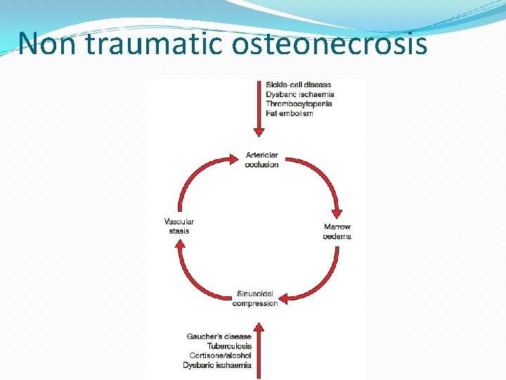
Pathways to necrosis �Vascular occlusion – direct trauma, stress fracture, SCD, venous stasis �Intravascular coagulation – hypercoaguable states �Primary cell death – alcohol, steroids, transplant patients
�Bone necrosis after 12 – 48 hrs of anoxia �Reactive new bone formation around necrotic bone �Granulation tissue over necrosed bone – sclerosis �Structural failure – subchondral fracture 1 st �Segmental collapse dependant on stress and area of necrosis
Aetiology � Trauma � Steroids � Alcohol abuse � CT diseases eg SLE � Hematologic (sickle cell disease, hemoglobinopathies, thrombophilia) � Metabolic (hyperlipidemia, gout, renal failure) � Orthopedic disorders (slipped capital femoral epiphysis, developmental dysplasia of the hip, Legg-Calve-Perthes disease) � Infection (osteomyelitis, HIV]) � Renal transplantation � Radiation therapy � Gaucher disease � Malignancy (marrow infiltration, malignant fibrous histiocytoma) � Caisson disease � Pregnancy � Bisphonate use
Trauma �Severance of blood supply – displaced femoral neck fractures �Scaphoid and talus – proximal osteonecrosis due to distal origin of vessels �Osteoarticular impact – localised osteonecrosis in convex surfaces (osteochondroses)
Non traumatic osteonecrosis
Presentation - History �Trauma �Corticosteroid use �Alcohol intake �Medical conditions – malignancy, thrombophilia, SLE, SCD �Pain – progressive, severity correlates with size of infarct �Deformity and stiffness – later stages
Presentation - examination �Limp �Antalgic gait �Restricted ROM �Tenderness around bone �Joint deformity �Muscle wasting
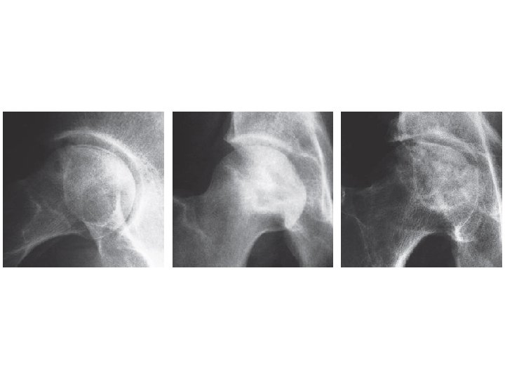
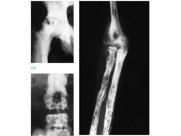
Imaging: X ray �Initially normal upto 3 months �Sclerosis �Flattening �Subchondral radiolucent lines (cresent sign) �Collapse of cortex �OA
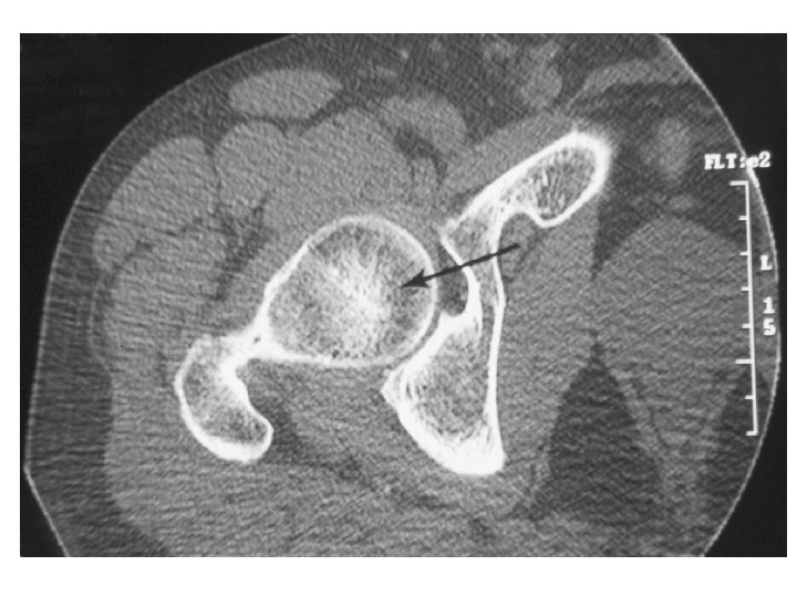
Imaging: CT scan �Used to assess extent of disease and calcification �Clearly shows articular deformity �Calcification and bone collapse �Central sclerosis in femoral head produces asterix sign
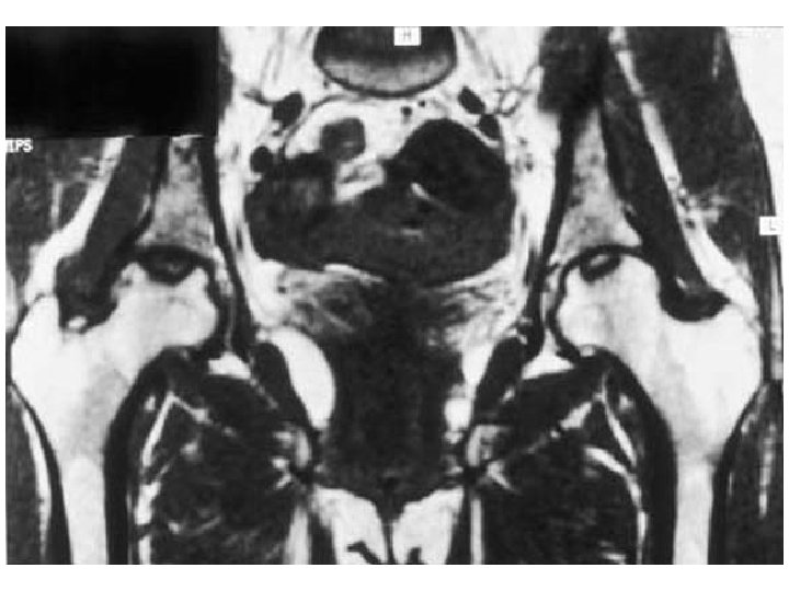
Imaging: MRI � 90% sensitive �Reduced subchondral intensity on T 1 representing boundary between necrotic and reactive bone �Low signal on T 1 and high signal on T 2 – reactive zone (diagnostic) �Changes detected early
Radionuclide scan �Donut sign – central reduced uptake with surrounding rim of increased uptake �More sensitive than plain films in early AVN �Less sensitive than MRI �Necrotic zone surrounded by reactive new bone formation
Histology �Definitive diagnosis �Usually retrospective/confirmatory during surgery for treatment �Occasionally biopsy of sclerotic lesion �Necrosis of cortical bone is followed by a regenerative process in surrounding tissues. �Increased osteoclastic activity to remove necrotic bone and increased osteoblastic activity as a reparative process
Intramedullary pressures �Cannula into metaphysis �Measure at rest and after saline injection �Femoral head: � 10 – 20 mm. Hg, increasing by 15 mm. Hg after saline �Markedly increased values in AVN (3 to 4 fold) �Less marked increase in OA
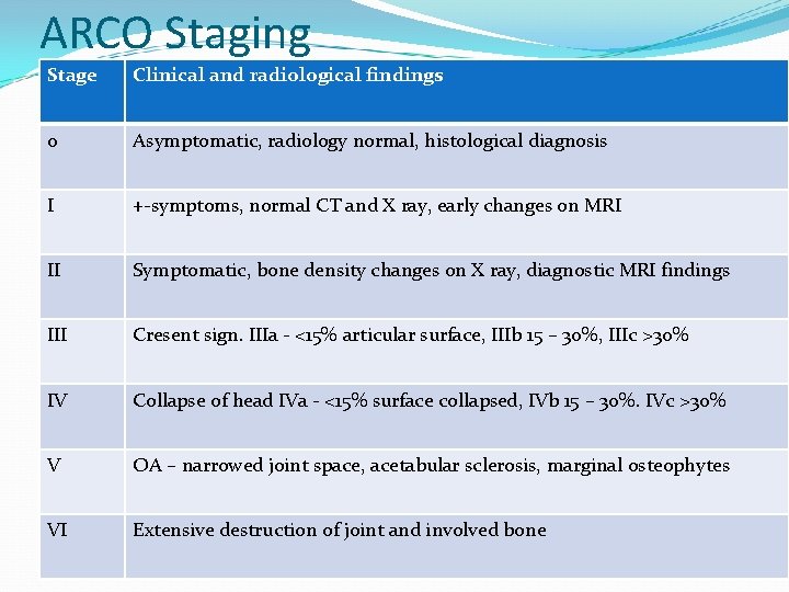
ARCO Staging Stage Clinical and radiological findings 0 Asymptomatic, radiology normal, histological diagnosis I +-symptoms, normal CT and X ray, early changes on MRI II Symptomatic, bone density changes on X ray, diagnostic MRI findings III Cresent sign. IIIa - <15% articular surface, IIIb 15 – 30%, IIIc >30% IV Collapse of head IVa - <15% surface collapsed, IVb 15 – 30%. IVc >30% V OA – narrowed joint space, acetabular sclerosis, marginal osteophytes VI Extensive destruction of joint and involved bone
Management principles �Early stages (I & II): �Bisphonates prevent collapse �Unloading osteotomies �Medullary decompression + bone grafting �Intermediate stage (III & IV): �Realignment osteototmies, decompression �Arthrodesis �Late stage (V & VI): �Analgesia, activity modification �Arthrodesis �Arthroplasties
Management - conservative �Offloading affected joints with use of crutches �Immobilisation �Analgesia �Bisphonates to delay femoral head collapse �Statins in patients on high dose corticosteroids – reduced lipid deposition
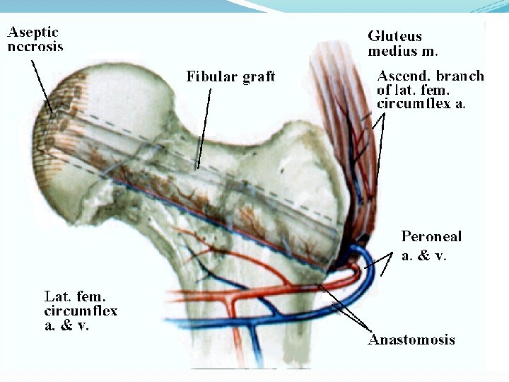
Core decompression �Indicated in ARCO I and II � 8 – 10 mm anterolateral core of bone �Filled with bone graft (vascularised/non vascularised) �Decompresses medullary cavity, reduces pain �Cortical (osteoconductive) or cancellous(osteoinductive) bone graft �Vascularised graft may reverse necrosis
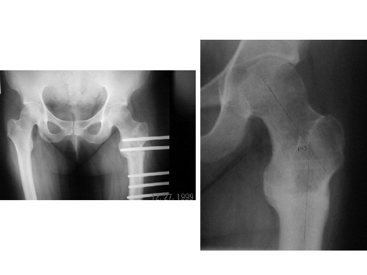
Realignment osteotomy �Indicated in ARCO III & IV �Used to relocate necrotic area from weight bearing portion of femoral head �Angular osteotomies more common �Multiple techniques for holding the fixation �Sugano intertrochanteric rotational osteotomy technically demanding but higher success rate
Arthroplasty �Indicated in ARCO IV onwards �Main aim is pain reduction �Young patients will need revision �Higher failure rates than in OA �Hemi arthroplasty an option
Eponymous syndromes �Kienbock’s disease – idiopathic avascular necrosis of the lunate bone that leads to collapse and progressive carpal arthritis. PRC as treatment �Legg-Calve-Perthes’s – idiopathic osteonecrosis of femoral capital epiphysis in children. Treated with orthotics, traction, surgery to rotate the femoral head �Preiser's disease – idiopathic osteonecrosis of scaphoid. Collapse with progressive arthritis. PRC, Excision and fusion,
THANK YOU