Annual Congress of National Cancer Institute Cairo University


- Slides: 55
Annual Congress of National Cancer Institute – Cairo University Oncoplastic Techniques For Partial Breast Reconstruction Cristiano Boneti, MD, FACS Plastic Surgery and Oncology Cancer Institute of America, Los Angeles
Disclosures • Consulting Agreement - Air. Xpanders • Consulting Agreement - Allergan
Delayed or Secondary Partial Breast Reconstruction • Defining The Challenge: Radiation Changes
Radiation Therapy • Alone Or In Combination With Other Treatments Responsible For Up To 40% Of Cancer Cures • 70% Of All Cancer Patients Receive Radiation Therapy • In The USA, Approx. 20% Of Population Has Or Will Receive Radiotherapy
Operative Risks After XRT • Complications After Radiation Therapy Occur In Up To 60 % Of Surgical Patients Marks JE, Freeman RB, Lee F, Ogura JH: Pharyngeal wall cancer: an analysis of treatment results complications and patterns of failure. Int J Radiat Oncol Biol Phys 1978, 4: 587 -593. 1016/0360 -3016(78)90179 -7 Tang Y, Shen Q, Wang Y, Lu K, Peng Y: A randomized prospective study of rehabilitation therapy in the treatment of radiation-induced dysphagia and trismus. Strahlenther Onkol 2011, 187: 39 -44. Melan'in VD, Rybak RF, Senkevich VM: Prophylaxis of the postop complications in combined treatment of patients with laryngeal cancer. Vestn Otorinolaringol 1998, 46: 8. Girod DA, Mc. Culloch TM, Tsue TT, Weymuller EA Jr: Risk factors for complications in clean-contaminated head and neck surgical procedures. Head Neck 1995, 17: 7– 13.
Radiation Therapy • Damage Caused At 3 Time Frames: • Physical • Chemical • Biological
Physical Stage • Involves “Ionization” where ionizing radiation is absorbed by tissues causing removal of an electron from atoms • Leads to break down of molecules in tissue • Extremely fast process (10^-12 sec) • Cellular to clinical level • Atom Molecule Tissue Organ Whole body
Chemical Stage • Ionization of water produces “free radicals” • Our bodies contain up to 80% water • Reaction that causes MAJORITY of damage in the human body • Reacts with DNA causing breaking of its structure interferes with cellular metabolism & reproduction
Biological Stage • The stage we see in clinical setting • This stage revealed when cells attempt to replicate • Fate of irradiated cells: • Immediate death (unusual) • DNA damage (eventual death vs mutation) • No effect
Biological Effects • Effect on cells depends on cell division • Cells that don’t divide are RESISTANT to radiation damage • Our body’s rapidly dividing cells MOST susceptible • • Blood (ie. platelets & WBC’s) Gastrointestinal Skin Central nervous system • LEAST sensitive tissues • Collagen, bone, muscle
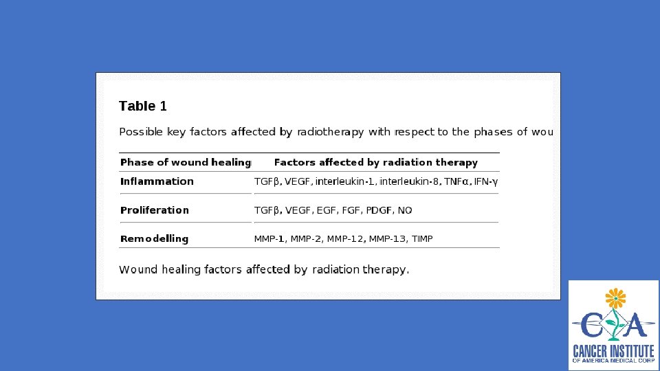
Effects on Wound Healing • Phases of wound healing • Complex process of cells & cytokines • Ordered sequence • RT disrupts this sequence “vicious cycle” inflammation & cell regeneration • Factors include: • • Time-dose-volume relationship Type Energy Part of body irradiated
Frank Haubner, Elisabeth Ohmann, Fabian Pohl, Jürgen Strutz and Holger G Gassner. Wound healing after radiation therapy: Review of the literature Radiation Oncology 20127: 162
Effects on Wound Healing • Reactions usually more severe at appositional skin sites: • Axilla, IMF, groin, perineum • Also involves tissue quality, patient age, medical comorbidities & nutritional status • Early vs Late side-effects
Early RT-Induced Skin Reactions • Occur during radiation therapy or within weeks after • Mainly effects proliferation phase • Single dose (5 -8 Gy) can induce transient erythema within 1 -2 days, due to capillary congestion • Leads to warm edema & dry or moist desquamation • Hyperpigmentation & hair loss • Due to effect on replicating basal layer of epidermis
Early RT-Induced Skin Reactions • These effects tend to reach a peak and begin to subside before treatment ends • Changes also occur in small vessels & lymphatics • These tend to persist leading to chronic reactions • Cytokine overexpression FIBROSIS
Delayed RT-Induced Reactions • RT has a lasting affect • Effect spans further than DNA level • Affects micro-environment & cell-cell interactions • “Chronic state of genomic instability” • Non-mutagenic effects • Fibrosis & vascular damage • Late findings include skin atrophy, dryness, telangiectasias, dyspigmentation, fibrosis & ulcers • Due to affect on remodeling phase ( MMF’s) Fibroblasts generate disorganized collagen
Reconstruction Options • Numerous Techniques Exist • Decision Of Which To Use Depends On Desired Breast Size, Severity of Radiation Changes, And Defect Location
Delayed or Secondary Partial Breast Reconstruction • Determine Patient Expectations: • Symmetry • Ideal Breast Volume (Reduction, Reshaping, Augmentation) • Radiation Changes (Mild, Moderate, Severe)
Reconstruction Options • Unfavorable Defects In Smaller Breasts Will Benefit From Volume Replacement Techniques: • Fasciocutaneous or Myocutaneous Flaps • Breast Implant • Fat Grafting • Moderate or Larger Breasts : • Volume Reduction • Volume Displacement Using Local Tissue Rearrangement • Often Requires A Contralatearl Procedure For Symmetry
Fasciocutaneous or Myocutaneous Flaps • Thoracoepigastric Flap • Latissimus /Split Latissimus Flap • Myocutaneous vs Muscle Alone • TRAM flap • SIEA/DIEP/Muscle Sparing TRAM/ Free TRAM
Thoracoepigastric Flap • It Is A Composed of Skin and Adipose Tissue Down To The Fascial Level • It Is An Axial Flap Based On Perforators From the Superior Epigastric Artery By Means Of The Rectus Muscle • It Is Designed Transversely To Take Advantage Of The Medial Blood Supply • Dissection From Lateral To Medial Stopping 2 Cm From Midline To Avoid Damage To Perforating Blood Supply • Most Reliable to the Anterior Axillary Line • Donor Site Can Usually Be Closed Primarily
Latissimus Dorsi Muscle Flap • Reliable, Large, Robust • Used For Large Soft Tissue Defects • Blood Supply From Terminal Branches Of Subscapular Vessels • Type 5 of Mathes and Nahai Flap Classification • May Be Partially Harvested for Smaller Defects • Flap Design May Vary Based On The Defect To Be Reconstructed.
Mathes And Nahai Flap Classification
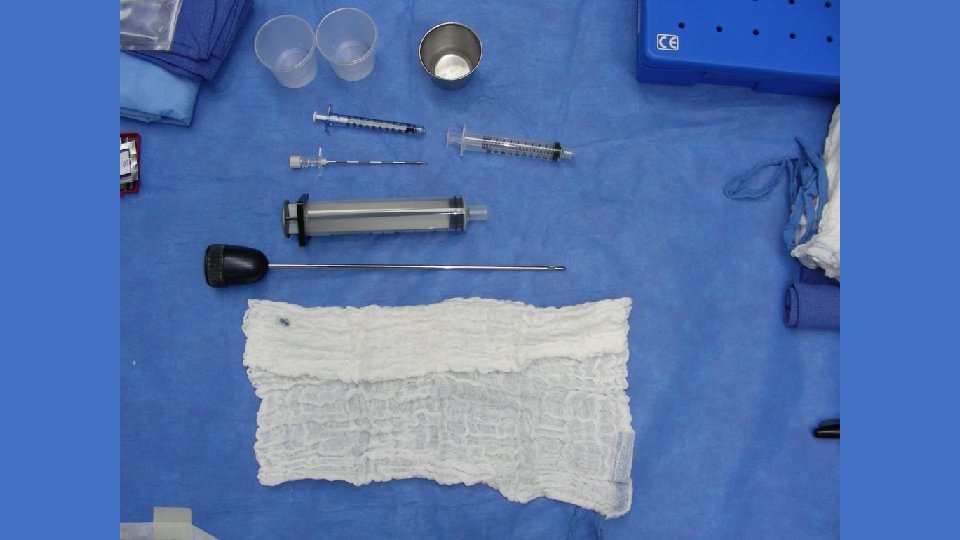
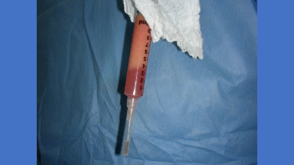
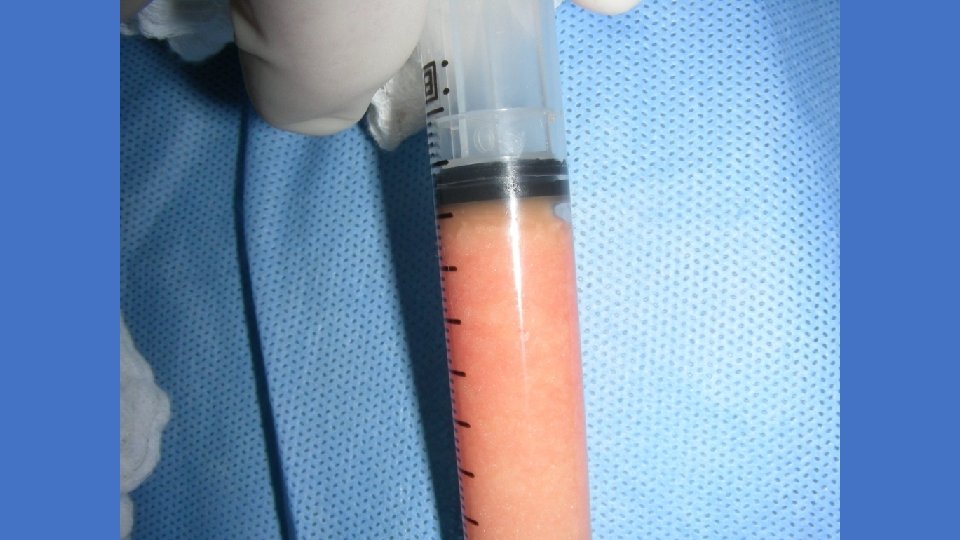
Fat Grafting • Fat Grafting Has Become Very Popular As We Strive For Better Aesthetic Outcomes And Patient Satisfaction • Most Women Will Tolerate Numerous Secondary Procedures To Achieve The Desired Result • Average Volume Injection Varies From 50 to 100 ml • Most Common Location are the Upper and Inner Quadrant • May Be Utilized To Restore The Entire Breast Volume Losken A, Pinell XA, Sikoro K, Yezhelyev MV, Anderson E, Carlson GW. Autologous fat grafting in secondary breast reconstruction. Ann Plast Surg. 2011 May; 66(5): 518 -22.
Fat Grafting
Safety of Lipofilling • Concern re. radiologic surveillance by mammography • Discerning malignant calcifications? • Issue largely resolved • Petit et al, Pulagam et al • Concern for oncologic transformation • Pearl et al (2011) reviews stem cell biology & i • ADMSC’s transform to variety of cell types & secrete angiogenic factors • VEGF known to promote tumor invasion and metastasis in various cancers • Manabe et al – adipocytes increase proliferation of breast cancer cells in vitro
Autologous fat grafting in onco-plastic breast reconstruction: A systematic review on oncological and radiological safety, complications, volume retention and patient/surgeon satisfaction JPRAS: Groen et al (2016) • Literature review • 43 studies, two level II, level III & IV, >6 K pts • Found no increased risk of cancer recurrence • LRR 2. 5% and DR 2. 0% • Were lower than cases of mastectomy w/o AFG or BCT w/ RT • Cancer recurrence SAME histology as primary • RESTORE-2 trial • 71 pts got fat grafting after BCT • NO recurrences at 1 year
• Lipofilling Of The Breast Does Not Increase The Risk Of Recurrence Of Breast Cancer: A Matched Controlled Study. Kronowitz SJ, Mandujano CC, Kuerer HM, Smith B, Velero V. PRS Feb 2016 • LRR 1. 3 % On Lipofilling Group vs 2. 4 On Control Group
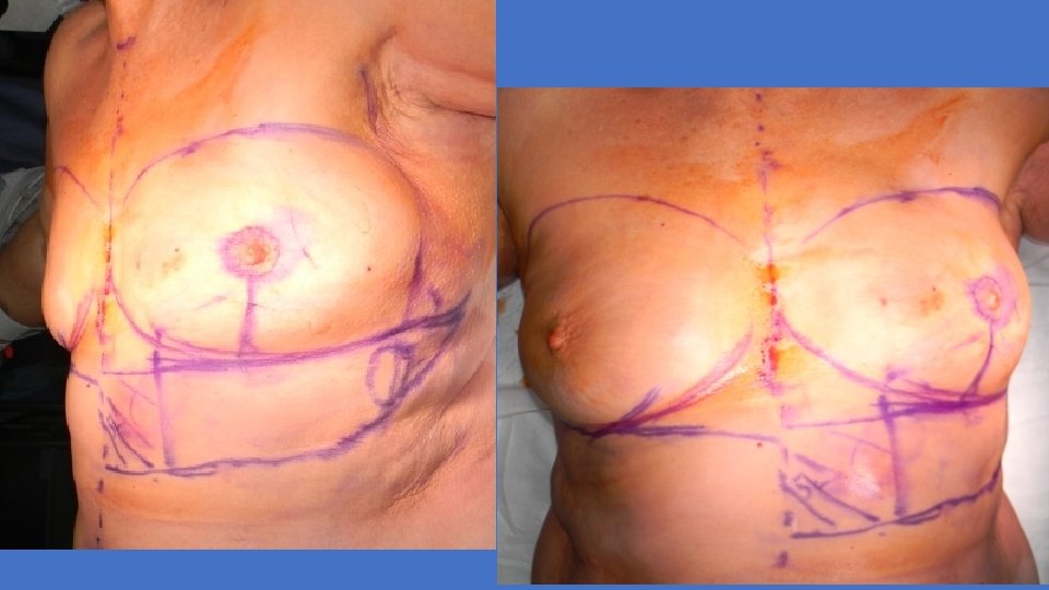
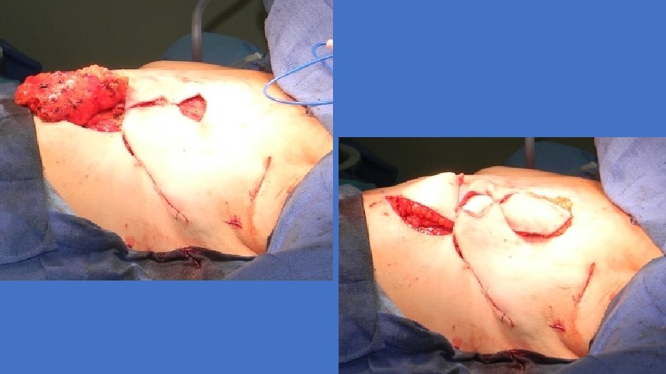
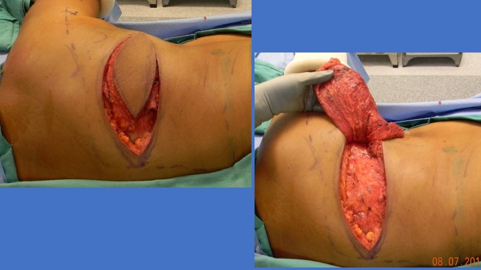
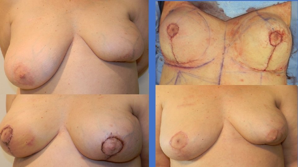
Case Scenarios • 50 y/o WF s/p Treatment Of A Left Breast Cancer at the 6 oc Position with Lumpectomy and Radiation
Preop Pictures
Operative Plan
Intra - Operative Pictures
Intra - Operative Pictures
1 Month Postop Pictures
6 Months Postop Pictures
Case Scenarios • 38 y/o WF With History Of Breast Augmentation Prior To Treatment Of A Right Central Breast Cancer with Lumpectomy and Radiation
Preop Pictures
Intra - Operative Pictures
1 Month Postop Pictures
6 Months Postop Pictures
Case Scenarios • 55 y/o WF With A New Left Breast Cancer
Lumpectomy + Local Tissue Rearrangement + Contralateral Reduction For Symmetry
Case Scenarios • 35 y/o WF With A Left Breast Phyllodes Tumor
Left Lumpectomy + LTR + Breast Augmentation
Conclusion • Understand Patient Expectations: • Symmetry • Ideal Breast Volume • Define Outcome Limitations: • Radiation Changes • Personalize Patient Care By Matching Reconstruction Options To Patients Desire
Thank You