Addressing Challenges in IUD InsertionsRemovals Tools for Teaching








- Slides: 29
Addressing Challenges in IUD Insertions/Removals: Tools for Teaching Residents Safely May 1, 2016 Seema Shah, MD (New York, NY) Carrie Pierce, MD (Klamath Falls, OR) James Becker, MD (New York, NY)
Disclosures • None
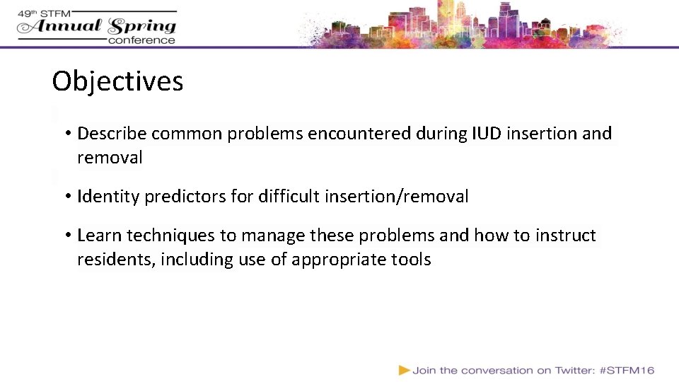
Objectives • Describe common problems encountered during IUD insertion and removal • Identity predictors for difficult insertion/removal • Learn techniques to manage these problems and how to instruct residents, including use of appropriate tools
(CDC Summary Chart of U. S. Medical Eligibility Criteria for Contraceptive Use, 2012)
Most Common Challenges Discomfort/difficulty with speculum placement Visualizing the cervix Extreme uterine position/ uterine distortion Pain and/or Vasovagal Symptoms Insertion: Tight cervical os Removal: Can’t see strings Removal: Embedded IUD, Malpositioned IUD
Teaching tips • Being able to verbalize steps for resident to follow • Hand-on-hand technique • Balancing patient comfort and resident learning • When to step in and when to step back out • Supervising check points for insertion: • sounding • insertion of device • final deployment of device • Others?
Case 1: Difficult Insertion • Cynthia, 38 year-old G 2 P 2 – 2 prior C-sections last 6 months prior, BMI 41, comes to clinic for a hormonal IUD. • What are aspects of the patient history that may make for challenges during insertion? • No prior vaginal deliveries • History of C-sections • BMI – body habitus • Experience of physician* (Bahamondes, 2011; Farmer, 2003)
Setting the Stage • § § Pre-meeting with Resident Assess level of knowledge Talk through insertion § • § § language, only necessary patient physical contact Consent & walk-through with patient Help her to know what to expect Ask how she usually does with pelvic exams, pain/vasovagal Set up the room Make environment comfortable (eg. music, support-person) Ensure equipment ready
Case 1: Difficult Insertion • During the procedure, resident attempts to place the speculum but cannot visualize the cervix. What to do? • Relaxation techniques • Have patient move further down – bottom off edge of table • Remove labial tissue from sides of speculum • Speculum handle close to perineum • Lateral wall traction (glove, condom, instrument) • Switching speculum size • Mc. Roberts maneuver Photo credit
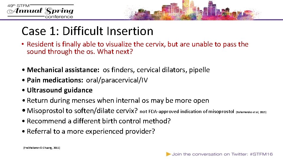
Case 1: Difficult Insertion • Resident is finally able to visualize the cervix, but are unable to pass the sound through the os. What next? • Mechanical assistance: os finders, cervical dilators, pipelle • Pain medications: oral/paracervical/IV • Ultrasound guidance • Return during menses when internal os may be more open • Misoprostol to soften/dilate cervix? not FDA-approved indication of misoprostol • Recommend a different birth control method? • Referral to a more experienced provider? (Prabhakaran & Chuang, 2011) (Bahamondes et al, 2015)
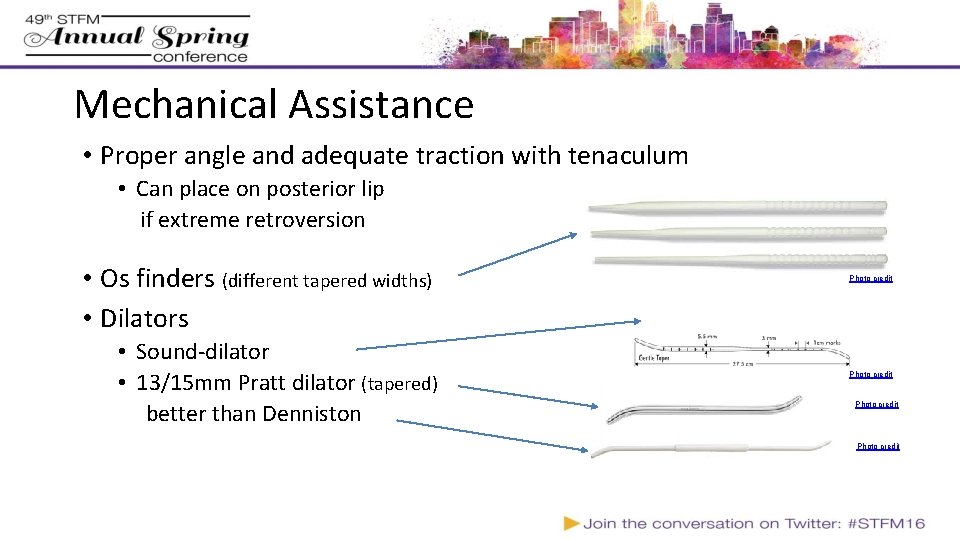
Mechanical Assistance • Proper angle and adequate traction with tenaculum • Can place on posterior lip if extreme retroversion • Os finders (different tapered widths) • Dilators • Sound-dilator • 13/15 mm Pratt dilator (tapered) better than Denniston Photo credit
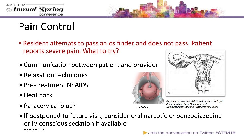
Pain Control • Resident attempts to pass an os finder and does not pass. Patient reports severe pain. What to try? • Communication between patient and provider • Relaxation techniques • Pre-treatment NSAIDS • Heat pack • Paracervical block • If postponed to future visit, consider oral narcotic or benzodiazepine or IV conscious sedation if available (Up. To. Date) (Bahamondes, 2014)
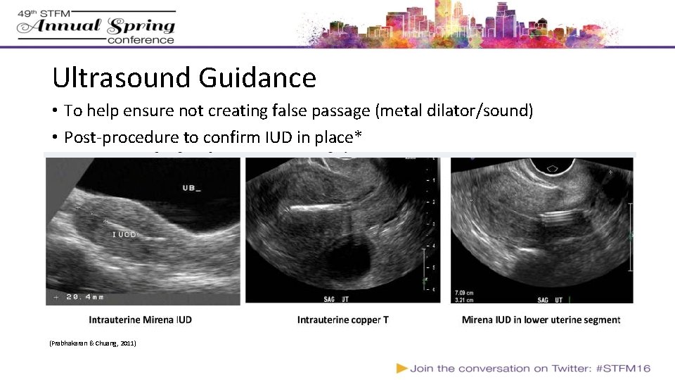
Ultrasound Guidance • To help ensure not creating false passage (metal dilator/sound) • Post-procedure to confirm IUD in place* (Prabhakaran & Chuang, 2011)
Case 1: Difficult Insertion • As you are about to step out of the room, the patient says she’s very lightheaded. What to do next?
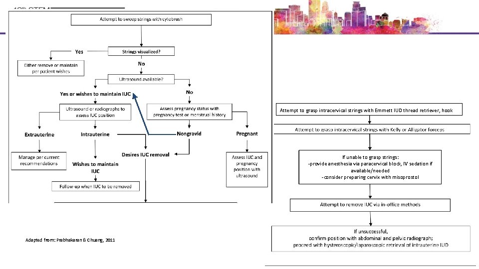
Case 2: Difficult Removal • 31 year-old G 0 presents for removal of Paragard IUD, because she is planning to get pregnant. • Resident reports not able to see strings on exam. • How common is this? • What is the differential? • What to do next?
Tools for Locating Strings/Removing IUD Aspiration 5 -6 mm cannula Photo credit (Prabhakaran & Chuang, 2011; Swenson, 2014)
Attempt to grasp intracervical strings with Emmett IUD thread retriever, hook If unable to grasp strings: -provide anesthesia via paracervical block, IV sedation if available/needed -consider preparing cervix with misoprostol Adapted from: Prabhakaran & Chuang, 2011
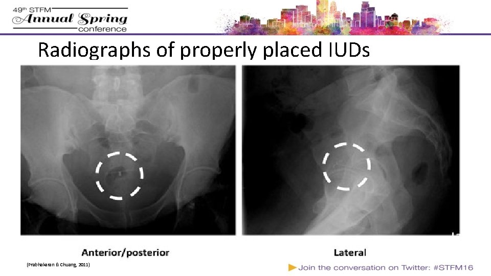
Radiographs of properly placed IUDs (Prabhakaran & Chuang, 2011)
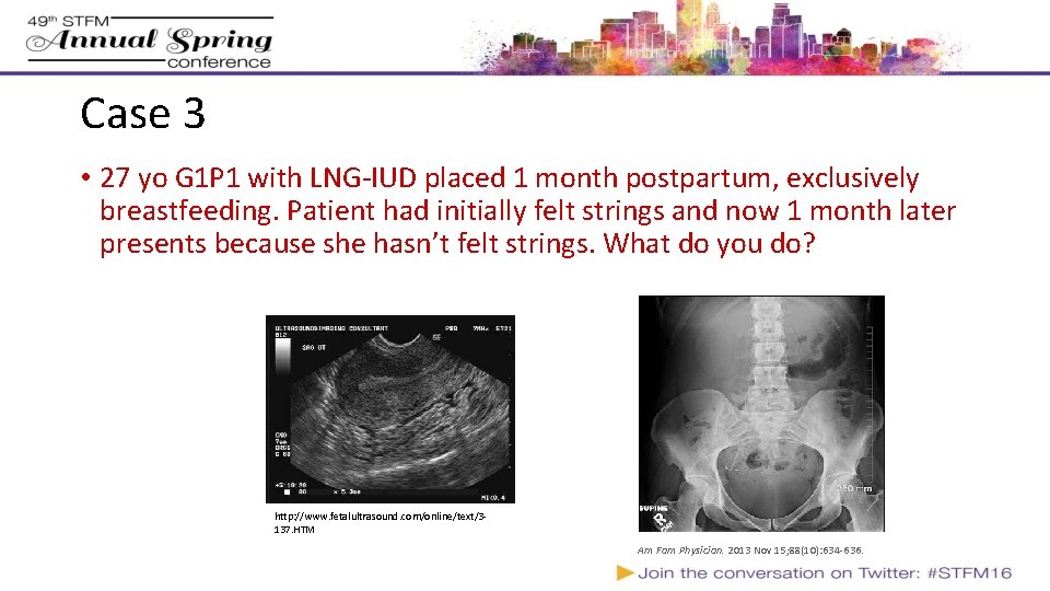
Case 3 • 27 yo G 1 P 1 with LNG-IUD placed 1 month postpartum, exclusively breastfeeding. Patient had initially felt strings and now 1 month later presents because she hasn’t felt strings. What do you do? http: //www. fetalultrasound. com/online/text/3137. HTM Am Fam Physician. 2013 Nov 15; 88(10): 634 -636.
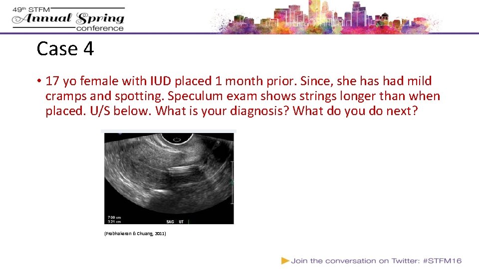
Case 4 • 17 yo female with IUD placed 1 month prior. Since, she has had mild cramps and spotting. Speculum exam shows strings longer than when placed. U/S below. What is your diagnosis? What do you do next? (Prabhakaran & Chuang, 2011)
Malposition statistics/risk factors 10% of IUDs malpositioned Risk Factors • More common if adenomyosis suspected OR 3. 04 (1. 08 -8. 52) – possibly related to change in contractility of uterus • Not associated with type of IUD, breastfeeding status, post-abortion or 6 -9 week postpartum insertion ** • Protective: prior vaginal delivery OR 0. 53 (0. 32 -0. 87) (Braaten, 2011)
The IUD Shuffle - Movement After Insertion • IUDs move up/down uterus after insertion (Shimoni et al, 2014) • Most commonly, IUDs shift towards fundus after insertion • This is particularly true in women with lower parity (Morales-Rosello 2005) • Around 2 of out 3 misplaced IUDs will move into proper position within 3 months without any intervention (Faundes 2000) • Watchful waiting is a reasonable option • Can repeat Ultrasound in 3 months to re-assess position • May want to cover with backup contraception in mean-time
The Low-down on Low-down IUDs: Malposition and IUD Failure • IUD not at fundus is at higher risk for expulsion (Petta, 1996) Copper IUD: • Odds ratio for pregnancy with intracervical insertion 13. 93 (95% CI 4. 13 - 48. 96) --- Absolute risk increase of ~1 -2% (Anteby, 1993) LNG IUD: • Intracervical versus fundal placement found no difference in failure between groups (Pakarinen, 2005)
So, What to do with an IUD in the Lower Uterine Segment? First, it depends on symptoms • Removal and reinsertion may resolve pain or bleeding Second, it depends on type of IUD • LNG IUD • Protects just as well against pregnancy in lower uterine segment versus fundus; so, if no symptoms let it be. • Copper IUD • May be less effective; best practice would be to replace • If replacement too costly, can advance with alligator forceps (if not in endocervical canal) • Even misplaced, still more effective than OCP’s (Braaten, 2012)
Summary • IUDs are great but there are some challenges for getting them in and out; you can increase your success rates by incorporating a few simple tools and techniques • It’s scary to let your residents take on the more challenging cases, but under appropriate supervision you can do it safely • Preparation is key- if you can identify risk factors for difficult insertion or removal, you can plan ahead and prep your learner, have extra staff, have extra equipment, etc to make the procedure go smoothly Contact: Seema Shah sshah@institute. org
AAFP. Reversible Contraception Methods. http: //www. aafp. org/about/policies/all/contraception-reversible. html. Published November 2, 2015. Accessed April 10, 2016. AAP Updates Recommendations on Teen Pregnancy Prevention. https: //www. aap. org/en-us/about-the-aap/aap-press-room/pages/AAP-Updates-Recommendations-on-Teen-Pregnancy-Prevention. aspx. Accessed April 10, 2016. ACOG. Increasing Access to Contraceptive Implants and Intrauterine Devices to Reduce Unintended Pregnancy - ACOG. http: //www. acog. org/Resources-And-Publications/Committee-Opinions/Committee-on-Gynecologic-Practice/Increasing-Access-to-Contraceptive-Implants-and-Intrauterine-Devices-to-Reduce-Unintended-Pregnancy. Accessed April 10, 2016. Anteby E, Revel A, Ben-Chetrit A, Rosen B, Tadmor O, Yagel S. Intrauterine device failure: relation to its location within the uterine cavity. Obstet Gynecol. 1993; 81(1): 112 -114. Bahamondes MV, Espejo-Arce X, Bahamondes L. Effect of vaginal administration of misoprostol before intrauterine contraceptive insertion following previous insertion failure: a double blind RCT. Hum Reprod. 2015; 30(8): 1861 -1866. Bahamondes MV, Hidalgo MM, Bahamondes L, Monteiro I. Ease of insertion and clinical performance of the levonorgestrel-releasing intrauterine system in nulligravidas. Contraception. 2011; 84(5): e 11 -e 16. Bahamondes L, Mansour D, Fiala C, Kaunitz AM, Gemzell-Danielsson K. Practical advice for avoidance of pain associated with insertion of intrauterine contraceptives. J Fam Plann Reprod Health Care. 2014; 40(1): 54 -60. Ber A, Seidman DS. Management of the malpositioned levonorgestrel-releasing intrauterine system. Contraception. 2012; 85(4): 369 -373. Boortz HE, Margolis DJA, Ragavendra N, Patel MK, Kadell BM. Migration of intrauterine devices: radiologic findings and implications for patient care. Radiozgraphics. 2012; 32(2): 335 -352. Braaten KP, Benson CB, Maurer R, Goldberg AB. Malpositioned intrauterine contraceptive devices: risk factors, outcomes, and future pregnancies. Obstet Gynecol. 2011; 118(5): 1014 -1020. Braaten KP, Goldberg AB. Malpositioned IUDs: when you should intervene (and when you should not). OBG Manag. 2012; 24(8): 39 -46. Cason P. No more fainting in your practice. Bedsiders Providers. http: //providers. bedsider. org/articles/no-more-fainting-in-your-practice. Accessed April 24, 2016. Caliskan E, Oztürk N, Dilbaz BO, Dilbaz S. Analysis of risk factors associated with uterine perforation by intrauterine devices. Eur J Contracept Reprod Health Care. 2003; 8(3): 150 -155. Centers for Disease Control and Prevention. Summary Chart of U. S. Medical Eligibility Criteria for Contraceptive Use. 2012. http: //www. cdc. gov/reproductivehealth/unintendedpregnancy/pdf/legal_summary-chart_english_final_tag 508. pdf. Farmer M, Webb A. Intrauterine device insertion-related complications: can they be predicted? J Fam Plann Reprod Health Care. 2003; 29(4): 227 -231. Faúndes D, Perdigão A, Faúndes A, Bahamondes L, Petta CA. T-shaped IUDs accommodate in their position during the first 3 months after insertion. Contraception. 2000; 62(4): 165 -168. Glantz JC, Shomento S. Comparison of paracervical block techniques during first trimester pregnancy termination. Int J Gynaecol Obstet. 2001; 72(2): 171 -178. Gill RS, Mok D, Hudson M, Shi X, Birch DW, Karmali S. Laparoscopic removal of an intra-abdominal intrauterine device: case and systematic review. Contraception. 2012; 85(1): 15 -18. Guttmacher Institute. Use of Long-Acting Reversible Contraceptive Methods Continues to Increase in the United States. https: //www. guttmacher. org/news-release/2015/use-long-acting-reversible-contraceptive-methods-continues-increase-united-states. Published October 8, 2015. Accessed April 10, 2016. Harrison-Woolrych M, Zhou L, Coulter D. Insertion of intrauterine devices: a comparison of experience with Mirena and Multiload Cu 375 during post-marketing monitoring in New Zealand. N Z Med J. 2003; 116(1179): U 538. Heinemann K, Reed S, Moehner S, Minh TD. Risk of uterine perforation with levonorgestrel-releasing and copper intrauterine devices in the European Active Surveillance Study on Intrauterine Devices. Contraception. 2015; 91(4): 274 -279. Heinemann K, Westhoff CL, Grimes DA, Moehner S. Intrauterine Devices and the Risk of Uterine Perforations: Final Results From the EURAS-IUD Study. Obstetrics & Gynecology. 2014; 123: 3 S. Kaislasuo J, Suhonen S, Gissler M, Lähteenmäki P, Heikinheimo O. Intrauterine contraception: incidence and factors associated with uterine perforation—a population-based study. Hum Reprod. 2012; 27(9): 2658 -2663. Kaislasuo J, Suhonen S, Gissler M, Lähteenmäki P, Heikinheimo O. Uterine perforation caused by intrauterine devices: clinical course and treatment. Hum Reprod. 2013; 28(6): 1546 -1551. Marchi NM, Castro S, Hidalgo MM, et al. Management of missing strings in users of intrauterine contraceptives. Contraception. 2012; 86(4): 354 -358. Morales-Roselló J. Spontaneous upward movement of lowly placed T-shaped IUDs. Contraception. 2005; 72(6): 430 -431. Pakarinen P, Luukkainen T. Five years’ experience with a small intracervical/intrauterine levonorgestrel-releasing device. Contraception. 2005; 72(5): 342 -345. Petta CA, Faúndes D, Pimentel E, Diaz J, Bahamondes L. The use of vaginal ultrasound to identify copper T IUDs at high risk of expulsion. Contraception. 1996; 54(5): 287 -289. Peipert JF, Madden T, Allsworth JE, Secura GM. Preventing unintended pregnancies by providing no-cost contraception. Obstet Gynecol. 2012; 120(6): 1291 -1297. Prabhakaran S, Chuang A. In-office retrieval of intrauterine contraceptive devices with missing strings. Contraception. 2011; 83(2): 102 -106. Renner R-M, Nichols MD, Jensen JT, Li H, Edelman AB. Paracervical block for pain control in first-trimester surgical abortion: a randomized controlled trial. Obstet Gynecol. 2012; 119(5): 1030 -1037. Russo JA, Miller E, Gold MA. Myths and misconceptions about long-acting reversible contraception (LARC). J Adolesc Health. 2013; 52(4 Suppl): S 14 -S 21. Shimoni N ’a, Davis A, Westhoff C. Can ultrasound predict IUD expulsion after medical abortion? Contraception. 2014; 89(5): 434 -439. Swenson C, Royer PA, Turok DK, Jacobson JC, Amaral G, Sanders JN. Removal of the LNG IUD when strings are not visible: a case series. Contraception. 2014; 90(3): 288 -290. Van Houdenhoven K, Van Kaam K, Van Grootheest AC. Uterine perforation in women using a levonorgestrel-releasing intrauterine system. Contraception. 2006. http: //www. sciencedirect. com/science/article/pii/S 0010782405003215. van Grootheest PK, Sachs B, Harrison-Woolrych M, Caduff-Janosa P, van Puijenbroek E. Uterine Perforation with the Levonorgestrel-Releasing Intrauterine Device. Drug-Safety. 2012; 34(1): 83 -88.
Please evaluate this presentation using the conference mobile app! Simply click on the "clipboard" icon on the presentation page.