1 6 Cell division Essential idea Cell division


- Slides: 38
1. 6 Cell division Essential idea: Cell division is essential but must be controlled. Edited by Eran Earland Acknowledgements to Chris Paine and Bioninja By Chris Paine https: //bioknowledgy. weebly. com/ http: //www. flickr. com/photos/pulmonary_pathology/4388301142/
Understandings Statement 1. 6. U 1 Mitosis is division of the nucleus into two genetically identical daughter nuclei. 1. 6. U 2 Chromosomes condense by supercoiling during mitosis. 1. 6. U 3 Cytokinesis occurs after mitosis and is different in plant and animal cells. 1. 6. U 4 Interphase is a very active phase of the cell cycle with many processes occurring in the nucleus and cytoplasm. 1. 6. U 5 Cyclins are involved in the control of the cell cycle. 1. 6. U 6 Mutagens, oncogenes and metastasis are involved in the development of primary and secondary tumours. Guidance The sequence of events in the four phases of mitosis should be known. To avoid confusion in terminology, teachers are encouraged to refer to the two parts of a chromosome as sister chromatids, while they are attached to each other by a centromere in the early stages of mitosis. From anaphase onwards, when sister chromatids have separated to form individual structures, they should be referred to as chromosomes.
Applications and Skills Statement Guidance 1. 6. A 1 The correlation between smoking and incidence of cancers. 1. 6. S 1 Identification of phases of mitosis in cells viewed Preparation of temporary mounts of root with a microscope or in a micrograph. squashes is recommended but phases in mitosis can also be viewed using permanent slides. 1. 6. S 2 Determination of a mitotic index from a micrograph.
Link to Previous Topics • Recall: Cell size is limited due to the Surface Area to Volume ratio of cells If an organisms is to grow larger, it needs to produce more cells, and each of those cells need a copy of the organism’s DNA CELL DIVISION Cell division allows for GROWTH Cell DIFFERENTIATION
In summary any time new cells are required, mitosis is required: Growth: Multicellular organisms increase their size by increasing their number of cells through mitosis Asexual reproduction: Tissue Repair: Certain eukaryotic organisms may reproduce asexually by mitosis (e. g. vegetative reproduction) Damaged tissue can recover by replacing dead or damaged cells Embryonic development: A fertilised egg (zygote) will undergo mitosis and differentiation in order to develop into an embryo http: //www. haroldsmithlab. com/images/pg_He. La_cell_division. jpg
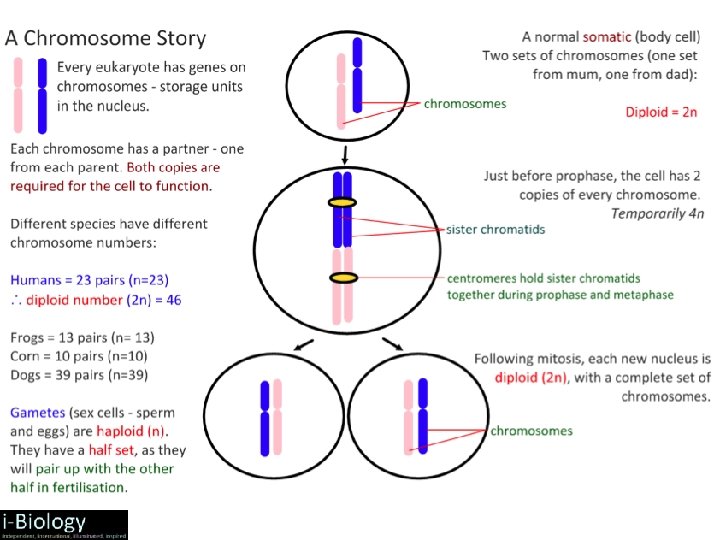
1. 6. U 1 Mitosis is division of the nucleus into two genetically identical daughter nuclei. Get the terminology right centromere is the part of a chromosome that links sister chromatids centrioles organise spindle microtubules Spindle microtubules (also referred to as spindle fibres) In animal cells two centrioles are held by a protein mass referred to as a centrosome Sister chromatids are duplicated chromosomes attached by a centromere After anaphase when the sister chromatids separate they should then be referred to as chromosomes It is easy to misuse the terms chromatid and chromosome. It is even easier to confuse the terms centromere, centriole and centrosome due to their similar spelling. Keep the terms clear in your mind to avoid losing marks. http: //commons. wikimedia. org/wiki/File: Chromosome. svg http: //commons. wikimedia. org/wiki/Mitosis#mediaviewer/File: Mitosis_cells_sequence. svg
1. 6. U 1 Mitosis is division of the nucleus into two genetically identical daughter nuclei. Use the animated tutorials to learn about mitosis http: //highered. mheducation. com/sites/0072495 855/student_view 0/chapter 2/animation__mitosis _and_cytokinesis. html http: //www. johnkyrk. com/mitosis. html http: //www. sumanasinc. com/webcontent/animations/conten t/mitosis. html http: //outreach. mcb. harvard. edu/animations/cellcycle. swf
• Mitosis is the process of nuclear division, whereby duplicated DNA molecules are arranged into two separate nuclei • Mitosis is preceded by interphase and is divided into four distinct stages: prophase, metaphase, anaphase, telophase • The division of the cell in two (cytokinesis) occurs concurrently with the final stage of mitosis (telophase)
1. 6. U 1 Mitosis is division of the nucleus into two genetically identical daughter nuclei. Prophase The centrosomes move to opposite poles of the cell and spindle fibres begin to form between them *supercoling is dealt with in more detail by 1. 6. U 2 DNA supercoils* chromatin condenses and becomes sister chromatids, which are visible under a light microscope The nuclear membrane is broken down and disappears http: //www. microscopy-uk. org. uk/mag/artnov 04 macro/jronionroot. html http: //commons. wikimedia. org/wiki/Mitosis#mediaviewer/File: Mitosis_cells_sequence. svg
1. 6. U 1 Mitosis is division of the nucleus into two genetically identical daughter nuclei. Metaphase Spindle fibres from each of the two centrosomes attach to the centromere of each pair of sister chromatids Contraction of the microtubule spindle fibres cause the sister chromatids to line up along the centre of the cell. http: //www. microscopy-uk. org. uk/mag/artnov 04 macro/jronionroot. html http: //commons. wikimedia. org/wiki/Mitosis#mediaviewer/File: Mitosis_cells_sequence. svg
1. 6. U 1 Mitosis is division of the nucleus into two genetically identical daughter nuclei. Anaphase Continued contraction of the microtubule spindle fibres cause the separation of the sister chromatids The chromatids are now referred to as chromosomes Chromosomes move to the opposite poles of the cell http: //www. microscopy-uk. org. uk/mag/artnov 04 macro/jronionroot. html http: //commons. wikimedia. org/wiki/Mitosis#mediaviewer/File: Mitosis_cells_sequence. svg
1. 6. U 1 Mitosis is division of the nucleus into two genetically identical daughter nuclei. Telophase Chromosomes arrive at the poles. The chromosomes uncoil decondense to chromatin (and are no longer visible under a light microscope). Microtubule spindle fibers disappear New nuclear membranes reform around each set of chromosomes Now cytokinesis begins! http: //www. microscopy-uk. org. uk/mag/artnov 04 macro/jronionroot. html http: //commons. wikimedia. org/wiki/Mitosis#mediaviewer/File: Mitosis_cells_sequence. svg
1. 6. U 1 Mitosis is division of the nucleus into two genetically identical daughter nuclei. Prophase Metaphase Anaphase Telophase http: //www. flickr. com/photos/chuckp/252924532/
1. 6. U 1 Mitosis is division of the nucleus into two genetically identical daughter nuclei. People Meet And Talk http: //www. flickr. com/photos/chuckp/252924532/
1. 6. U 2 Chromosomes condense by supercoiling during mitosis. Why supercoil chromosomes? Human cells are on average 10μm in diameter and the nucelus within each is less than 5 μm in diameter. Human chromosomes are 15 mm to 85 mm (15, 000μm to 85, 000 μm) in length. Chromatin fibres Chromosomes need to be stored compactly to fit within the nuclei of cells. This problem becomes more acute during mitosis when chromosomes need to be short and compact enough that they can be separated and moved to each end of the cell. http: //www. genome. gov/dmd/img. cfm? node=Photos/Graphics&id=85282
1. 6. U 2 Chromosomes condense by supercoiling during mitosis. Chromatin versus Chromosome Chromatin: • DNA is usually loosely packed within the nucleus as unravelled chromatin • In this unravelled form, the DNA is accessible to transcriptional machinery and so genetic information can be translated • DNA is organised as chromatin in all non-dividing cells and throughout the process of interphase Chromosome: • DNA is temporarily packaged into a tightly wound and condensed chromosome prior to division (via supercoiling) • In this condensed form, the DNA is able to be easily segregated however is inaccessible to transcriptional machinery • DNA is organised as chromosomes during the process of mitosis (condense in prophase, decondense in telophase)
• A chromosome is the condensed form of DNA which is visible during mitosis (via microscopy) • As the DNA is replicated during the S phase of interphase, the chromosome will initially contain two identical DNA strands = sister chromatids and are held together by a central region called the centromere • When these chromatids separate during mitosis, they become independent chromosomes, each made of a single DNA strand • The cell cycle is an ordered set of events which culminates in the division of a cell into two daughter cells. It can be roughly divided into two main phases:
The cell cycle is the series of events through which cells pass to divide and create two identical daughter cells. http: //upload. wikimedia. org/wikipedia/commons/thumb/7/71/Diagram_of_mitosis. svg/800 px-Diagram_of_mitosis. svg. png
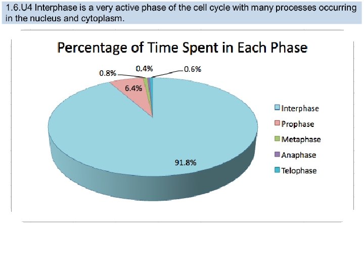
1. 6. U 4 Interphase is a very active phase of the cell cycle with many processes occurring in the nucleus and cytoplasm. Interphase consists of the parts of the cell cycle that don’t involve cell division. G 1 (Gap 1) • Increase the volume of cytoplasm • Organelles produced • Proteins synthesised n. b. cells can also be said to be in G 0 (Gap 0). This is a ‘resting’ phase where the cell has left the cycle and has stopped dividing. Cells in G 0 still carry out all their normal functions. http: //gardeningstudio. com/wp-content/uploads/the-cell-cycle-diagram-318. jpg S (Synthesis) • DNA replicated G 2 (Gap 2) • Increase the volume of cytoplasm • Organelles produced • Proteins synthesised
1. 6. U 4 Interphase is a very active phase of the cell cycle with many processes occurring in the nucleus and cytoplasm. Interphase Cells spend the majority of their time in interphase. It is a very active phase of the cycle. Interphase is an active period in the cell cycle when many metabolic reactions occur Many events need to occur in interphase to prepare the cell for successful division These key processes include: • DNA replication – DNA is copied during the S phase of interphase • Organelle duplication – Organelles must be duplicated for twin daughter cells • Cell growth – Cytoplasmic volume must increase prior to division • Transcription / translation – Key proteins and enzymes must be synthesised • Obtain nutrients – Vital cellular materials must be present before division • Respiration (cellular) – ATP production is needed to drive the division process Mnemonic: DOCTOR
1. 6. U 3 Cytokinesis occurs after mitosis and is different in plant and animal cells. Urrrmm, we have only divided the nucleus … what about the rest of the cell? • Cytokinesis is the process of cytoplasmic division, whereby the cell splits into two identical daughter cells • Cytokinesis occurs concurrently with the final stage of mitosis (telophase) and is different in plant and animal cells http: //www. haroldsmithlab. com/images/pg_He. La_cell_division. jpg
1. 6. U 3 Cytokinesis occurs after mitosis and is different in plant and animal cells. Animal Cells • After anaphase, microtubule filaments form a concentric ring around the centre of the cell • The microfilaments constrict to form a cleavage furrow, which deepens from the periphery towards the centre • When the furrow meets in the centre, the cell becomes completely pinched off and two cells are formed • Because this separation occurs from the outside and moves towards the centre, it is described as centripetal
1. 6. U 3 Cytokinesis occurs after mitosis and is different in plant and animal cells. Plant Cells • After anaphase, carbohydrate-rich vesicles form in a row at the centre of the cell (equatorial plane) • The vesicles fuse together and an early cell plate begins to form within the middle of the cell • The cell plate extends outwards and fuses with the cell wall, dividing the cell into two distinct daughter cells • Because this separation originates in the centre and moves laterally, it is described as centrifugal
1. 6. U 5 Cyclins are involved in the control of the cell cycle. Cyclins are a family of proteins that control the progression of cells through the cell cycle 1 2 Cells cannot progress to the next stage of the cell cycle unless the specific cyclin reaches it threshold. Cyclins bind to enzymes called cyclin-dependent kinases 4 3 These kinases then become active and attach phosphate groups to other proteins in the cell. The attachment of phosphate triggers the other proteins to become active and carry out tasks (specific to one of the phases of the cell cycle). http: //upload. wikimedia. org/wikipedia/commons/thumb/9/99/Protein_CCNE 1_PDB_1 w 98. png/800 px-Protein_CCNE 1_PDB_1 w 98. png
Cyclin concentrations need to be tightly regulated in order to ensure the cell cycle progresses in a proper sequence • Different cyclins specifically bind to, and activate, different classes of cyclin dependent kinases • Cyclin levels will peak when their target protein is required for function and remain at lower levels at all other times
1. 6. U 5 Cyclins are involved in the control of the cell cycle. Progression through parts of the cell cycle are affected in various ways by specific cyclins Triggers cells to move from G 0 to G 1 and from G 1 into S phase. prepares the cell for DNA replication in S phase. activates DNA replication inside the nucleus in S phase. promotes the assembly of the mitotic spindle and other tasks in the cytoplasm to prepare for mitosis. http: //upload. wikimedia. org/wikipedia/commons/thumb/9/99/Protein_CCNE 1_PDB_1 w 98. png/800 px-Protein_CCNE 1_PDB_1 w 98. png
1. 6. U 6 Mutagens, oncogenes and metastasis are involved in the development of primary and secondary tumours. Tumours are abnormal cell growths resulting from uncontrolled cell division and can occur in any tissue or organ Diseases caused by the growth of tumours are collectively known as cancers http: //youtu. be/8 BJ 8_5 Gyhg 8 http: //www. e-learningforkids. org/health/lesson/cancer/ What causes cancer? http: //www. cancerresearchuk. org/cancerinfo/cancerandresearch/all-about-cancer/what-is-cancer/ http: //www. cancer. gov/cancertopics/types/commoncancers
Mutagens A mutagen is an agent that changes the genetic material of an organism (either acts on the DNA or the replicative machinery) Mutagens may be physical, chemical or biological in origin: • Physical – Sources of radiation including X-rays (ionising), ultraviolet (UV) light and radioactive decay • Chemical – DNA interacting substances including reactive oxygen species (ROS) and metals (e. g. arsenic) • Biological – Viruses, certain bacteria and mobile genetic elements (transposons) Mutagens that lead to the formation of cancer are further classified as carcinogens
1. 6. U 6 Mutagens, oncogenes and metastasis are involved in the development of primary and secondary tumours. If a mutation occurs in an oncogenes it can become cancerous. In normal cells oncogenes control of the cell cycle and cell division. mutation in a oncogene malfunction in the control of the cell cycle uncontrolled cell division tumour formation http: //en. wikipedia. org/wiki/Oncogene#mediaviewer/File: Oncogenes_illustration. jpg
1. 6. U 6 Mutagens, oncogenes and metastasis are involved in the development of primary and secondary tumours. Several mutations must occur in the same cell for it to become a tumour causing cell. The probability of this happening in a single cell is extremely small. Factors (other than exposure to mutagens) that increase the probability of tumour development include: • The vast number of cells in a human body – the greater the number of cells the greater the chance of a mutation. • The longer a life span the greater the chance of a mutation. http: //en. wikipedia. org/wiki/Oncogene#mediaviewer/File: Oncogenes_illustration. jpg
1. 6. U 6 Mutagens, oncogenes and metastasis are involved in the development of primary and secondary tumours. The development of a primary tumours (cancers) have been outlined. Below is how a primary tumor can become a secondary tumour. A primary tumor is a malignant tumor growing at the site where the abnormal growth first occurred. Metastasis is the movement of cells from a primary tumour to set up secondary tumours in other parts of the body. The circulating cancerous cells invade tissues at a different locations and develop, by uncontrolled cell division, into a secondary tumours. Cancerous cells can detach from the primary tumour. Some cancerous cells gain the ability to penetrate the walls of lymph or blood vessels and hence circulate around the body http: //en. wikipedia. org/wiki/Neoplasm#mediaviewer/File: Colon_cancer_2. jpg
1. 6. A 1 The correlation between smoking and incidence of cancers. a. b. c. d. There are many other similar surveys in different countries, with different demographics that show similar results. Along with lung cancer, cancers of mouth and throat are very common as these areas are in direct contact with the smoke too. It might surprise you that the following cancers are also more common in smokers: • Head and neck • Bladder • Kidneys • Breast • Pancreas • Colon Describe the relationship shown. What type of correlation is shown How strong is the correlation? Justify your answer by discussing the evidence. The correlation shown here is lagged. A lag is a time gap between the factors. Estimate the size of the lag between cigarette consumption and lung cancer death. http: //en. wikipedia. org/wiki/File: Smoking_lung_cancer. png
1. 6. A 1 The correlation between smoking and incidence of cancers. There are many other similar surveys in different countries, with different demographics that show similar results. Along with lung cancer, cancers of mouth and throat are very common as these areas are in direct contact with theusmoke too. It : d n o f e v a that the following ions hyou t a might surprise g i t s e v n i rs incommon in oratory cancersuare e b c a l n a r c e v d e also more e s w ion, ho ave ca t h a s o u c c a a c b ≠ o t n smokers: und in o f s Correlatio l a c i m e ch fied as i 0 t 2 n e n d e s i h n t n a • Head and neck e e m e r /or hu • mo have b d o n c a c s a l a b o m t i n n i a • Bladder found s l a c i laboratory m e h c other 0 4 • Kidneys n a h t e r • Mo • Breast s carcinogen • Pancreas • Colon a. Describe the relationship shown. b. What type of correlation is shown c. How strong is the correlation? Justify your answer by discussing the evidence. d. The correlation shown here is lagged. A lag is a time gap between the factors. Estimate the size of the lag between cigarette consumption and lung cancer death. http: //en. wikipedia. org/wiki/File: Smoking_lung_cancer. png
1. 6. S 1 Identification of phases of mitosis in cells viewed with a microscope or in a micrograph. 1. 6. S 2 Determination of a mitotic index from a micrograph. http: //www. nuffieldfoundation. org/practical-biology/investigating-mitosis-allium-root-tip-squash A very good, well explained lab outline for creating slides and calculating the mitotic index. An excellent online alternative if resources don’t permit students to create and view their own slides http: //www. biology. arizona. edu/cell_bio/activities/cell_cycle. html
It is neat to do a root tip squash preparation in onion or garlic roots. They have a small number (8) of large chromosomes which are visible during mitosis. The cells must be carefully teased apart and the DNA stained up. It is fun but tricky to accomplish successfully so many labs do not bother! Here is what a preparation might look like:
Bibliography / Acknowledgments Jason de Nys