1 6 CELL DIVISION Cell division is essential


 ■ DNA is")


. – Chromosomes condense")


")


 or spread")

- Slides: 39
1. 6 CELL DIVISION Cell division is essential but must be controlled
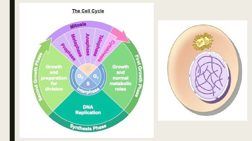
Cell Cycle ■ The cell cycle is an ordered set of events which culminated in the division of a cell into two daughter cells. ■ It can be roughly divided into two main phases: – Interphase – M Phase
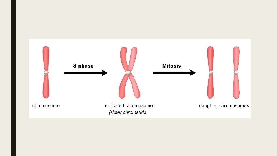
Interphase ■ The largest part of the cell cycle is interphase. ■ The stage in the development of a cell between two successive divisions. ■ This phase of the cell cycle is a continuum of three distinct stages. – G 1 -First intermediate gap stage in which the cell grows and prepared for DNA replication. – S- Synthesis stage in which DNA is replicated – G 2 - Second intermediate gap stage in which the cell finishes growing and prepares for cell division.
■ Interphase is a very active period in the cell cycle with many processes occurring in the nucleus and cytoplasm. (U. 4) ■ Many events need to occur in interphase to prepare the cell for successful division. – DNA Replication – Organelle duplication – Cell growth – Transcription/translation – Obtain nutrients – Respiration (cellular) – *DOCTOR
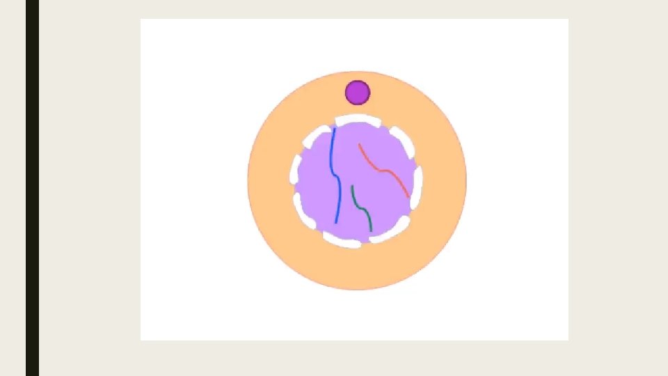
■ DNA is present as uncondensed chromatin (not visible under microscope) ■ DNA is contained within a clearly define nucleus ■ Centrosomes and other organelles have been duplicated ■ Cell is enlarged in preparation for division.
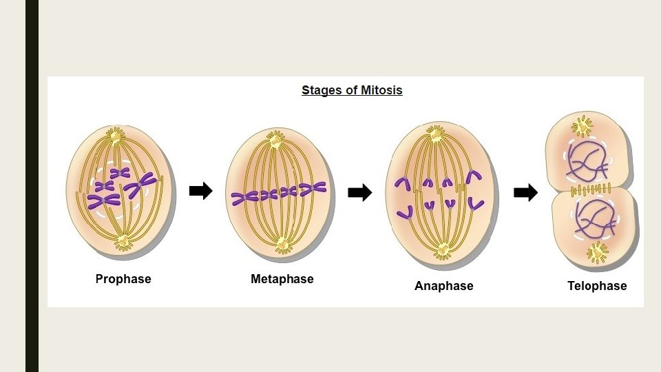
Mitosis ■ Mitosis is the division of the nucleus into two genetically identical daughter nuclei. (U. 1) ■ Mitosis is preceded by interphase and is divided into four distinct stages: – Prophase – Metaphase – Anaphase – Telophase
Before we begin……vocab ■ Chromatin: unraveled, loosely packed DNA within the nucleus – In this unraveled form, the DNA is accessible to be transcribed (copied) ■ Chromosome: DNA tightly wound and condensed (via supercoiling) – In this form, DNA is able to easily segregate, however it is inaccessible to transcription.
Prophase ■ DNA supercoils and chromosomes condense (becoming visible under microscope). – Chromosomes condense by supercoiling during mitosis. (U. 2) ■ Chromosomes are comprised of genetically identical sister chromatids (joined at a centromere) ■ Paired centrosomes move to the opposite poles of the cell and form microtubule spindle fibers ■ The nuclear membrane breaks and the nucleus dissolves.
Metaphase ■ Microtubule spindle fibers from both centrosomes connect to the centromere of each chromosome ■ Microtubule depolymerisation causes spindle fibers to shorten in length and contract. ■ This causes chromosomes to align along the center of the cell (metaphase plate)
Anaphase ■ Continued contraction of the spindle fibers causes genetically identical sister chromatids to separate. ■ Once the chromatids separate, they are each considered an individual chromosome. ■ The genetically identical chromosomes move to the opposite poles of the cell.
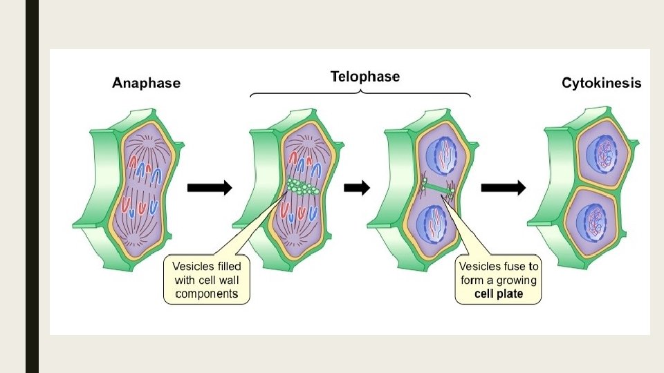
Telophase ■ Once the two chromosome sets arrive at the poles, spindle fibers dissolve ■ Chromosomes decondense (no longer visible under light microscope) ■ Nuclear membranes reform around each chromosome set ■ Cytokinesis occurs concurrently, splitting the cell into two.
Cytokinesis ■ Cytokinesis in the process of cytoplasmic division, where the cell splits into two identical daughter cells. ■ Cytokinesis occurs after mitosis and is different in plant and animal cells. (U. 3)
Cytokinesis: Animal Cells ■ After anaphase, microtubule filaments form a concentric ring around the center of the cell. ■ The microfilaments constrict to form a cleavage furrow, which deepens from the periphery towards the center. ■ When the furrow meets in the center, the cell becomes completely pinched off and two cells are formed. ■ Because this separation occurs from the outside and moves towards the center, it is described as centripetal.
Cytokinesis: Plant Cell ■ After anaphase, carbohydrate-rich vesicles form in a row at the center of the cell (equatorial plane). ■ The vesicles fuse together and an early plate begins to form within the middle of the cell. ■ The cell plate extends outwards and fuses with the cell wall, dividing the cell into two distinct daughter cells. ■ Because this separation originates in the center and moves laterally, it is described as centrifugal.
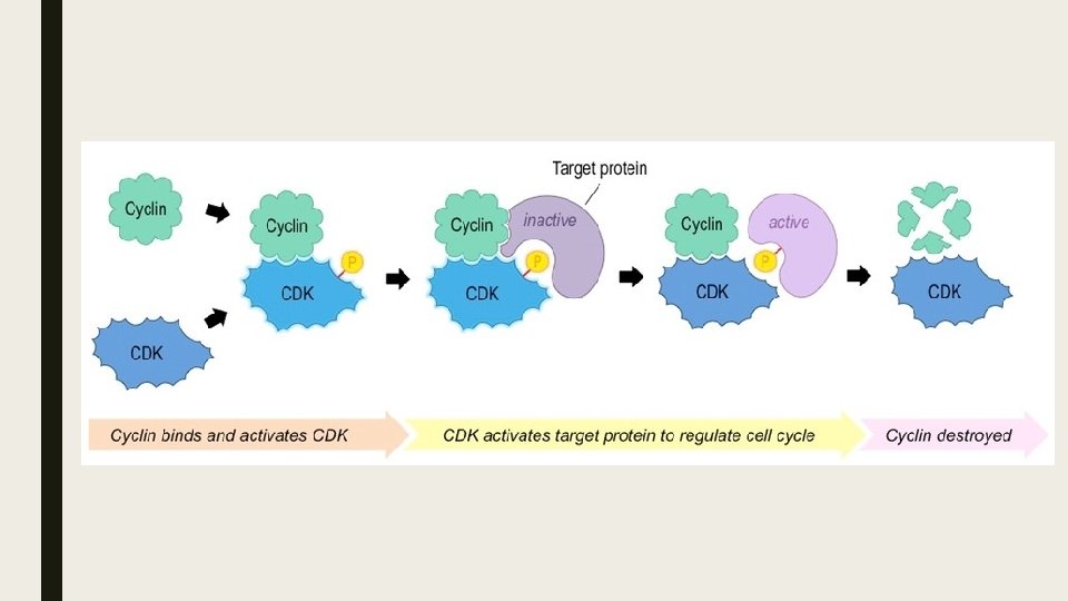
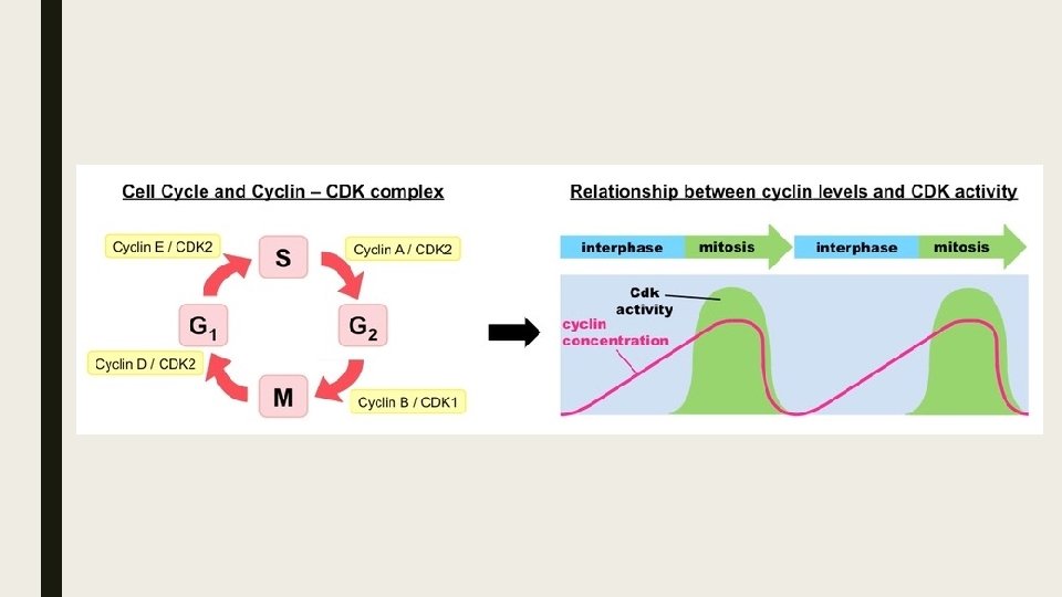
Cyclins ■ Cyclins are involved in the control of the cell cycle. (U. 5) ■ Cyclins are a family of regulatory proteins that control the progression of the cell cycle. ■ Cyclins activate cyclin dependent kinases (CDKs), which control cell cycle processes through phosphorylation. – When a cyclin and CDK form a complex, the complex will bind to a target protein and modify it via phosphorylation. – The phosphorylated target protein will trigger some specific event within the cell cycle (centrosome duplication, etc) – After the event has occurred, the cyclin is degraded and the CDK is rendered inactive again.
Cyclin Expression Patterns ■ Cyclin concentrations need to be tightly regulated in order to ensure the cell cycle progresses in a proper sequence. ■ Different cyclins specifically bind to, and activate, different classes of cyclin dependent kinases. ■ Cyclin levels will peak when their target protein in required for function and remain at lower levels at all other times.
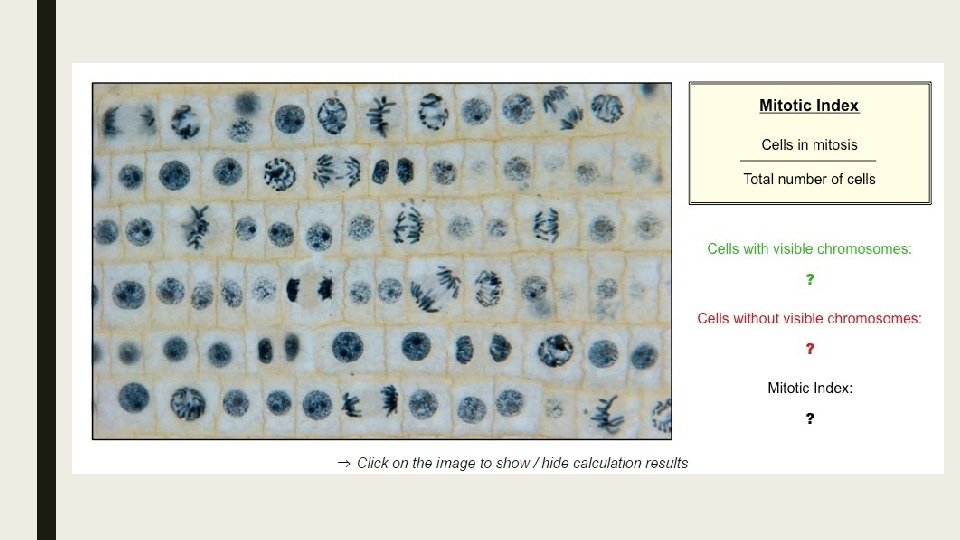
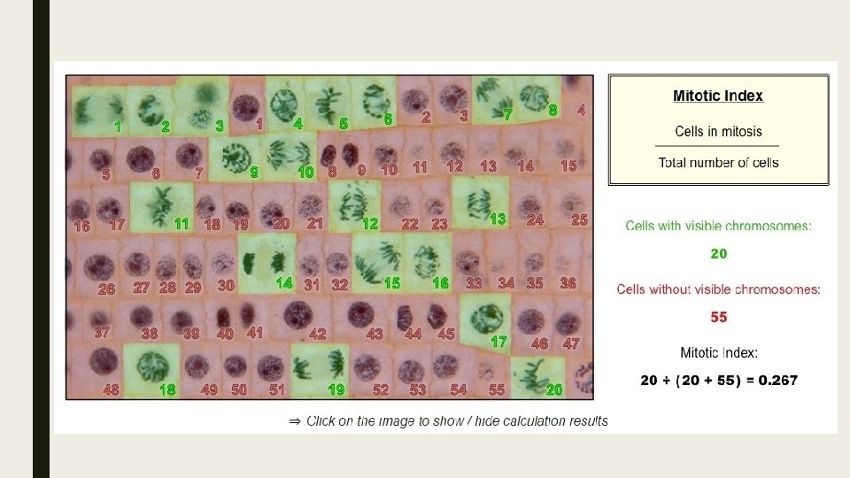
Mitotic Index ■ Identification of phases of mitosis in cells viewed with a microscope or in a micrograph. (S. 1) ■ The mitotic index is a measure of the proliferation status of a cell population (i. e. the proportion of dividing cells) ■ The mitotic index may be elevated during processes that promote division, such as normal growth or cellular repair. – It also functions as an important prognostic tool for predicting the response of cancer cells to chemotherapy.
Identifying Mitotic Cells ■ Cells undergo mitosis will lack a clearly defined nucleus and posses visibly condensed chromosomes. – Prophase- Chromosomes condensed but still confined to a nuclear region. – Metaphase- Chromosomes aligned along the equator of the cell. – Anaphase- Two distinct clusters of chromosomes apparent at poles of the cell. – Telophases- Two nuclear regions present within a single cell (difficult to see as cytokinesis occurs
Calculating Mitotic Index ■ The mitotic index is the ration between the number of cells in mitosis and the total number of cells. ■ It can be determined by analyzing micrographs and counting the relative number of mitotic cells versus non-dividing cells.
Cancer Development ■ Mutagens, oncogenes and metastasis are involved in the development of primary and secondary tumors. (U. 6) ■ Tumor are abnormal cell growths resulting from uncontrolled cell division and can occur in any tissue or organ. ■ Diseases caused by the growth of tumors are collectively known as cancers.
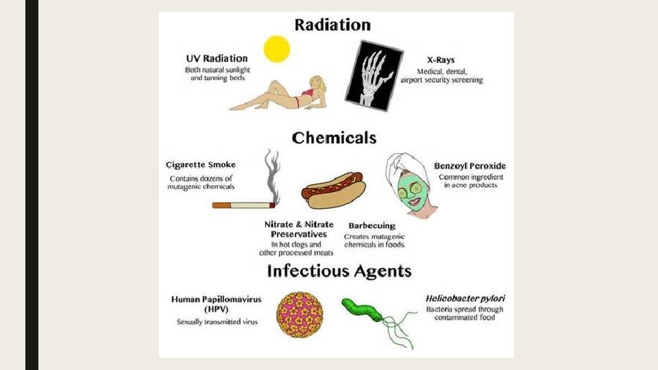
Mutagens ■ A mutagen is an agent that changes the genetic material of an organism (either acts on the DNA or the replicative machinery). ■ Mutagens may be physical, chemical, or biological in origin: – Physical: Sources of radiation including X-rays, ultraviolet (UV) light and radioactive decay. – Chemical: DNA interacting substances including reactive oxygen species and metals (arsenic) – Biological: Viruses, certain bacteria and mobile genetic elements.
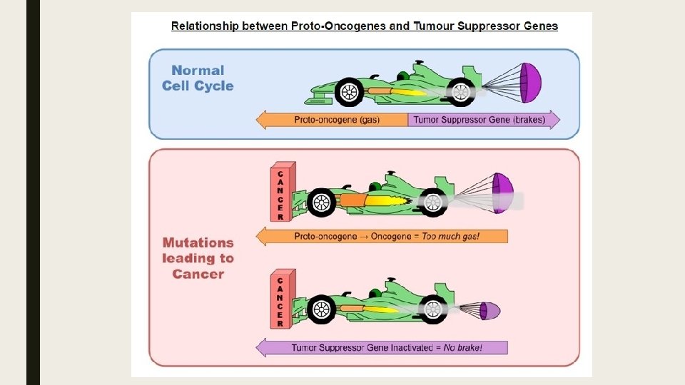
Oncogenes ■ An oncogenes is a gene that has the potential to cause cancer. ■ Most cancers are caused by mutations to two basic classes of genes – proto-oncogenes and tumor suppressor genes. – Proto-oncogenes code for proteins that stimulate the cell cycle and promote cell growth and proliferation. – Tumor suppressor genes code for proteins that repress cell cycle progression and promotes apoptosis. ■ When a proto-oncogene is mutated or subjected to increased expression it becomes a cancer-causing oncogene. ■ Tumor suppressor genes are sometimes referred to an antioncogenes, as their normal function prevents cancer.
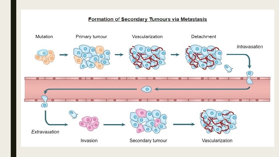
Metastasis ■ Tumor cells may either remain in their original location (benign) or spread and invade neighboring tissue (malignant) ■ Metastasis is the spread of cancer from one location (primary tumor) to another, forming a secondary tumor. ■ Secondary tumors are made up of the same type of cell as the primary tumor – this affects the type of treatment required. – E. g. If breast cancer spread to the liver, the patient has secondary breast cancer of the liver (treat with breast cancer drugs).
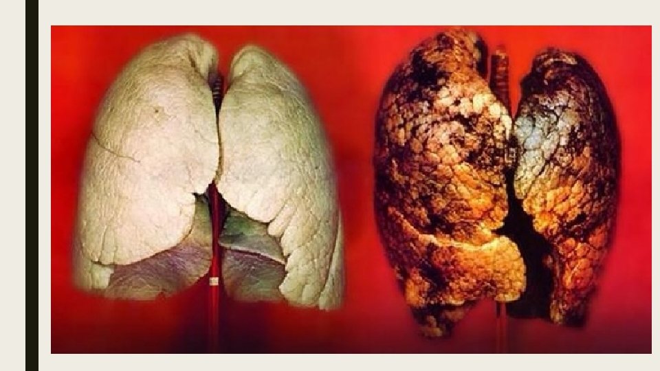
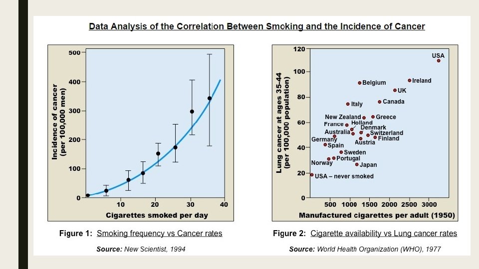
Smoking and Cancer ■ A significant body of scientific literature exists which provides a strong link between smoking and the incidence of cancers. ■ Cigarette smoke contains over 4, 000 chemical compounds, over 60 of which are known to be carcinogenic. ■ There appears to be a strong positive correlation between the frequency of smoking and the development of cancer. ■ The risk of lung cancer is strongly correlated with smoking, with about 90% of such cancers attributable to tobacco use. ■ Smoking also increases the risk of over a dozen other cancers, including mouth, stomach, liver, pancreas and bowel. ■ The correlation between smoking and incidence of cancers. (A. 1)