uterine leiomyomas fibroids Uterine leiomyomas fibroids or myomas
 • Uterine leiomyomas (fibroids or myomas) are the most common pelvic")
uterine leiomyomas (fibroids) • Uterine leiomyomas (fibroids or myomas) are the most common pelvic tumor in women. They are benign monoclonal tumors arising from the smooth muscle cells of the myometrium. • A hysterectomy study found myomas in 77 percent of uterine specimens • Myomas are clinically apparent in approximately 12 to 25 percent of reproductive age women Symptomatic

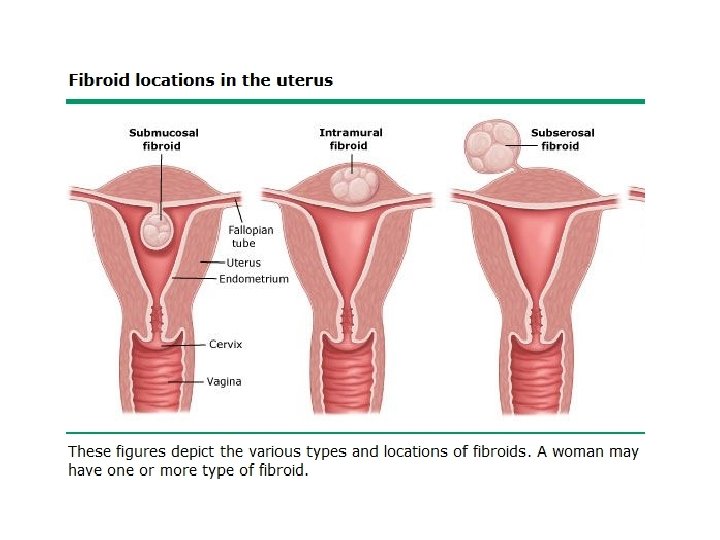
TERMINOLOGY AND LOCATION • Intramural myomas – These leiomyomas develop from within the uterine wall. They may enlarge sufficiently to distort the uterine cavity or serosal surface. Asymptomatic • Submucosal myomas – These leiomyomas derive from myometrial cells just below the endometrium. These neoplasms often protrude into the uterine cavity Bleeding • Subserosal myomas – These leiomyomas originate from the myometrium at the serosal surface of the uterus. They can have a broad or pedunculated base and may be intraligamentary (ie, extending between the folds of the broad ligament). 1. Torsion> may undergo necrosis > severe pain • Cervical myomas – These leiomyomas are located in the cervix, rather than the uterine corpus. 2. Pressure symptoms

RISK FACTORS • Race — The incidence rates of fibroids are typically found to be two- to three-fold greater in black women than in white women. The etiology of the increased incidence of leiomyomas in black women is unknown. The natural history of leiomyomas also differs by race. Most white women with symptomatic fibroids are in their 30 s or 40 s; however, black women develop symptoms on average four to six years younger and may even present with disease in their 20 s. Compared with white women, black women experience more severe disease • Black nullipara , early menarche , FHx , HTN (obese) , alcoholic , infections , early age , more severe
 is associated")
• Menstrual history and parity — Early menarche (<10 years old) is associated with an increased risk of developing fibroids. • Parity (having one or more pregnancies extending beyond 20 weeks) decreases the chance of fibroid formation • Hormonal contraception — Use of low dose oral contraceptives (OCs) does not cause fibroids to grow, therefore administration of these drugs is not contraindicated in women with fibroids • Long acting progestin-only contraceptives (eg, depot medroxyprogesterone) protect against development of leiomyomas. • Decreased risk –parity –progestin –smoking • NO risk–caffeine - OCPs

• Obesity — Most studies show a relationship between fibroids and increasing body mass index • Diet, caffeine, and alcohol use — Significant consumption of beef and other reds meats (1. 7 -fold) or ham (1. 3 -fold) is associated with an increased relative risk of fibroids and consumption of green vegetables (0. 5 -fold) and fruit (especially citrus fruit) with a decreased risk. Dietary vitamin A from animal sources may also be associated with decreased fibroid risk. Increases in dietary glycemic index or load are associated with a weak increase in fibroid risk in some women.

• Consumption of alcohol, especially beer, appears to increase the risk of developing fibroids. • Caffeine consumption is not a risk factor. • Smoking — Smoking decreases the risk of having fibroids through an unknown mechanism. • Heredity — Studies imply a familial predisposition to leiomyomas in some women. • Other factors — Hypertension is associated with an increased leiomyoma risk • Several measures of uterine infection appear to be associated with an increased risk of leiomyomas.

CLINICAL MANIFESTATIONS • Although the majority of myomas are small and asymptomatic, many women with fibroids have significant problems that interfere with some aspect of their lives and warrant therapy. These symptoms are related to the number, size, and location of the neoplasms. • Hx-PE-imaging - > US, HYSTEROSCOPE MRI HSG

• Abnormal uterine bleeding any type of bleeding • Pelvic pressure and pain - Dysmenorrhea -Dyspareunia -Leiomyoma degeneration or torsion Infrequently, fibroids cause acute pain from degeneration (eg, carneous or red degeneration) or torsion of a pedunculated tumor. Pain may be associated with a low grade fever, uterine tenderness on palpation, elevated white blood cell count, or peritoneal signs • Reproductive dysfunction miscarriage-infertility – due to near location to tubes
 , vitals BP , abdominal > if it >12 w")
DIAGNOSIS • General (pallor) , vitals BP , abdominal > if it >12 w maybe palpable • Pelvic examination — A thorough pelvic examination should be performed. On bimanual pelvic examination, an enlarged, mobile uterus with an irregular contour is consistent with a leiomyomatous uterus. • Infrequently, on speculum exam, a prolapsed submucosal fibroid may be visible at the external cervical os. Or other causes of bleeding Imaging • Ultrasound — Transvaginal ultrasound has high sensitivity (95 to 100 percent) for detecting myomas in uteri less than 10 weeks' size. Localization of fibroids in larger uteri or when there are many tumors is limited. • Saline infusion sonography (sonohysterography) improves characterization of the extent of protrusion into the endometrial cavity by submucous myomas and allows identification of some intracavitary lesions not seen on routine ultrasonography (

• Diagnostic hysteroscopy — Diagnostic hysteroscopy can be performed in the office with a flexible or rigid hysteroscope. + biopsy in >40 YO • Magnetic resonance imaging — Magnetic resonance imaging is the best modality for visualizing the size and location of all uterine myomas and can distinguish among leiomyomas, adenomyosis, and adenomyomas. Due to the expense of this modality, its use is best reserved for surgical planning for complicated procedures. It may also be useful in differentiating leiomyomas from leiomyosarcomas, and before uterine artery embolization since imaging patterns predict uterine artery embolization outcome. • Hysterosalpingography — A hysterosalpingogram (HSG) is a good technique for defining the contour of the endometrial cavity. It has poor ability to visualize the rest of the myometrium and can falsely identify an intramural fibroid impinging on the uterine cavity as a submucosal fibroid. It is typically used to visualize myomas only when a HSG is needed to evaluate tubal patency in women with infertility.
 EXPECTANT MANAGEMENT • expectant management is for asymptomatic women,")
treatment of uterine leiomyomas (fibroids) EXPECTANT MANAGEMENT • expectant management is for asymptomatic women, except in the case of a woman with moderate or severe hydronephrosis or a woman with a hysteroscopicallyresectable submucous leiomyoma who is pursuing pregnancy

MEDICAL THERAPY OCP-IUCD-POP-NSAIDS Gn. Rh analougues – PRM- Raloxifene – aromatase inhibitor –danazol gestrinone • Hormonal therapies • strogen-progestin contraceptives — Many texts continue to suggest that estrogen-progestin contraceptive pills (OC) are contraindicated in women with uterine leiomyomas. However, clinical experience suggests some women with heavy menstrual bleeding associated with leiomyomas respond to OC therapy • Levonorgestrel-releasing intrauterine system —. Observational studies and systematic reviews have shown a reduction in uterine volume and bleeding, and an increase in hematocrit after placement of this IUS. The presence of intracavitary leiomyomas amenable to hysteroscopic resection is a strong relative contraindication to use. A second advantage of this treatment is that it provides contraception for women who do not desire pregnancy.

• Progestin implants, injections, and pills — As with OCs, it is difficult to discern the effectiveness of progestin-only contraceptive steroids specifically for treatment of leiomyomas. As with the breast, progesterone is a growth factor for myomas and may even be more critical than estrogen. That being said, progestin-only contraceptives cause endometrial atrophy and thus provide relief of menstrual bleeding-related symptoms. They can be considered for treatment of mild symptoms, especially for women who need contraception. There is also consistent evidence from cohort studies that these agents are associated with a decreased risk of leiomyoma formation.
 agonists are")
Gonadotropin-releasing hormone agonists • Gonadotropin-releasing hormone agonists — Gonadotropin-releasing hormone (Gn. RH) agonists are the most effective medical therapy for uterine myomas. These drugs work by initially increasing the release of Up to 6 gonadotropins, followed by desensitization and downregulation to a months hypogonadotropic, hypogonadal state that clinically resembles menopause. Most women will develop amenorrhea, improvement in anemia (if present), and a significant reduction (35 to 60 percent) in uterine size within three months of initiating this therapy, • However, there is rapid resumption of menses and pretreatment uterine volume after discontinuation of Gn. RH agonists. In addition, significant symptoms can result from the severe hypoestrogenism that accompanies such therapy, including hot flashes, sleep disturbance, vaginal dryness, myalgias and arthralgias, and possible impairment of mood and cognition. Bone loss leading to osteoporosis after long-term (12+ months) use is the most serious complication and most often limits therapy.

• Because of the rapid rebound in symptoms and side effects, Gn. RH agonists are primarily used as preoperative therapy. Gn. RH agonists are approved for administration for three to six months prior to leiomyoma-related surgery in conjunction with iron supplementation to facilitate the procedure and enable correction of anemia. Reduction in uterine size can facilitate subsequent surgery by reducing intraoperative blood loss and by increasing the number of women who are candidates for a vaginal procedure, a transverse (rather than vertical) abdominal incision, or a minimally-invasive procedure. • The side effects of long-term Gn. RH agonist administration can be minimized during therapy by giving add-back therapy with low dose estrogen-progestin after the initial phase of downregulation. • Rarely, Gn. RH-agonists are used to provide short-term relief to women close to menopause or with acute medical contraindications to surgery.

• Gonadotropin-releasing hormone antagonists — Similar clinical results have been achieved with Gn. RH antagonists, which compete with endogenous Gn. RH for pituitary binding sites. The advantage of antagonists over agonists is the rapid onset of clinical effects without the characteristic initial flare-up observed with Gn. RH agonist treatment. • Antiprogestins and progesterone receptor modulators PRM ---- increase risk of endometrial hyperplasia or cancer. - Mifepristone (antiprogestin) - Ulipristal acetate (PRM)

• Raloxifene is a selective estrogen receptor modulators • Aromatase inhibitors tranexemic acid • Antifibrinolytic agents: useful in the treatment of idiopathic menorrhagia (35% reduction) • Nonsteroidal antiinflammatory drugs — NSAIDs do not appear to reduce blood loss in women with myomas, but they decrease painful menses. • Danazol and gestrinone — Androgenic steroids may be an effective treatment of leiomyoma symptoms in some women, but are associated with frequent side effects. Danazol is a 19 -nortestosterone derivative with androgenic and progestin-like effects. Side effects are common (eg, weight gain, muscle cramps, decreased breast size, acne, hirsutism, oily skin, decreased high density lipoprotein levels, increased liver enzymes, hot flashes, mood changes, depression). Gestrinone, decreases myoma volume and induces amenorrhea in women with leiomyomas

SURGERY • Indications — Surgery is the mainstay of therapy for leiomyomas. Hysterectomy is the definitive procedure; myomectomy by various techniques, endometrial ablation, uterine artery embolization (UAE), magnetic resonanceguided focused-ultrasound surgery (MRg. FUS), and myolysis are alternative procedures. • The following are indications for surgical therapy: 1. Abnormal uterine bleeding or bulk-related symptoms 2. Infertility or recurrent pregnancy loss
 women with acute hemorrhage who do not respond to otherapies; •")
Hysterectomy • 1) women with acute hemorrhage who do not respond to otherapies; • (2) women who have completed childbearing and have current or increased future risk of other diseases (cervical intraepithelial neoplasia, endometriosis, adenomyosis, endometrial hyperplasia, or increased risk of uterine or ovarian cancer) that would be eliminated or decreased by hysterectomy; • (3) women who have failed prior minimally invasive therapy for leiomyomas; and • (4) women who have completed childbearing and have significant symptoms, multiple leiomyomas, and a desire for a definitive end to symptomatology. • The main advantage of hysterectomy over other invasive interventions is that it eliminates both current symptoms and the chance of recurrent problems from leiomyomas

Myomectomy • Myomectomy is an option for women who have not completed childbearing or otherwise wish to retain their uterus. Although myomectomy is an effective therapy for menorrhagia and pelvic pressure, the disadvantage of this procedure is the risk that more leiomyomas will develop from new clones of abnormal myocytes. • The classic approach to removing subserosal or intramural myomas has been through a laparotomy incision, laparoscopic and robotic-assisted procedures are becoming more common. However, there is a small number of reports of pregnancy following robotic-assisted myomectomy. • Hysteroscopic myomectomy is the procedure of choice for removing intracavitary myomas (submucosal and intramural myomas that protrude into the uterine cavity). When a fibroid prolapses through the cervix, myomectomy can be performed vaginally

Endometrial ablation • In women who have completed childbearing, endometrial ablation, either alone or in combination with hysteroscopic myomectomy, is an option for management of bleeding abnormalities. Since intramural and subserosal leiomyomas are not affected by this procedure, bulk or pressure symptoms are unlikely to improve. MYOLYSIS • Myolysis refers to laparoscopic thermal coagulation or cryoablation (cryomyolysis) of leiomyoma tissue. This technique is easier to master than myomectomy, which requires suturing. However, localized tissue destruction without repair may increase the chance of subsequent adhesion formation or rupture during pregnancy. It is used infrequently in current practice, however, a new device utilizing radiofrequency ablation has recently been published
, or uterine fibroid embolization (UFE), is")
Uterine artery embolization • Uterine artery embolization (UAE), or uterine fibroid embolization (UFE), is a minimally invasive option for management of leiomyoma -related symptoms; excellent technical and clinical success has been reported. It is an effective option for women who wish to preserve their uterus and are not interested in optimizing future fertility. UFE results in shrinkage of myomas of approximately 30 to 46 percent. • Systematic review of randomized trials concluded that women undergoing UAE have a shortened hospital stay, less pain than following surgery and a quicker return to work than those undergoing hysterectomy or myomectomy. However, they have more complications, unscheduled visits, and readmissions. Data also suggest that women with larger uteri and/or more leiomyomas at baseline are at greater risk of failure.
")
Magnetic resonance guided focused ultrasound • Magnetic resonance guided focused ultrasound surgery (MRg. FUS) is a more recent option for the treatment of uterine leiomyomas in premenopausal women who have completed childbearing. This noninvasive thermoablative technique converges multiple waves of ultrasound energy on a small volume of tissue, which leads to its thermal destruction, and can be performed as an outpatient procedure. • Magnetic resonance imaging gives good visualization of the anatomic structures and provides real-time thermal monitoring to optimize tissue destruction. Symptomatic improvement is observed within the first three months postprocedure, and this improvement has been maintained at least through 24 to 36 months follow-up, with more complete ablation leading to better outcomes.

Uterine adenomyosis • Adenomyosis refers to a disorder in which endometrial glands and stroma are present within the uterine musculature (uterine adenomyomatosis). The ectopic endometrial tissue appears to induce hypertrophy and hyperplasia of the surrounding myometrium, which results in a diffusely enlarged uterus (often termed "globular" enlargement) analogous to the concentric enlargement of the pregnant uterus

EPIDEMIOLOGY AND RISK FACTORS • The incidence of adenomyosis has not been determined accurately since the diagnosis can only be made with certainty by microscopic examination of the uterus, typically following hysterectomy. • generally estimated: 20 percent of women, Presenting age is around 42 years old. • Adenomyosis appears to be more common in parous than nulliparous women. However, a greater number of pregnancies are not associated with a higher risk of the disease. The relationship between parity and adenomyosis may be biased since the diagnosis is typically made only at hysterectomy. Interestingly, the situation is just the opposite in women with leiomyomas, in whom parity is associated with a decreased risk of disease. • Several studies have reported that prior uterine surgery may also be a risk factor for adenomyosis.

PATHOLOGY • Just as endometriosis can be localized or diffuse, adenomyosis can be present diffusely throughout the myometrium, or confined to a discrete area termed an adenomyoma. • On gross inspection, the uterus with diffuse adenomyosis is uniformly enlarged and boggy, in contrast to the irregular and firm appearance of the fibroid uterus. • Adenomyomas can clinically resemble leiomyomas. • Adenomyomas are not easily excised, unlike myomas, which can be readily shelled out from the surrounding myometrium. A plane is not easily developed and sharp dissection is required. • Microscopic appearance — The pathognomonic feature of adenomyosis is the presence of endometrial tissue within the myometrium at a distance of at least one low power field (some authorities insist on two low power fields) from the endomyometrial junction.

CLINICAL MANIFESTATIONS • Heavy menstrual bleeding and painful menstruation are the major symptoms of adenomyosis, occurring in approximately 60 and 25 percent of women, respectively. • Chronic pelvic pain may occur. Symptoms typically are reported to develop between the ages of 40 and 50 years; • Approximately one-third of women are asymptomatic. • Physical examination usually reveals diffuse uterine enlargement (often termed "globular" enlargement), generally not exceeding the size of a pregnant uterus at 12 weeks of gestation. However, some women have a normal sized uterus while others develop nodules (termed adenomyomas), which clinically resemble leiomyomas. • The uterus may be tender. • There is no evidence that uterine adenomyosis affects the risk of spontaneous abortion or other obstetric outcomes

DIAGNOSIS • • • A definitive diagnosis of adenomyosis can only be made from histological examination of a hysterectomy specimen. The preoperative diagnosis is suggested by characteristic clinical manifestations (ie, menorrhagia and dysmenorrhea with a uniformly enlarged uterus) in the absence of endometriosis or leiomyomas. MRI is clearly the best imaging technique, but is expensive. Transvaginal ultrasound, although less accurate than MRI, can be useful for assessment of a woman with suspected adenomyosis Sensitivity of needle biopsy depends upon several factors, including the extent of disease, number of biopsy specimens obtained, sampling site, needle gauge, and operator experience. Needle biopsy is not a common practice and is reserved for clinical situation in which a malignancy needs to be excluded.

TREATMENT • The only guaranteed treatment for adenomyosis is total hysterectomy. Since disease is confined to the uterus, ovarian conservation can be employed unless there are other contraindications. • There are no large or controlled studies of medical or limited surgical therapy for this disease. • Hormonal manipulation with progestins (including the levonorgestrelreleasing intrauterine contraception [IUC]), gonadotropin releasing hormone analogs, or aromatase inhibitors may be effective for reducing menorrhagia and dysmenorrhea, as in endometriosis • While estrogen-progestin contraceptives are frequently used as primary treatment for dysmenorrhea, the efficacy of these contraceptives specifically for adenomyosis is not known. • Conservative surgery using endomyometrial ablation or resection, laparoscopic myometrial electrocoagulation, or excision of adenomyosis has been helpful in some patients, although follow-up has been restricted to three years.
 – non cystic")
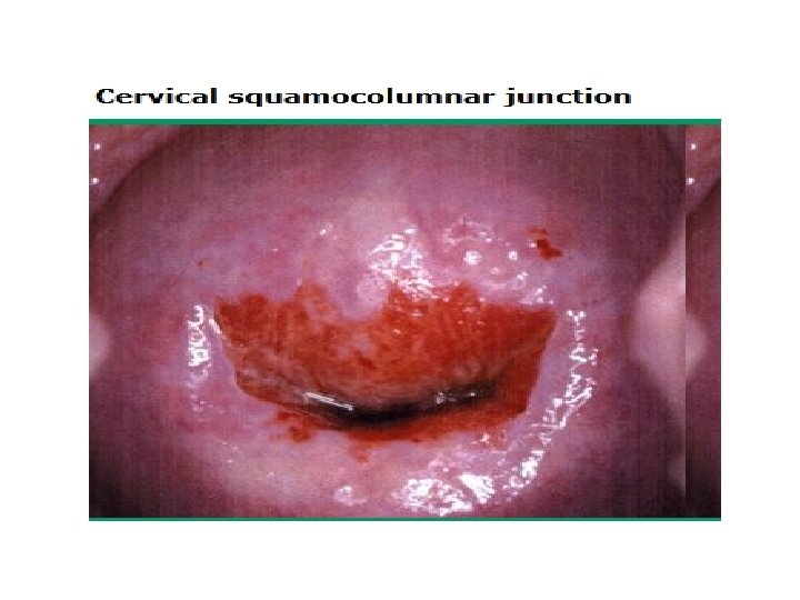
benign cervical lesions- stenosis – ectropion – cervicitis- cystic (nabothian, mesonephric) – non cystic (polyps , warts) ECTROPION Ectropion occurs when eversion of the endocervix exposes columnar epithelium to the vaginal milieu. The everted epithelium has a reddish appearance similar to granulation tissue, and may be covered by a yellow turbid discharge. • Ectropion is common in adolescents. After adolescence, it may be observed in women who are pregnant or taking estrogen-progestin contraceptives or who had a cervical laceration during labor and delivery. • Ectropion has also been called erosion, which is a poor term because it implies that the superficial squamous cells have eroded away to expose underlying tissue. • Ectropion should not be treated except in the rare occurrence of excessive mucous When discharge or spotting that is very bothersome to the woman. In such cases, malignancy to treat should be excluded before undertaking any treatment. • An ablative procedure using cryocautery or electrocautery is effective, but is invasive and will result in copious vaginal discharge until healing is completed, which may take weeks. Ablative treatment can also result in cervical stenosis, which can adversely affect future fertility and, if pregnancy is achieved, labor and delivery. As an alternative, we suggest a two-week trial of an acidifying agent, such as boric acid suppositories 600 mg vaginally at bedtime, which may be effective. Use of deoxyribonucleic acid 5 mg vaginal suppositories has also been reported. • •

• CERVICITIS • Cervicitis is classified as either acute or chronic. • The most common symptom, and often the only one, is purulent vaginal discharge. • The appearance of the acutely inflamed cervix varies greatly, depending upon the degree of involvement and the infecting organism. Mucopurulent cervical discharge, cervical friability, and cervical edema are characteristics of both gonococcal and chlamydial cervicitis; the latter is more common. • Punctate hemorrhages on the vagina and cervix suggest trichomonas infection. • Vesicular or ulcerative lesions suggest infection with herpes simplex virus (HSV).

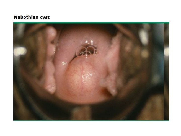
CYSTIC LESIONS • Cervical cysts are usually asymptomatic, but may cause dyspareunia or a pressure sensation. • Nabothian cysts — Nabothian cysts (also called mucinous retention cysts, epithelial inclusion cysts) are discrete cystic structures that form when a cleft of columnar epithelium becomes covered with squamous cells and the columnar cells continue to secrete mucoid material. The cysts vary from microscopic to several centimeters in size; the larger ones project above the surface of the portio. They may appear translucent or opaque. Nabothian cysts may occur following minor trauma or childbirth. The only indication for treatment is relief from pain or a bothersome feeling of fullness in the vagina. Ablation of the cyst using electrocautery is the usual approach; however, if the diagnosis is uncertain, excision to evaluate histopathology is advised. The main disadvantage to surgical treatment is the possibility of causing scar tissue, which itself can lead to dyspareunia.
 duct may be found")
• Mesonephric cysts Microscopic remnants of the mesometanephric (Wolffian) duct may be found deep within the cervical stroma when tissue removed at conization is examined pathologically. Occasionally, one or more of these remnants form a cyst, which may be confused with Nabothian cysts. Mesonephric cysts seldom reach a size greater than 2. 5 cm, and are usually located at the three and nine o'clock points, near the base of the cervix. (ducts sites) Intervention should be avoided in asymptomatic women. Treatment similar to that recommended above for Nabothian cysts is indicated if the patient has bothersome symptoms. A large cyst can be marsupialized

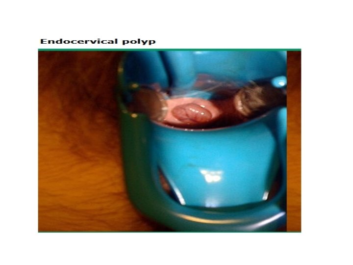
NONCYSTIC LESIONS • • Polyps Cervical polyps commonly occur during the reproductive years, especially after age 40 years. The etiology is unknown. Chronic inflammation of the cervical canal may play a role, as may hormonal factors, since endometrial hyperplasia and polyps coexist more frequently than one would expect by chance alone. Differential diagnosis includes leiomyoma and endometrial polyps Single or multiple cervical polyps usually arise from the endocervical canal, but can also originate from the portio. The tear-shaped or lobular structures appear red, purple, or flesh-colored, depending upon the vascularity and congestion present, and usually look succulent and glistening. The size is typically less than 3 cm in diameter; however, polyps large enough to fill the vagina or presenting at the introitus have been described. The pedicle is usually long and thin, but may be short and broad-based. Histologically, cervical polyps are characterized by vascular connective tissue stroma covered by epithelium, which may be columnar, squamous, or squamocolumnar. Polyps should be removed when symptomatic (eg, bleeding, excessive discharge), large (≥ 3 cm), or appearing atypical. Polypectomy can usually be accomplished by grasping the base of the polyp with forceps and twisting it off. Malignancy is rarely found in a cervical polyp;

Warts • Vulvar, perineal, and anogenital warts are frequently associated with vaginal and cervical lesions. Single or multiple condylomata acuminata due to HPV infection may be visible grossly, and are asymptomatic. These warts are often flat, in contrast to condyloma in other areas of the anogenital region. They become white, and thus more visible to the naked eye when swabbed with a solution of 3 to 5 percent acetic acid. Cytology/colposcopy may reveal squamous intraepithelial neoplasia. These abnormalities are discussed separately.

CERVICAL STENOSIS • Cervical stenosis is usually caused by contraction of scar tissue, agglutination of raw surfaces within the endocervical canal, or obliteration of the endocervical canal by tumor (eg, endometrial or cervical cancer), although some cases are congenital. • Cervical surgery and radium application are common causes of scarring; secondary dysmenorrhea or amenorrhea after cervical procedures strongly suggests cervical stenosis has developed. • The hypoestrogenic state of menopause induces endocervical changes favorable for agglutination. • Diagnosed when cervical canal does not permit the insertion of an instrument, such as a Hegar dilator (2. 5 mm in diameter).

• The diameters of the cervical canal in nulliparous and multiparous women are approximately 5 mm and 8 mm, respectively. • Potential consequences of cervical stenosis include: 1. Impeded menstrual flow — Menses may be scant, painful, or prolonged. In severe stenosis, hematometra can develop. 2. Impeded access to the endocervical canal and endometrial cavity for diagnostic and therapeutic procedures. 3. Cervical dystocia during labor. • Treatment of cervical stenosis consists of dilatation of the cervix to allow access to and drainage from the uterine cavity.
- Slides: 42