UTERUS DR SAVITA KADAM KHISTE ASSOCIATE PROFESSOR MGM
 ASSOCIATE PROFESSOR MGM MEDICAL COLLEGE AURANGABAD")


 • Flexion")


 •")


- Slides: 66
UTERUS DR. SAVITA KADAM (KHISTE) ASSOCIATE PROFESSOR MGM MEDICAL COLLEGE AURANGABAD
Female reproductive system • External genital organs • Internal genital organs
Internal genital organs consist of – • Pair of fallopian tubes • Pair of ovaries • Uterus • Vagina
INTRODUCTION • The uterus is a dynamic female reproductive organ responsible for several reproductive functions including menses , implantation, gestation, labour and delivery. • It remains in a relatively quiescent state during the prepubertal and postmenopausal years.
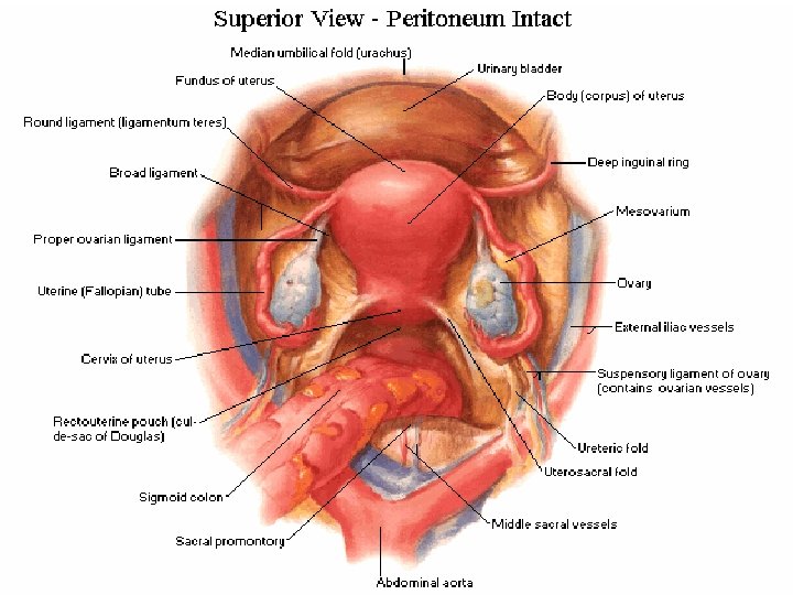
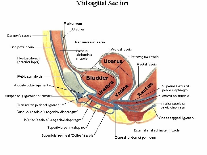
LOCATION • Uterus is situated in pelvic cavity with bladder in front and rectum behind • Thick walled muscular organ and firm in consistency Three layers • Endometrium • Myometrium • Perimetrium
SHAPE AND SIZE • Pyriform / pear shaped • Total length – 7. 5 cm • Breadth – 5 cm • Thickness – 2. 5 cm • Weight – 30 -40 gms
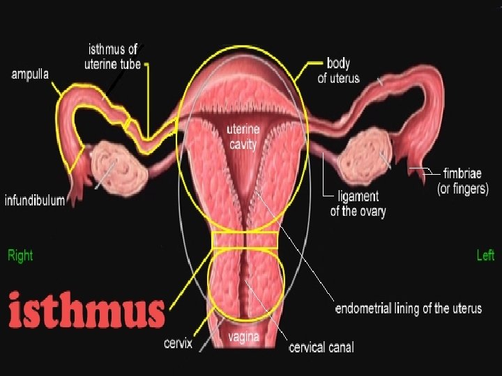
PARTS OF UTERUS Two parts 1. Body 2. Cervix Junction of body and cervix Isthmus (corresponds with internal os of cervical canal)
PRESENTING PARTS OF BODY 1. Fundus 2. Body proper FUNDUS –Expanded upper part above opening of uterine tubes BODY– extends from fundus—isthmus has cavity Two surfaces → ant /vesical ↘ post /intestinal Two lateral borders
Sagittal and coronal section of uterus
Cornu of uterus
Cervix of uterus • Cylindrical part • Less mobile and more fixed • Fusiform shaped • Length— 2. 5 cm • Two parts → Supra-vaginal ↘Vaginal Relations Anteriorly: bladder Posteriorly: rectouterine pouch On sides: ureter and uterine A.
Vaginal Part • Conical in shape • Rest on posterior vaginal wall • Divides vagina into fornices
Nalliparous cervix Multiparous cervix Arbor vitae uteri
• In adult body and cervix ratio is 2: 1 • In new born body and cervix ratio is 1: 2
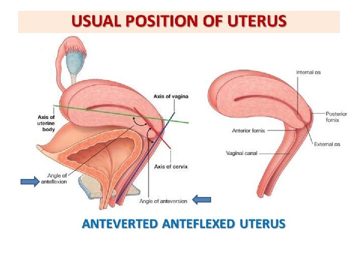
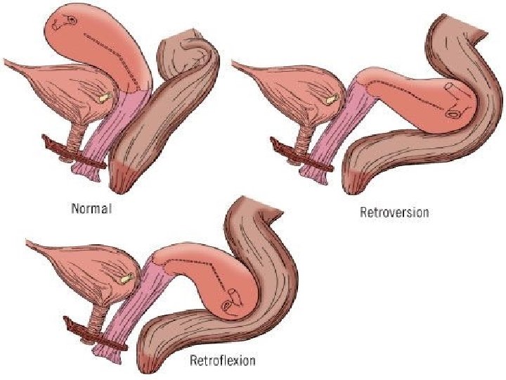
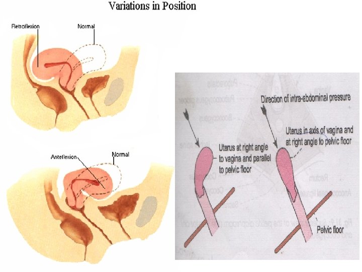
NORMAL POSITION • AVAF Provided rectum and bladder are empty • Long axis of vagina corresponds to axis of pelvic outlet • Long axis of uterus corresponds to axis of pelvic inlet
Antevertion • Vertion is relation of long axis of cervix with vagina • It occurs around transverse axis passing through ex. os • It is maintained by 1. Forward pull on fundus by traction of Round Ligament 2. Backward pull on cervix by traction of Uterosacral Ligament
• Ante flexion forward angle bet body and cervix at isthmus(125) • Flexion of uterus takes place around transverse axis passing through the internal os • When uterus rotated anteriorly –Anteflexion • When rotated posteriorly –Retroflexion
• Fundus tilted to Rt side –Dextro-rotation • Fundus tilted to Lt –Levo -rotation
LIGAMENTS OF UTERUS 1. Peritonial 2. Fibromuscular
PERITONIAL LIGAMENTS • Do not support • Anterior false ligament (uterovesical fold ) • Posterior false ligament (rectovaginal fold ) • Broad ligament
BROAD LIGAMENT • Fold of peritoneum • Extends from lateral border of uterus to lateral pelvic wall • Divides pelvic cavity into • Anterior compartment for bladder • Posterior compartment for rectum and sigmoid colon
SUB-DIVISIONS 1 Meso-salpinx 2 Meso-metrium 3 Meso –ovarium 4 Suspensory ligament of ovary
CONTENTS OF BROAD –LIGAMENT One tube : Uterine tube Two ligaments : 1. Round ligament of uterus 2. Ligament of ovary Two vessels : 1. Uterine vessels 2. Ovarian vessels Two plexus of nerves : 1. Utero-vaginal plexus 2. Ovarian plexus Three embryological remnants 1. Epo-ophoron 2. Par-ophoron 3. Vesicular appendicies Other structures : 1. Lymph vessel and lymphnodes 2. Parametrium on sides of uterus
FIBRO-MUSCULAR LIGAMENTS • Round ligament • Uterosacral ligament • Mackenrodt’s ligament • Pubocervical ligament
ROUND LIGAMENT • Fibro muscular band • 10 -12 cm • Extent–cornu of Uterus passes through inguinal canal splits into fibrous threads and attaches to labia majora • Developementaly remnant of distal part of gubernaculam of ovary • Pulls fundus forward
UTEROSACRAL LIGAMENT • Condensation of endopelvic fascia • Extend –back of cervix to periosteum of sacrum (S 2 , S 3) • Pulls cervix backward
MACKENRODT’S LIGAMENT • Fan shaped fibromuscular band • Extent –cervicovaginal junction to lateral pelvic wall • Keep cervix in median position and prevent downward displacement of uterus.
PUBOCERVICAL LIGAMENT • Connect anterior surface of cervix to inner surface of pubic symphysis • Keep the cervix in midline
uterosacral Cardinal Lig pubocervical
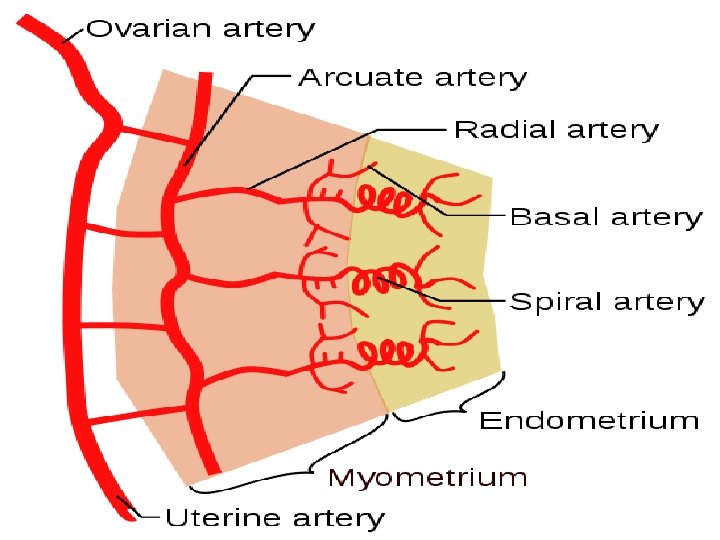
BLOOD SUPPLY • Uterine artery • Ovarian artery
VENOUS DRAINAGE Internal iliac veins
NERVE SUPPLY • SYMPATHETIC NERVES –T 12 -L 1 • Causes uterine contraction and vasoconstriction • PARASYMPATHATIC NERVES –S 2 3 4 • Causes vasodilatation and uterine inhibition • Pain sensation from body is carried by sympathetic • Pain sensation from cervix is carried by parasympathetic
LYMPHATIC DRAINAGE • Consist of three intercommunicating plexus • Endometrial • Myometrial • Subperitonial • Upper part—aortic LN • Middle part– external iliac nodes • Lower part—external, internal and superficial inguinal nodes
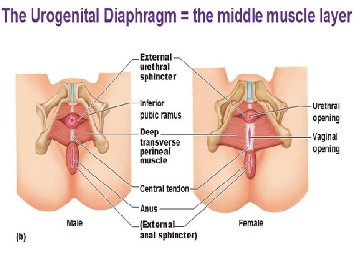
SUPPORTS OF UTERUS • SECONDARY SUPPORTS PRIMARY SUPPORTS MUSCULAR/ ACTIVE -PELVIC DIAPHRAGM -UROGENITAL DIAPHRAGM -PERINEAL BODY FIBROMUSCULAR/ MECHANICAL -UTERINE AXIS -PUBOCERVICAL LIGAMENT -MACKENRODT LIGAMENT -UTROSACRAL LIGAMENT -ROUND LIGAMENT -BROAD LIGAMENT -UTROVASICALE FOLD -RECTOVAGINAL FOLD
Resists increase intra-abdominal pressure
PERINEAL BODY Acts as anchor for pelvic diaphragm Maintains integrity of pelvic floor
FUNCTIONAL SUPPORTS Acting from above-Peritonial folds Acting from below-Levator Ani Acting from sides- Mackenrodt’s Maintaining the Anteversion and Anteflexion- Round, Utrosacral and pubocervical ligaments • Loose packing Material –Parametric tissue • •
INVESTIGATIONS • PELVIC ULTRASOUND • HYSTEROSALPHYNGOGRAM • MRI • LAPROSCOPY • HYSTEROSCOPY • COLPOSCOPY
USG
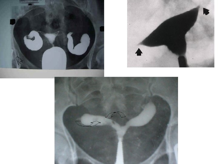
HYSTROSALPHINGOGRAM • Defination • H. S. G. is a radiological procedure to visualise interior of the uterus and fallopian tube to access its patency. • Indications– Infertility
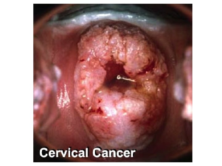
APPLIED ANATOMY • • Endometritis IUCD D AND C ENDOMETRIOSIS PID HYSTEROTOMY HYSTERECTOMY • • COLPOTOMY PROLAPSE UTERUS UTERINE FIBROIDS CARCINOMAS
ENDOMETRIOSIS • Endometriosispresence and proliferation of endometrial tissue outside the uterine cavity • Major symptomsdysmenorrhoea, infertility
IUCD
PID
HYSTEROTOMY
HYSTERECTOMY
COLPOTOMY
UTERINE FIBIROIDS leiomyomas –myometrial –submucosal, –subserosal –Pedenculated
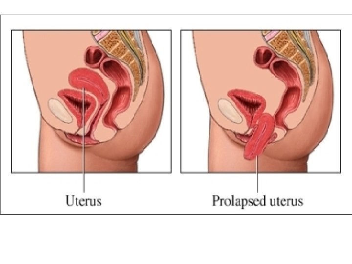
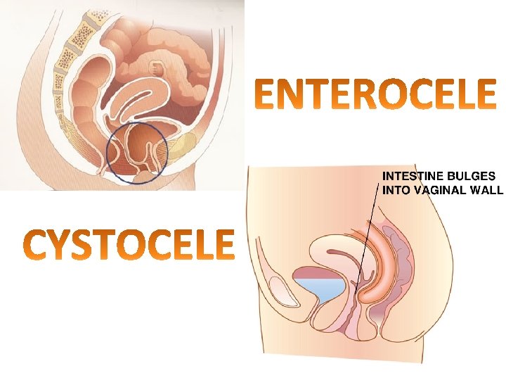
PROLAPSED UTERUS
ENDOMETRIAL CANCER
L. S. C. S
• At the end of lecture students should able to 1. enumerate functions of uterus 2. Enumerate parts of uterus 3. Describe the position of uterus with its applied importance 4. Explain the peritonial reflections 5. Clinical importance of POD 6. Enumerate various supports of uterus 7. Describe HSG 8. Describe blood supply , nerve supply 9. Describe lymphatic drainage with its clinical importance 10. Enumerate clinical conditions of uterus