Urogenital flagellates Trichomonas vaginalis TRICHOMONIASIS Trichomonas vaginalis A

 q. Trichomonas vaginalis has a world-wide distribution; incidence is")


 v. Rarely Infants may")


 or cell mouth is a part")


 7. Colitis produced by Balantidium coli often is")


- Slides: 25
Urogenital flagellates Trichomonas vaginalis
TRICHOMONIASIS Trichomonas vaginalis (A Flagellate) q. Trichomonas vaginalis has a world-wide distribution; incidence is as low as 5% in normal females and as high as 70% among Females expressing illegal sexual activity. q. It is estimated by WHO annually 170 million cases is recorded over the world and q 8 millions in USA annually, 13% of black women are affected compared with 1. 8% of White women.
q The infection by T. vaginalis it is high percentage among persons with multiple sex partners, exchanging sex for payment, or a history of STD). q Vaginitis with a purulent (Pus- filled)discharge is the main symptom, and can be accompanied by vulvar and cervical lesions, abdominal pain, dysuria and dyspareunia. q In men, the infection is frequently asymptomatic; occasionally, urethritis, epididymitis, and prostatitis can occur. q Confusing diagnosis with double stranded RNA virus called T. vaginalis virus (TVV) q jerky movement
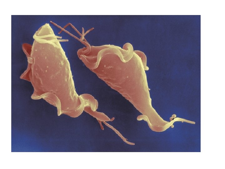
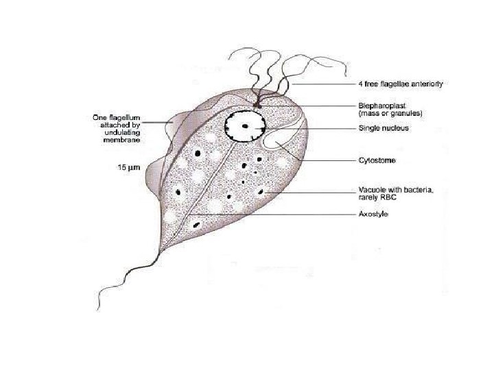
v Morphology The Trophozoite form is 15 to 18 micrometers in diameter and is half pear shaped with a single nucleus, Four anterior flagella and a lateral flagellum attached by an undulating membrane. Axostyles are arranged asymmetrically. The organism does not Encyst while it has psedocyst in alkaline environment. v Life cycle: T. vaginalis colonizes the vagina of women and the urethra (sometimes prostate) of men. Infection occurs primarily via sexual contact, although non-venereal infections are possible. The are divides by binary fission which is favored by low acidity (p. H > 5. 9; the normal p. H is 3. 5 to 4. 5). The women become Reservoir host while men are carrier host
LIFE CYCLE
Pathology v T. vaginals causes contact-dependent damage to the epithelium of the infected organ. v. Role of infertility for men and women(tubal infertility). v. Role of cervical cancer. v. Increased in immunocomparised cases. v. Increased in preterm birth, and other adverse pregnant women. v. T vaginalis infection is associated with increased risk for PID v. T. vaginalis increase with other STDS cases.
v. Infections can be transmitted by fomites (toilet articles, clothing) v. Rarely Infants may be infected by passage through the mother’s infected birth canal. (not congenital)
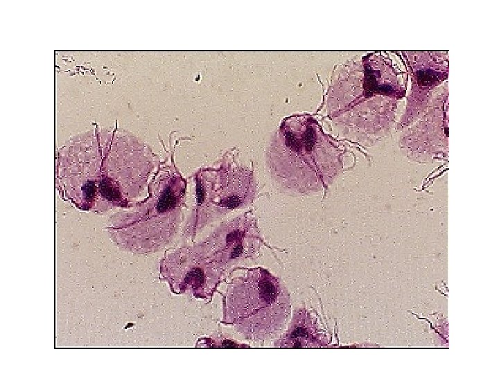
Symptoms T. vaginalis infection is rarely symptomatic in men, although it may cause mild urethritis or occasionally prostatitis. In women, it is often asymptomatic, but heavy infections in a high p. H environment may cause mild to severe vaginitis with foul-smelling , malodorous, or yellow-green with or without vulvar irritation. . LAB DIAGNOSIS 1. vaginal swab(wet-mount) directly or finding the organism in Giemsa-stained smears of vaginal discharge. 1 S (35 -80%) compared with 2 2. Cultivation of a swab sample in Diamond's broth medium. 2 S (38 -82%) compared with 7&8
Note : A. Trophozoites must be distinguished from the non-pathogenic flagellate Trichomonas hominis. in mixed or contaminated samples. B. Confusing between T. vaginalis and T. vaginalis viruse(TVV) 3. Detection psedocyst in semen by Seminal fluid Analysis(SFA) &Post Coital Test (PCT). 4. Pap smear for cervical cancer 5. In sedimentation of urine. 6. El. ISA in vaginal secretion
7. NAAT is highly sensitive, often detecting three to five times more T. vaginalis infections than wet-mount microscopy, and assay called (Hologic Gen-Probe, San Diego, CA) which is FDA-cleared for detection of T. vaginalis from Vaginal, Endocervical, or Urine specimens from women. This assay detects RNA by transcription-mediated amplification with a gold sensitivity Among women, by vaginal swab and urine. In men this assay can be used with urine or urethral swabs. 8. Convertial and real time PCR
Trichomonas hominis The trophozoites live in the caecal area of the large intestine and feed on bacteria. It is considered to be nonpathogenic, although it is often recovered from diarrheic stools. Since there is no known cyst stage Trichomanas tenax was first recovered from the mouth, specifically in tartar from the teeth. There is no known cyst stage. The trophozoite has a pyriform shape and is smaller and more slender than that of T. hominis. Diagnosis is based on the recovery of the organism from the teeth, gums, or tonsillar crypts, and no therapy is indicated.
Ciliates Only example in Human Balantidium coli
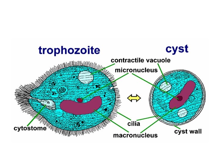
Balantidium coli v IS only ciliate infecting humans v Is the largest pathogenic protozoan v Defining characteristic is nuclear Dimorphism v Micronucleus & Macronucleus v Disease: Balantidiasis, Balantidial dysentery v Infective stage: Cysts, infection most commonly occur in Rural Dwellers. v Mode of transmission: Fecal-oral route v Habitat: Large intestine (colon) of human and some animals e. g. Pig, cow and horses.
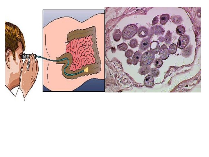
v. Acytostome (from cyto-, cell and stome-, mouth) or cell mouth is a part of a cell specialized for phagocytosis, usually in the form of a microtubule-supported funnel or groove. Food is directed into the cytostome, and sealed into vacuoles.
B. coli trophozoites are characterized by their large size (40 to more than 70µ; the presence of cilia on the cell surface which are particularly visible in (B); a cytostome (arrows); a bean-shaped macronucleus than is often visible, see (A); and a smaller, less conspicuous micronucleus. Balantidium coli cyst in stool preparation
Life Cycle: Direct.
CLINICAL MANIFESTATIONS: 1. The pathogenesis of Balantidiasis are similar to those seen in Entamoebiasis including intestinal erosion Most human infections are asymptomatic. 2. Acute infection is characterized by rapid onset of nausea, vomiting. 3. Abdominal discomfort or pain, and bloody or watery mucoid diarrhea. 4. Infected patients can develop chronic intermittent(coming and going at intervals) & episodes of diarrhea. 5. Rarely, organisms spread to mesenteric nodes, pleura, or liver.
6. Inflammation of the gastrointestinal tract(GIT) 7. Colitis produced by Balantidium coli often is indistinguishable from that produced by Entamoeba histolytica. 8. Fulminant disease can occur in malnourished or otherwise debilitated(weak) patients. Fulminant: process that occurs suddenly and escalates quickly, and is intense and severe to the point of lethality
DIAGNONSIS A: Microscopic examination of a patient’s feces. Stool sample is collected and a wet mount is prepared. Cysts or trophozoites can be detected in feces B: Trophozoites can also be detected in the tissue of rectum and the sigmoid colon by Sigmoidoscopy during looking for bleeding, ulcers, and inflammation by physicians then take a tissue biopsy for inspection. C. Molecular and Immunological techniques
Summary Organism Transmission Symptoms Diagnosis Oro-fecal Dysentery with blood and necrotic tissue. Chronic: abscesses Stool: cysts with 1 -4 nuclei and/or trophs. Trophs in aspirate. Giardia lamblia Oro-fecal Fowl-smelling, bulky and lipidemia diarrhea; blood or necrotic tissue rare. Stool : troph. and/or cyst. Iodoquinol or Metronidazole. Balantidium coli Oro-fecal; zoonotic Dysentery with blood and necrotic tissue but no abscesses. Stool: trophs and/or cysts. Iodoquinol or Metronidazole. Trichomonas vaginalis Sexual Vaginitis; occasional urethritis/prostatitis. in vaginal (or in urethral smear. Mebendazole; vinegar douche; metronidazole Entameba histolytica Treatment GI: Iodoquinol or Metronidazole