TISSUES TISSUES DEFINITION A GROUP OF SIMILAR CELLS


.")


 ?")


")
 Dense CT")


 Not quite as strong as bone LOCATIONS: external ear and epiglottis,")


 provides support for soft tissues of the body LOCATION: BONES")

 Transports oxygen, carbon dioxide, nutrients, wastes, antibodies, and hormones There")

 • Lymph")


- Slides: 47
TISSUES
TISSUES DEFINITION: A GROUP OF SIMILAR CELLS THAT ARE SPECIALIZED TO PERFORM A SPECIFIC FUNCTION. 4 PRIMARY TISSUE TYPES: 1. 2. 3. 4. EPITHELIAL(covers and lines organs) CONNECTIVE (connects parts of the body) MUSCLE (movement) NERVOUS
Tissue Outline • 1. Epithelial – Simple squamous epithelium – Simple cuboidal epithelium – Simple columnar epithelium – Pseudostratified columnar epithelium – Stratified squamous epithelium – Stratified cuboidal epithelium – Stratified columnar epithelium – Transitional epithelium – Glandular epithelium
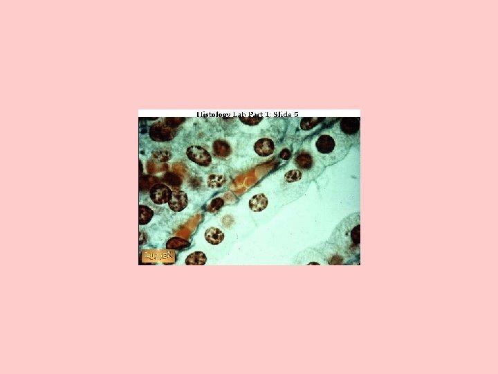
2. Connective Tissue – Connective Tissue Proper (loose/areolar CT, adipose, reticular CT, Dense CT, Elastic CT) – Cartilage – Bone – Blood – Lymph (book doesn’t classify)
3. Muscle – Striated – Smooth – Cardiac
4. Nervous – (no sub tissue just lots of cells like neurons, neuroglial etc).
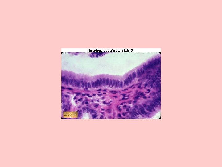
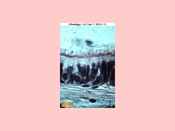
1. Epithelium Covers or lines organs.
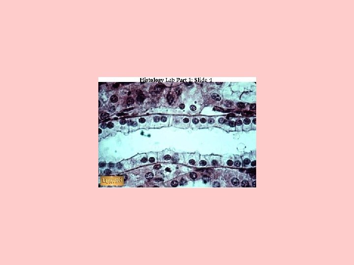
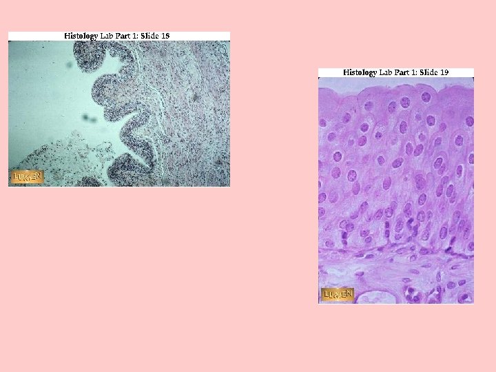
2 WAYS TO CLASSIFY EPITHELIA BY NUMBER OF LAYERS: SIMPLE HAS ONE LAYER STRATIFIED HAS 2 OR MORE LAYERS Skin is an example
BY THE SHAPE OF THE CELL: SQUAMOUS - FLATTENED CUBOIDAL- CUBE SHAPED
COLUMNAR - BRICK SHAPED
Identify the tissue type. (See next slide for answer) ?
Stratified cuboidal epithelium.
2. Connective Tissue Connects the parts of the body.
CONNECTIVE TISSUE • THE MOST ABUNDANT OF THE PRIMARY TISSUES FUNCTIONS: • BINDING & SUPPORT, PROTECTION, INSULATION, TRANSPORTATION
CONNECTIVE TISSUE SUBCLASSES a. CONNECTIVE TISSUE PROPER (Binding, Supporting, Protection, Insulation and Transportation CT) b. CARTILAGE (Supporting CT) c. BONE (Supporting CT) d. BLOOD (Fluid CT) e. LYMPH (Fluid CT)
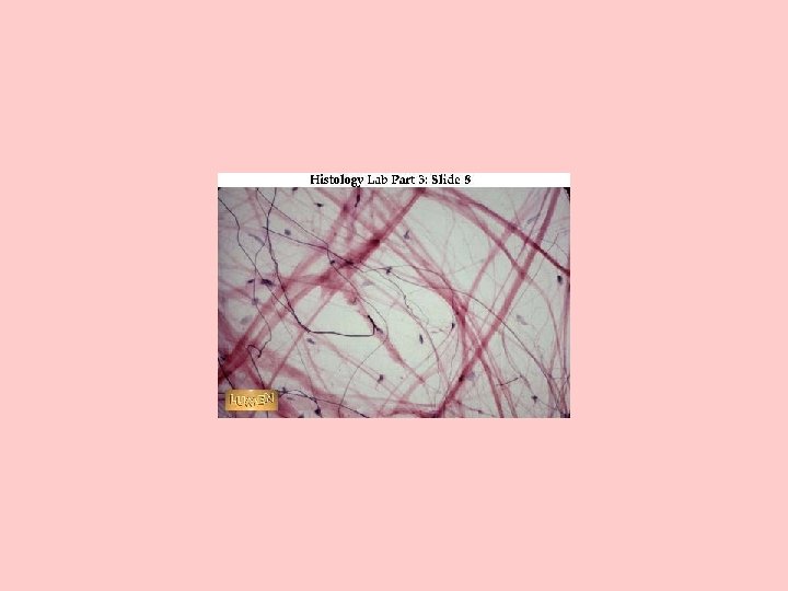
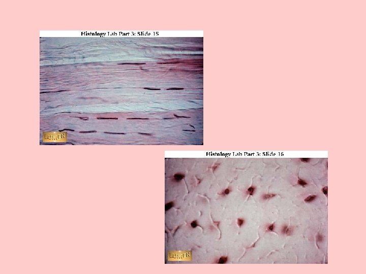
CONNECTIVE TISSUE PROPER Types - Review • • • Loose CT (aerolar) Dense CT Adipose Reticular CT Elastic Ct
CONNECTIVE TISSUE PROPER Loose – Fibers create loose, open framework between organs, beneath the skin and between muscles.
• Dense – Fibers densely packed, found in tendons and ligaments
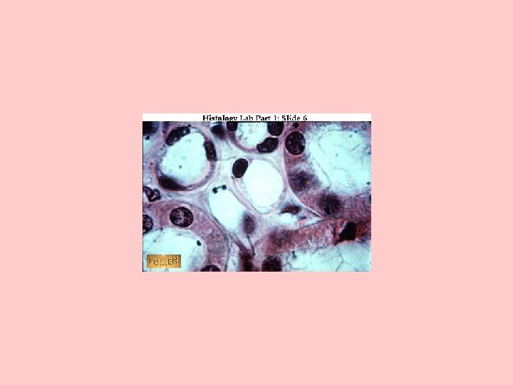
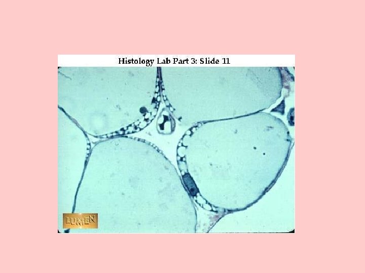
• Adipose – Cells called Adipocytes. They store lipids which are a reserve of energy. Collectively they assist in thermoregulation (body temperature) and in some places like around the kidney/behind the eyeball provide some cushioning.
adipose tissue stored fat nucleus
• Reticular CT – Consists of thin, branched collagenous fibers. This tissue supports the walls of the liver, spleen, and lymphatic organs.
• Elastic CT – composed of elastic fibers. It gives an elastic property to hollow internal organs (ex. Blood vessels and lungs)
Connective Tissue Proper – The Cells – Fibroblasts – – Macrophages– Adipocytes– Mesenchymal cells– Melanocytes • Melanin- – Mast cells– Lymphocytes– Microphages-
Connective Tissue Proper – The Tissue Fibers • • Collagen fibers. Reticular Fibers. Elastic Fibers. Ground Substance-
CARTILAGE (supporting CT) Not quite as strong as bone LOCATIONS: external ear and epiglottis, fetal skeleton, growth plates, larynx, trachea, rib cage.
CARTILAGE
Types of Cartilage • Hyaline cartilage • Elastic Cartilage • Fibrocartilage-
BONE (supporting CT) provides support for soft tissues of the body LOCATION: BONES
HAVERSIAN CANAL HAVERSIAN SYSTEM LAMELLAE OSTEOCYTE
BLOOD (Fluid Connective tissue) Transports oxygen, carbon dioxide, nutrients, wastes, antibodies, and hormones There are 3 types of blood cells: RBC- carries oxygen WBC- functions in immunity Platelet- important in blood clotting LOCATION: BLOOD VESSELS
RBC red blood cell PLATELET WBC White blood cell
Lymph (Fluid Connective tissue) • Lymph
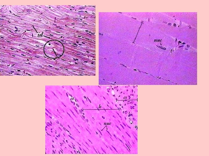
Muscle • Striated Muscle- Also called skeletal muscle – attached to bone – for movement -usually voluntary • Smooth Muscle- in the walls of hollow organs ex. Intestine - involuntary • Cardiac Muscle-only in the heart. It is striated also but connected by intercalated discs. Involuntary also.
Nervous
• Complete text questions pg. 143 -162 Wed. • Complete text questions for pg. 163 -165 for Friday.
• http: //www. siumed. edu/~dking 2/intro/ct. ht m#adipocytes • http: //www. meddean. luc. edu/lumen/meded/ histo/frames/h_frame 3. html • http: //www. austincc. edu/histologyhelp/tissu es/tn_elas_ct. html