THROAT THORAX INJURIES Unit 3 Understanding Injuries to


- Slides: 40
THROAT & THORAX INJURIES Unit 3: Understanding Injuries to the Axial Region
Objectives Understand the basic anatomy of the throat and thorax Understand how to prevent injuries of the throat and thorax Know the necessary care to treat an injury to the throat or thorax Understand the implications of illness or injury related to specific organs in the thorax
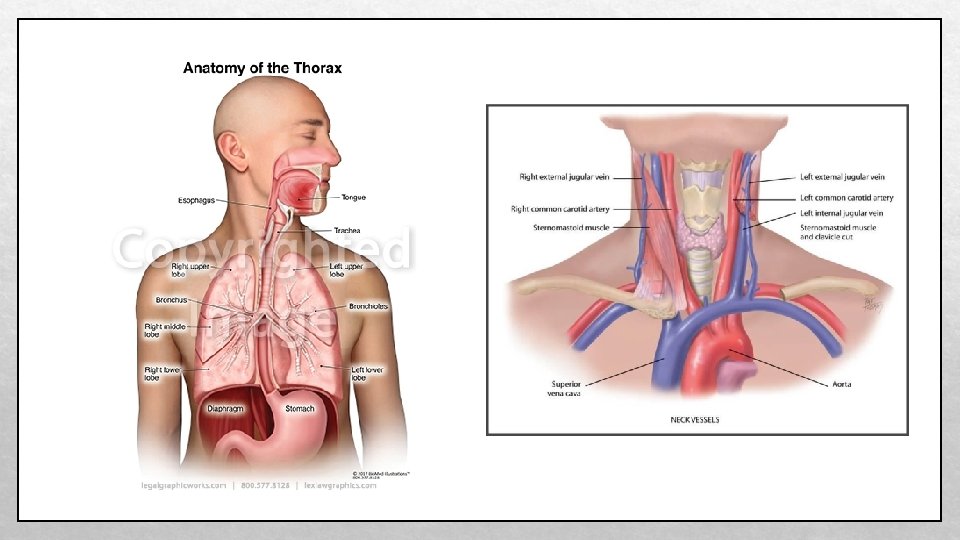
Anatomy of the Throat • The throat contains the carotid arteries, jugular veins, larynx, trachea, and esophagus • These structures are very sensitive and vital to life, therefore, the AT must understand their purpose • Esophagus: passageway for food going from the mouth to the stomach • Lies in front of the cervical vertebrae and behind the trachea and larynx • Trachea: made up of circular rings of cartilage and is the main trunk of the system of tubes through which air passes to and from the lungs, also known as the windpipe • Larynx: an enlarged tube at the top of the trachea made of cartilage and muscles and contain the vocal cords; also known as the voice box
Carotid artery: one of the two vessels that run from the aorta through the neck and supply the head with oxygenated blood Anatomy of the Throat Jugular vein: one of two vessels that takes unoxygenated blood from the head to the heart Severing one of these vessels can cause death in a short time, so protection of the neck is vital in sports like ice hockey and field hockey
Anatomy of the Thorax • The bony structure of the thorax is made up of the thoracic vertebrae posteriorly, 12 ribs on each side, and the sternum anteriorly • These bones protect the sensitive organs in the thorax • The two lowest ribs do not attach to the sternum and are called floating ribs
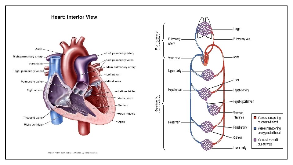
Heart and Lungs • The heart is about the size of a fist and is responsible for pumping blood to all parts of the body • The blood carries nutrients and oxygen to cells and carbon dioxide and waste products away from cells • The heart is divided into 4 chambers: the upper chambers, left and right atria and the lower chambers, the left and right ventricles • Atria: upper chambers of the heart that receive blood from the veins and force it into the ventricles • Ventricles: lower chambers of the heart that receive blood from the corresponding atrium and from which blood is forced into the arteries
Heart and Lungs • The ventricles are generally larger and have thicker walls than the atria because they pump the blood throughout the body • Exercising the heart muscle makes it larger and more efficient at pumping • Enlarged heart can also be a sign of heart disease • The heart pumps blood into the lungs and around the body • The right atrium fills with blood from a vein, which is carrying waste products and CO 2, the right ventricle receives blood from the right atrium and pumps it to the lungs to get rid of CO 2 and pick up O 2, the left atrium fills with oxygenated blood from the lungs, the left ventricle, which is the largest chamber of the heart, received the oxygenated blood from the left atrium and pumps it throughout the body
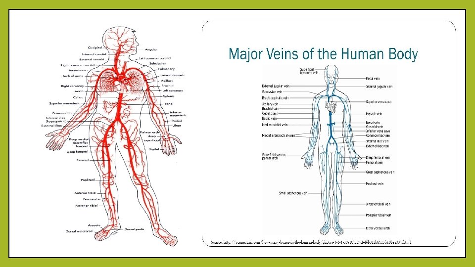
Heart and Lungs • Aorta: main artery leaving the heart • The aorta travels downward throughout the chest and abdomen, and other large arteries branch off to the head (carotid), arms (brachial) and legs (femoral) • After oxygen-rich blood has been delivered and used by the tissues, the deoxygenated blood returns to the heart through major veins • Two electrical nodes in the right atrium begin a contraction • A slight delay in impulses conducted from the nodes through the heart allows blood to be squeezed from one chamber to another • Injured or diseased electrical nodes cause the heart to stop or to beat ineffectively
Heart and Lungs • The lungs, which are located on each side of the heart, exchange oxygen and carbon dioxide and dissipate body heat • The trachea divides into two bronchi; the two bronchi further subdivide into bronchioles, and each bronchiole ends in an alveolus • Alveoli: lung tissue where the gas exchange of oxygen and carbon dioxide occurs • Bronchi are filled with cilia, which are small, hair like projections that help remove foreign substances • Coughing and sneezing help to keep the trachea and bronchi clear and remove phlegm and allergy-causing agents • Phlegm: thick mucous in the respiratory passages in response to allergy or infection • The lung tissue is divided into 3 lobes in the right lung and 2 lobes in the left lung • Lung capacity is hampered primarily by smoking, pollution, and lung disease
Heart and Lungs • Lung function and breathing rate are controlled by CO 2 receptors. When receptors register presence of too much CO 2, inhalation occurs • Exercise increases cell metabolism and causes cells to need more O 2 and eliminate more CO 2 • The increased cellular need increases the number of breaths per minute • Over time, ability of the lungs to exchange air effectively, increases as the athlete breaths become deeper and more forceful • A conditioned athlete will return to a normal breathing rate more quickly after exercising compared to someone who is out of shape
Diaphragm • Diaphragm: muscle that separates the thorax and the abdominal cavity • The diaphragm contracts and pulls down to assist in inhalation and moves upward to push air out of the lungs during exhalation • The diaphragm has 3 openings to allow passage of the esophagus, abdominal aorta, and inferior vena cava
Preventing Throat and Thorax Injuries • Protective equipment and rules in athletic contests are both designed to prevent injuries in the throat and thorax because these areas contain organs that are vital to life • Throat protectors, shoulder pads, chest protectors, and sternal pads • If a ball strikes an athlete in the chest, it could cause the heart to beat irregularly or even stop • A safe distance between the boundary of a playing surface and objects, such as bleachers, fences, scoring tables, and spectators (15 ft. is suggested) • Walls, tables, and fences could be padded • Make sure equipment is certified
Treating Throat Injuries and Conditions • Throat injuries can be simple or devastating • Most injuries that occur to the throat are contusions caused by blows from sticks, feet, or arms • Contusions can be treated by applying ice • In any type of throat injury, the general response by the athlete is coughing, spitting, difficulty breathing, and pain
Throat Laceration • Can occur when one player's ice skate goes across another player's throat • Lacerations that are not deep can be handled with direct pressure • Deep lacerations, or those that affect a jugular vein or carotid artery, are medical emergencies and require immediate action and treatment • Apply direct pressure over the site of the laceration and treat for shock until EMS arrive on scene • Vessels in the neck are large and massive amount of blood will be lost rapidly, so the AT must respond quickly to save the athlete's life
Cartilage Fracture • A severe blow to the throat can result in a fracture of the circular. cartilaginous rings of the trachea and can be life threatening • The athlete will have difficulty breathing and gasp for air, spit up blood, complain of pain, have difficulty talking, and be very anxious • Skin may turn bluish due to lack of oxygen • The AT must use extreme caution because the trauma also may cause a fracture to the cervical spine and is a medical emergency • The AT will place the athlete on a backboard for transportation to the hospital and apply ice to the area to reduce swelling; will also keep the airway free of blood and ensure medical care is on the way • Anyone treating the athlete must remain calm to keep the athlete calm
Treating Thorax Injuries • Many people suffer from conditions such as asthma or experience hyperventilation • If the thorax is not adequately protected, it is vulnerable to blunt trauma that can result in fractures of the ribs or sternum • Severe trauma may cause lung injury
• Hyperventilation: quick, deep breathing at a rate of more than 24 breaths per minute, which leads to abnormal loss of CO 2 from the blood • Can be caused by an athlete getting too excited or an underlying illness, such as diabetes • If the athlete does not get their breathing under control, they will experience lightheadedness, numbing of the fingers, toes, and lips, and loss of consciousness • Muscular contractions will occur in the limbs • Treatment: AT should speak calmly to athlete and encourage them to control breathing rate Hyperventilation
Exercise-Induced Asthma • Asthma: chronic inflammation of the breathing passageways to the lungs • There can be many triggers • The bronchi spasm and narrow the breathing passageways and causes the athlete to wheeze and struggle with taking a full breath • Been compared to breathing through a straw • Athlete may experience coughing, tightness in the chest, and mucous production • Immediate treatment is cease activity, sit upright, and breathe in through the nose and out through the mouth • The athlete should take medication if it has been prescribed before the attack gets worse • If the athlete is unable to breathe or progressively getting worse, EMS must be called • Medication can be used as a preventative measure 20 minutes before exercise
Jogger's Nipple • Most commonly found among male athletes and occurs when the shirt rubs against the nipple repetitively • Called jogger's nipple because long-distance runners are the most inclined to suffer from this condition • The nipple becomes irritated, sometimes to the point where application of a lubricant or bandages become necessary • Prevention involves using lubricant or applying an adhesive bandage over the nipple • If irritation does not subside with minimal treatment a physician may have to evaluate the athlete to determine if there is infection
Blow to the Solar Plexus • Typically caused by a blow to the area of the diaphragm that hits the nerve of the solar plexus • When this occurs, the athlete will struggle to take a breath because the diaphragm spasms • After a short span of time, the diaphragm relaxes and the athlete is able to breathe again normally • The AT should reassure the athlete while they recover
Pulmonary Contusion • Bruise of the lung due to an impact, such as a baseball to the chest or a tackle • The contusion results in an accumulation of blood and other fluids within the lung tissue • The accumulated fluid keeps the lung from exchanging O 2 and CO 2 • The larger the contusion, the more serious the injury • The athlete will have difficulty breathing and may have a bluish skin color • Application of ice may be helpful, but EMS should be called immediately
Myocardial Contusion • Impact to the chest over the heart and the heart becomes bruised • A typical impact would be from a ball or shoulder to the chest • An athlete may experience pain in the chest, especially over the sternum, and rapid heart rate • EMS should be called. In the meantime, the athlete may need to be treated for shock
• Caused by the same impact or compression of a rib fracture, but the force does not cause a fracture • There is pain and signs and symptoms may be similar to rib fracture • When the AT evaluates the athlete, there is pain over the site of the impact, but not on compression away from the site • Treatment involves application of ice and rest • An athlete may participate as pain allows • The area may need to be padded to allow participation Rib Contusion
Ruptured Diaphragm • Infrequently in athletics does the diaphragm rupture as a result of a blow in the general area of the diaphragm • The athlete will present with difficulty breathing with no real trauma to the chest • The athlete may have other internal injuries, most likely in the abdominal region • If the AT auscultates the chest, bowel sounds may be heard • Treatment is to care for the presenting signs or symptoms • If the athlete is having difficulty breathing, elevating the head will be helpful • Treat the athlete for shock • This injury can be hard to diagnose, so if difficulty with breathing continues, call EMS
Sternal Fracture • Occurs because of a direct impact to the sternum that causes a fracture • Should also expect other internal injuries; heart and lungs may be involved • A suspected fracture is treated with application of ice and referral to a hospital • If the sternum is only contused, the athlete could possibly return to activity with a special sternal pad
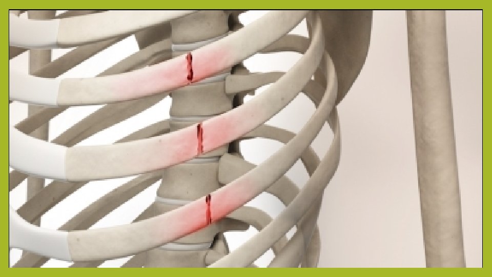
Rib Fracture • A rib fracture is caused by a direct impact or chest compression • On rare occasions, a sudden, violent muscular contraction, such as throwing a baseball, will cause the rib to fracture • Blow to the front or the back of rubs generally do not result in inward displacement of the fractured rib • Blows to the lateral aspect are more likely to lead to inward penetration, causing complications, such as internal bleeding or a punctured lung • An athlete with fractured ribs experience pain and difficulty breathing • The pain increases with inhalation and the athlete usually holds a hand over the injured area in an effort to support the ribs • The area may be deformed due to swelling • A key to determining if a rib is fractured or severely contused is to note whether the athlete experiences increased pain with inhalation • If they have pain during inhalation and exhalation, they more like have a contusion • Treatment for the uncomplicated rib fracture is applying ice and sending the athlete for X-Rays • Physician will restrict an athlete's physical activity until inhalation is no longer painful • Contact sport should be stopped for a minimum of 6 weeks
Flail Chest • Flail chest: occurs when several consecutive ribs are fractures in two or more places • The injury occurs from impact directly to the ribs and the entire fractured portion moves in and out when the athlete breathes • The portion moves opposite to the normal breathing pattern • The movement creates extreme pain and difficulty breathing. The athlete will also feel anxious and may present with a bluish skin tone • Treatment includes decreasing movement of the fractured ribs by placing an object, such as a sandbag or pillow, over the fractured segment • The athlete can be placed on their injured side as a way of controlling the movement of flail chest and may also need to be treated for shock • Medical emergency and requires rapid medical care
Pneumothorax • Pneumothorax: presence of air in the pleural cavity, commonly known as a collapsed lung, which can occur as a result of a trauma or without trauma • A traumatic pneumothorax can occur from a rib puncturing the lung, gunshot wound, or severe laceration • A nontraumatic pneumothorax occurs due to weakness of the lung tissue • When a pneumothorax occurs, the injured lung moves toward the center of the chest, which put pressure on the heart and other lung • Because only one lung is functioning, the athlete will experience difficulty breathing and will gasp for air • As the athlete continues to breathe, air goes through the hole in the lung and into the chest cavity, which causes the collapsed lung to compress the heart and opposite lung even further
• When there is an imperfection in the tissue of the lung, it can break and cause the lung to collapse • There doesn’t have to be any impact or illness Spontaneous Pneumothorax associated; the athlete may appear healthy in the past and had no previous signs of illness • The athlete will experience difficulty breathing, chest pain, and possibly bluish color of the skin if breathing is poor • The athlete should be placed so that the side with the injured lung is closest to the ground • The AT will treat the athlete for shock and get them to a hospital • Generally will heal itself without surgical interventions
Tension Pneumothorax • An athlete with a pneumothorax may develop a more serious problem called tension pneumothorax • As air leaks out of the collapsed lung and into the chest cavity, it forces the lung to press against the other lung and the heart • If the AT observes the trachea deviated to the side of the throat, they should suspect tracheal shifting • As pressure builds in the chest, the trachea moves away from the side of the pneumothorax • If the trachea moves, the athlete will experience severe respiratory distress • As more air enters the chest cavity, move pressure builds against the heart and uninjured lung • As the pressure mounts, the heart begins to labor as blood flow and breathing are impeded • Death can occur if the athlete is not treated rapidly • If the athlete has an external puncture wound, partially cover it, leaving one side unsealed • Sealing the wound entirely will prevent the inner air from escaping, worsening the condition • With tension pneumothorax, the athlete will experience respiratory distress, absent breath sounds on the injured side, anxiety, and bluish skin color • His pulse will be rapid and weak and blood pressure will drop • As the pneumothorax, worsens, tracheal deviation and neck vein distension will occur, as will bulging of the muscles between the ribs • The AT should place the athlete so that the side with the injured lung is closest to the ground; treat them for shock, and get them to a hospital; put in chest tube and possible surgical intervention
Sucking Chest Wound • Sucking chest wound: a puncture injury to the wall of the chest that causes air to be drawn noisily into the chest cavity • The lung is not punctured, however, the air that is being sucked into the chest cavity applies pressure on the lungs and heart, causing distress • The athlete will have difficulty breathing, and circulation may become impaired, resulting in a bluish skin color • The athlete is best treated by sealing the wound with a cellphone wrap or a piece rom a plastic bag • EMS must be called immediately
Hemothorax • Hemothorax: blood in the chest cavity • The bleeding can occur from an internal injury, such as a ruptured lung or blood vessel • May also occur from an external wound that penetrates the chest, such a javelin into the chest • Similar to a pneumothorax in that bloods puts pressure on the hear and lungs, which decreases their ability to function normally • As blood fills the chest cavity, the athlete will have difficulty breathing, may turn blue from lack of O 2, may become unconscious, will have a rapid weak pulse, will sweat, and will go into shock • Breath sounds may be absent on the side of the bleeding • Bleeding into the chest cavity is serious an requires immediate care to prevent death • The AT must call for immediate transportation to the hospital and control the bleeding as best they can • The athlete may require CPR
Cardiac Tamponade • Cardiac tamponade: a critical injury whereby fluid fills the sac surrounding the heart • Blows to the thorax can affect not only the lungs, but also the heart • There is a thin pericardial sac around the heart and when fluid fills the sac, it places pressure on the heart to the point where it may stop beating • An injury to the heart increases fluid in the sac • Most likely caused by a blow to the. Chest • Medical emergency that will cause death if not diagnosed and treated quickly • The athlete will be in shock, with a narrowing of pulse pressure • EMS must be called and oxygen administered • A need will be inserted in the chest to remove fluid
Dorsal Aortic Rupture • Dorsal aortic rupture: a rupture of the aorta at the dorsal aspect of the heart • The aorta can be ruptured with a severe deceleration force to the chest over the dorsal aorta • Most commonly seen in car accidents when the strap of the seat belt tightens rapidly • In sport, a hit in football • The aorta commonly tears away from the heart and the athlete most often bleeds to death in seconds • Those who do not die may have a partial tear and can bleed to death more slowly • They will show signs of shock and will be anxious • The deceleration may be the most revealing aspect of the injury • EMS must be called immediately and athlete must not be moved • Movement can cause the aorta to shift, causing immediate death
Source • Fundamentals of Athletic Training