Throat and Tongue 1 NB The throat is

Throat and Tongue 1 NB The throat is the front part of the neck including the pharynx and larynx

Muscles grouped for function • • Facial Expression Eye movements Move the lower jaw Soft palate Pharynx Larynx The tongue – Extrinsic – move the tongue – Intrinsic – inside the tongue

Previous weeks • Facial Expression • Eye movements • Move the lower jaw • Hyoid muscles – Suprahyoid – Infrahyoid

Relationship to each other • Soft Palate • Pharynx – Nasopharynx – Oropharynx – Laryngopharynx (Hypopharynx) • Larynx • Tongue

Reminder skull

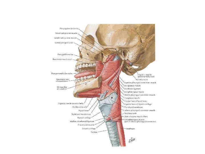
Soft Palate • Also known as the velum • Posterior muscular extension of the hard palate which together make up the palate (roof) of the oral cavity (mouth) and the floor of the nasal cavity (nose). • Multi-functional and – Aids speech – pronunciation of velar consonants (k , g and n) along with the dorsum of the tongue – Swallowing – closes the nasal passages to prevent any food or liquid from entering into the nasal cavity – Sneezing – closes the larynx when sneezing – Uvula helps produce the gag reflex when touched • 1/3 of the palate – posterior portion • Formed by 5 muscles: – – – Musculus uvulae Tensor veli palatini Levator veli palatini Palatopharyngeus muscle Palatoglossus muscle.

• Musculus uvulae – – – • From the auditory tube and temporal bone to palatine aponeurosis and fibres of opposite muscle Elevates and pulls back the soft palate helping to close the nasopharynx during swallowing Palatopharyngeus muscle – – • From auditory tube (Eustachian) and sphenoid bone to palatine aponeurosis Broadens the soft palate by pulling it laterally Levator veli palatiniarises – – • From nasal spine and palatal aponeurosis to fibres on opposite side Elevates uvula and slight lateral movement Tensor veli palatini – – • Soft Palate Muscles From posterior border of the hard palate and palatine aponeurosis to posterior thyroid cartilage of larynx Helps close the nasopharynx and raises larynx and pharynx Palatoglossus muscle – – From palatine aponeurosis to lateral aspect of the tongue where its fibres intermingle with the intrinsic musculature of the tongue Narrows the oropharyngeal isthmus during digestion and elevates the tongue.

Pharynx • A muscular column that runs between the nasal and oral cavitites and the oesophagus • All 3 cavities of the nose, mouth and larynx open posteriorly into the pharyngeal tube. • 3 main sections known as: – Nasopharynx • Base of skull to upper surface of soft palate • Eustachian (auditory/pharyngotympanic) tube opens into here • Passage mainly for air – Oropharynx • Uvula to hyoid bone • Isthmus faucium narrow opening between mouth and pharynx • Passage for air and food – Laryngopharynx • Hyoid bone to where it diverges into the respiratory (laryngeal) and digestive (oesophageal) pathways where it is continuous with the oesophagus posteriorly • Roughly level with C 4 -6 • During swallowing food has right of way (epiglottis closes to prevent aspiration)

Pharynx – posterior view

Pharyngeal muscles • Form the walls of the pharynx • Outer layer – – Three pharyngeal muscles form a sleeve • Superior constrictor • Middle constrictor • Inferior constrictor – Contract involuntarily in a sequence known as peristalsis pushing the food down from the oral cavity to the oesophagus (occurs during and immediately after swallowing) • Inner layer – Three longitudinal paired muscles. • Stylopharyngeus • Palatopharyngeus • Salpingopharingeus – Act as a group to elevate the larynx, shorten the pharynx during swallowing and speaking

Pharyngeal constrictor muscles • Superior constrictor – From pterygoid hamulus, pterygomandibular raphe and posterior end of mylohyoid line of mandible to pharyngeal tubercle and pharyngeal raphe – Constricts the upper pharynx. • Middle constrictor – From stylohyoid ligament and hyoid bone to pharyngeal raphe (blends with superior and inferior constrictors) – Constricts the middle pharynx. • Inferior constrictor – Thyropharyngeal - from the oblique line of the thyroid cartilage to pharyngeal raphe – Cricopharyngeal – from lateral aspect of the cricoid cartilage to blend with circular oesophageal fibres – Constricts lower pharynx

Longitudinal pharyngeal muscles • Stylopharyngeus – From styloid process to posterior thyroid cartilage and blends with constrictors – Elevates the pharynx and expands it laterally • Palatopharyngeus – From palatine aponeurosis to posterior thyroid cartilage – Elevates the pharynx to close off the nasopharynx during swallowing • Salpingopharingeus – From Eustachian tube in the nasal cavity blends with the palatopharyngeus muscle. – Raises the pharynx and larynx during swallowing and laterally draws the pharyngeal walls up – Opens the Eustachian tube during swallowing the equalization of pressure between the auditory canal (middle ear) and the pharynx (swallowing when ears pop)
- Slides: 13