THE NERVOUS SYSTEM Introduction Neurons masses of nerve


- Slides: 67
THE NERVOUS SYSTEM
Introduction Neurons: masses of nerve cells Nerves Structural and functional units of nervous system Specialized to react to physical and chemical changes Neuroglical Cells: provide neurons with physiological requirements Functions like connective tissue
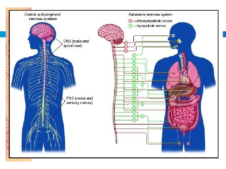
Introduction Nerve Impulse: information transmitted in form of electrochemical signal Travels along nerve fiber Functions: sensory, integrative, motor Central Nervous System: brain and spinal cord Peripheral Nervous System: nerves
Sensory Functions sensory receptors at the ends of peripheral neurons Detect changes outside the body Light and sound intensities Temperature and oxygen concentration Convert sensory information into nerve impulses Transmitted from peripheral nerves to CNS Integrative function: at CNS, signals are integrated sensation, adding to memory, or produce thoughts for perceptions
Sensory Functions Motor Functions: after making conscious or subconscious decisions, act upon perceptions � Effectors: signal from CNS travels to PNS to respond Muscles and glands that respond to nerve impulses Action counteracts the change detected Maintain homeostasis
Neuron Structure Cell Body: granular cytoplasm, cell membrane, and organelles Neurofibrils: network of fine threads which extend into nerve fibers Chromatophilic substance (Nissl Bodies): Scattered in cytoplasm Similar to rough ER protein synthesis Nucleus: Mature center of cell body neurons don’t divide
Neuron Structure Nerve Fibers: extend from cell body Dendrites: many Shorter and highly branched Main receptive surface for communication Axons: only one Conducts nerve impulse away from the body At end, fine extensions that contact the receptive surfaces of other cells
Axon Schwann Cell: enclosed axons � Wind tightly around axon Myelin: membrane layers of lipoproteins � Myelin sheath: layers with more lipids � Smaller axons lack a myelin sheath Neurilemma: surrounds myelin sheath Nodes of Ranvier: narrow gaps in the myelin sheath
TYPES OF NERVE CELLS
Classification of Neurons by Structure Bipolar Neurons: has only two nerve fibers Arise from each end 1 axon and 1 dendrite Eyes, nose, and ears
Classification of Neurons by Structure Unipolar Neuron: single fiber that extends from cell body that divides into two branches Dendrite: connects to peripheral body part Axon: enters the brain or spinal cord Ganglia: aggregation of unipolar cells Located outside brain and spinal cord
Classification of Neurons by Structure Multipolar Neurons: many nerve fibers Axon: only one Dendrite: remaining fibers Neurons in brain and spinal cord
Classification of Neurons by Function Sensory: afferent Unipolar or bipolar Carry impulses from PNS to CNS Specialized receptor ends at tips of dendrites Receptor cells: dendrites in skin or sensory organs Changes inside or outside the body Single travels to brain or spinal cord
Classification of Neurons by Function Interneurons: internuncial or association neuron multipolar Lie within brain or spinal cord Link other neurons Transmit impulses from one part of the brain or spinal cord to another Direct incoming sensory impulses for appropriate processing
Classification of Neurons by Function Motor Neurons: efferent neurons Multipolar Carry impulses out of the brain or spinal cord to effectors Stimulate muscle to contract and glands to release hormone
Classification of Neuroglical Cells Fill spaces, support neurons, provide structural frameworks, produce myelin, and carryon phagocytosis Outnumber neurons Schwann Cells Nicroglial Cells: scattered in CNS � Support neurons � phagocytize bacterial cells and cellular debris
Classification of Neuroglial Cells Oligodendrocytes: occur in rows along nerve fibers Form myelin within the brain and spinal cord Do not form sheaths
Classification of Neuroglical Cells Astrocytes: between neurons and blood vessels Structural support, join parts, regulate concentrations of nutrients and ions within tissue Form scar tissue in CNS
Classification of Neuroglical Cells Ependymal Cells form an epithelial like membrane that covers specialized brain parts Forms the inner lining that enclose spaces within the brain and spinal cord
NERVE IMPULSES
Cell Membrane Potential Surface of cell membrane is usually charge Polarized Unequal distribution of positive and negative ions between sides of the membrane Important in conducting nerve impulses
Distribution of Ion Determined by pores or channels in the membrane Some channels are always open Others can be closed Channels can be selective Only allow certain ions to pass K pass easier than Na, and less so than Ca
Resting Potential difference: difference in electrical charge between two regions Inside of cell: slightly negative Outside of cell: slightly positive Resting Potential: potential difference in a nerve cell
Potential Changes Nerve cell are excitable Respond to changes in their surroundings Temperature, light, or pressure Signals from other neurons Changes in resting potential Depolarizing: inside of membrane becomes less negative
Resting Potential Resting potential is graded Amount of change in potential is proportional to the intensity of stimulation Summation: accumulation of stimulation Threshold Action Na K potential Potential: 1/1000 th of a second diffuses freely inward Membrane becomes depolarized diffuses out of cell inside becomes negative repolarized
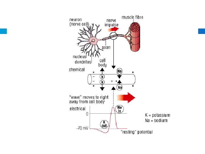
Nerve Impulse When an action potential occurs in one region, it causes the bioelectric current to flow to the adjacent portions of the membrane This stimulates the adjacent membrane Wave of action potentials down a nerve fiber
Impulse Conduction Unmyelinated fiber conducts an impulse over its entire surface Myelinated surface prevents ion flow from the insulated membrane If the sheath were continuous, no impulse travel Nerve impulse jumps from node of Ranvier to next node Contain Na and K channels
Impulse Conduction Impulse is much faster than on an unmyelinated nerve Speed of nerve impulse is proportional to the fiber diameter Skeletal Muscle fiber: 120 meters per second Sensory Fiber: 0. 5 meters per second Nerve stimulation is all-or-none response Amount of stimulation does not vary degree of response
The Synapse Junction between two neurons Not a direction connection Gap: synaptic cleft Synaptic Transmission: signal runs from dendrite of a neuron to the axon At axon, neurotransmitter are released Bind with receptor on the next neuron
Excitatory and Inhibitory Actions Excitatory: increases Na permeability Trigger nerve impulses Inhibitory: decrease Na permeability Lessens chance of nerve impulse
Neurotransmitters ~50 types have been identified Some neurons only release one type where others release two or three Acetylcholine: stimulates muscles Monoamines: epinephrine, norepinephrine, dopamine, serotonin Amino acids and peptides
Neurotransmitters Synthesized in the cytoplasm of the synaptic knob Stored in synaptic vesicles When action potential is reached, Ca ions are transported inward Neurotransmitters are released Afterwards, some are reabsorbed while others are broken down
Impulse Processing Neuronal Pools: receive impulses, processes them, and conducts impulses away Facilitation: neuron receives a subthreshold stimuli and becomes more excitable Convergence: two or more impulses from different sources on a single neuron Additive effect on neurons Divergence: impulses may pass into several output fibers Amplifies impulses
NERVE PATHWAYS, MENINGES, AND SPINAL CORD
Nerve Pathway Routes nerve impulses follow through nervous system Reflex arc: involuntary actions Begins with a receptor at the end of a sensory fiber Leads to interneurons in CNS, or reflex center Communicate with motor neurons to effectors Reflex Behavior; automatic subconscious responses to stimuli Heart rate, breathing, blood pressure, and digestion Swallowing, sneezing, coughing, and vomiting
Reflex Behavior Knee-jerk reflex: patellar tendon reflex � sensory neuron communicating directly to a motor neuron � Helps maintain upright posture
Reflex Behavior Withdrawal Reflex: touching a finger to something painful � Motor neurons in the flexor muscles are simulated � Arm jerks back � Signal is also sent to the brain to signal pain � Protective reflex
Meninges Membranes located between the bone and soft tissues the nervous system Protect the brain and spinal cord
Meninges: 3 layers Dura mater: outermost layer Tough, white fibrous connective tissue Contains many blood vessels and nerves
Meninges: 3 layers Arachnoid mater: thin, weblike membrane Lacks blood vessels Located b/w dura and pia maters
Meninges: 3 layers Pia mater: very thin layer Contains many nerves and blood vessels Nourish the cells of the brain and spinal cord
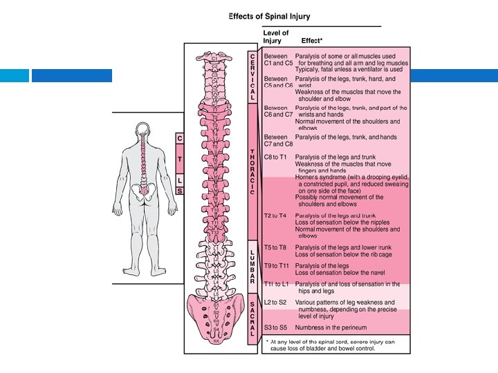
Spinal Cord Slender nerve column that passes downward from the brain into the vertebral canal
Spinal Cord
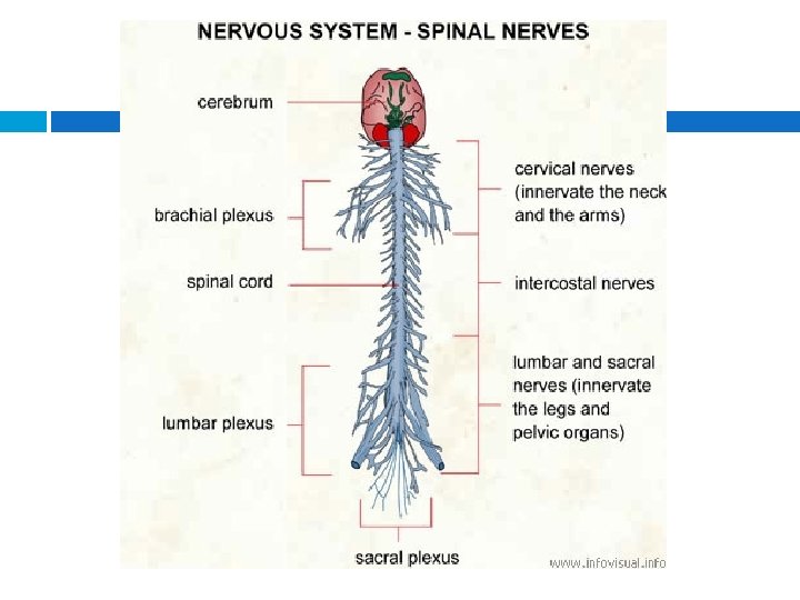
Structure of Spinal Cord 31 segments Each gives rise to a pair of spinal nerves Branch Cervical Nerves Lumbar Nerves to various parts of the body Enlargement: thickening in the neck to upper limbs Enlargement: thickening in lower back to lower limbs
Functions of the Spinal Cord Conducting nerve impulses Ascending tract: nerve tract or pathway that carries sensory information to the brain Descending tracts: conduct motor impulses form the brain to the muscles and glands Spinal reflexes: knee jerk and withdrawal Reflex cord arc passes through spinal
BRAIN
Brain Subdivides Cerebrum Cerebellum Brain Stem
Cerebrum Two cerebral hemispheres Connected by corpus callosum Cerebral Cortex: thin layer of grey matter near the surface White matter consists of myelinated nerve fibers that connect neurons within the nervous system and communicate with
Functions of Cerebrum Higher brain function Sensory, motor, and association areas One hemisphere usually dominates for certain intellectual functions
Diencephalon Thalamus: central relay station for incoming sensory impulses Hypothalamus: maintains homeostasis Limbic System: emotions and modifies behaviors
Brain Stem Midbrain, pons, and medulla oblongata Midbrain: reflex centers Head Pons: transmits impulses b/w cerebrum and other parts of nervous system Rate and eye movement and depth of breathing Medulla Oblongata: all ascending and descending impulses Several vital and nonvital reflex centers
Brain Stem Cerebellum: two hemispheres Reflex center for integrating sensory information coordination of skeletal muscle movements Maintenance of equilibrium
PERIPHERAL NERVOUS SYSTEM AND AUTONOMIC NERVOUS SYSTEM
PNS Cranial and spinal nerves Bunch form the brain and spinal cord to all body parts Somatic and autonomic systems
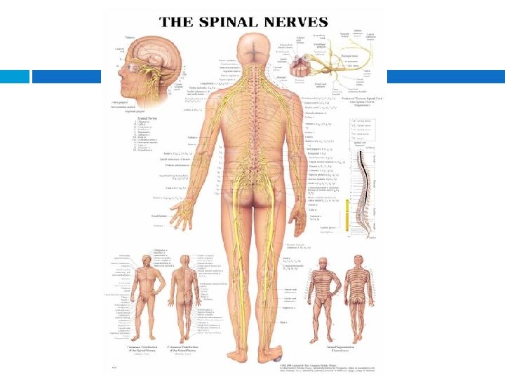
Cranial Nerves 12 pairs of cranial nerves connect the brain to parts of the head, neck, and trunk Some sensory or primarily motor Mostly mixed Some are somatic, others autonomic
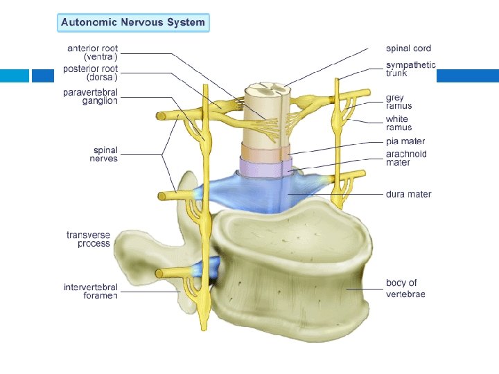
Spinal Nerves 31 -pairs originate from the spinal cord Mixed nerves provide 2 -way communication b/w spinal cord and upper and lower limbs, neck, and trunk Grouped according to the levels from which they arise Emerges from a dorsal and ventral root Divides into branches beyond its foramen Most combine to form plexuses � Nerve fibers are sorted and recombined so that those associated with a particular region reach it together
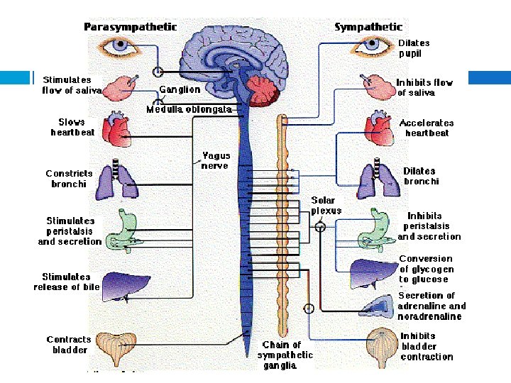
Autonomic Nervous System Functions without conscious effort Regulates visceral activities maintaining homeostasis Autonomic functions are reflexes controlled from nerve centers in the brain and spinal cord Sympathetic and parasympathetic Parasympathetic is most active
Autonomic Nerve Fibers Motor fibers Sympathetic fibers leave the spinal cord and synapse in paravertebral ganglia Parasympathetic fibers begin n the brain stem and sacral region of the spinal cord and synapse in ganglia near viscera
Autonomic Neurotransmitters Differences in neurotransmitters different effects of the autonomic divisions Sympathetic and parasympathetic preganglionic fibers secrete acetylcholine Parasympathetic postganglionic fibers secrete acetylcholine Sympathetic postganglionic fibers secrete norepinephrine
Control of autonomic activity Somewhat independent Control centers in medulla oblongata & hypothalamus Utilize autonomic nerve pathways Limbic system and cerebral cortex control the autonomic system during emotional distress