Structure Functions of Chromatin Introduction Genetic material DNA


 of an unfolded set")


 Constitutive heterochromatin • (II)")


 at these")


 to package DNA into")
- Slides: 39
Structure & Functions of Chromatin
Introduction • Genetic material DNA in eukaryotic cells never found as a naked molecule but always found in association with: • • • i. iii. iv. v. vi. Histon proteins, HMG proteins, Residual proteins, Phosphoproteins, Different RNA species & Different lipid species • that constituting a complex structure called as chromatin.
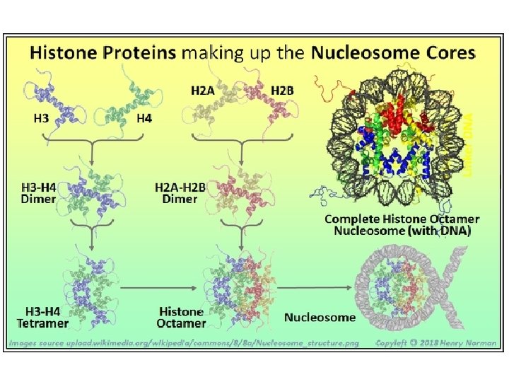
Histone proteins
• High-Mobility Group or HMG is a group of chromosomal proteins that are involved in the regulation of DNA-dependent processes such as transcription, replication, recombination, and DNA repair. • The HMG proteins are subdivided into 3 superfamilies each containing a characteristic functional domain: • HMGA – contains an AT-hook domain – HMGA 1 – HMGA 2 • HMGB – contains a HMG-box domain – – • HMGB 1 HMGB 2 HMGB 3 HMGB 4 HMGN – contains a nucleosomal binding domain – – HMGN 1 HMGN 2 HMGN 3 HMGN 4
• Beside the above said constituents, smaller molecules like steroid and thyroid hormones, vitamins A and D are also found within the cell nucleus bound to DNA or to one of the other chromatin macromolecules. • Foreign substances like antibiotics, dyes, enzymes and complex carbohydrates also penetrate the cell nucleus to the chromatin, where they also have some stimulatory or inhibitory effects on RNA or DNA synthesis.
• Thus, chromatin is the combination of DNA, histone, and other proteins that condense to form chromosomes. It is found inside the nuclear envelope of eukaryotic cells. • In simple words, Chromatin is DNA plus the proteins (and RNA) that package DNA within the cell nucleus.
• Walther Flemming in 1882 used this term for the first time. • The functions of chromatin are to package DNA into a smaller volume to fit in the cell, to strengthen the DNA to allow mitosis and meiosis, and to control gene expression and DNA replication.
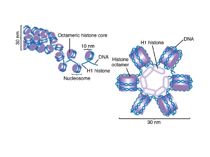
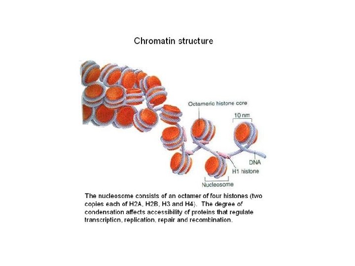
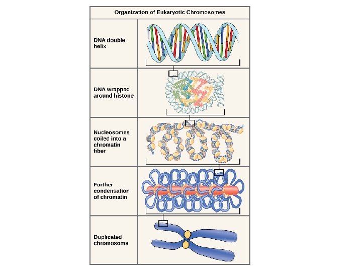
• Basic structure • In simple terms, there are three levels of organization: • At first level of organization, the DNA wraps around the histone octamer proteins forming nucleosomes. • Each histone octamer is composed of two copies each of the histone proteins H 2 A, H 2 B, H 3, and H 4. • Structurally it appears as "beads on a string“.
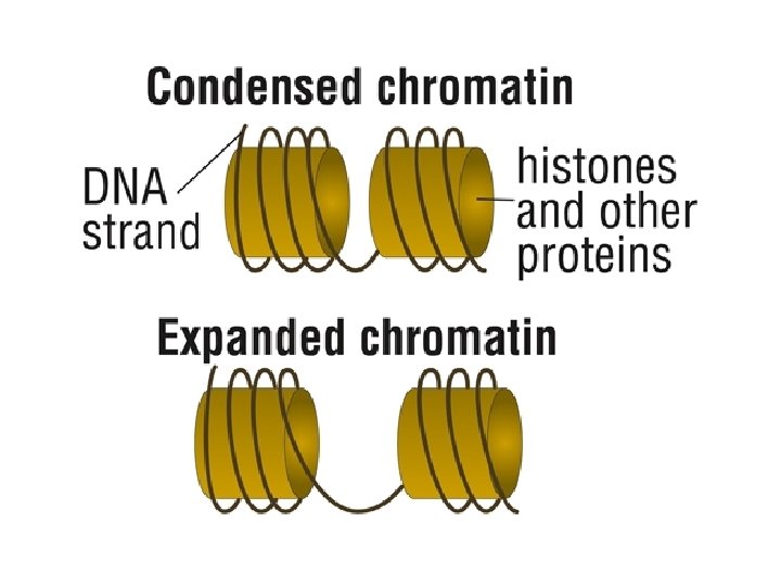
• The second level of organization is the formation of 30 nm condensed chromatin fiber consisting of nucleosome arrays in their most compact form. • The third level organization is higher-level where DNA packaging into the metaphase chromosome. • The structure of chromatin varies considerably as the cell progresses through the cell cycle. • The changes in structure of chromatin are required to allow the DNA to be used and minimizing the risk of damage.
First level organization Second level organization
IIIrd level Ist level organization IInd level organization
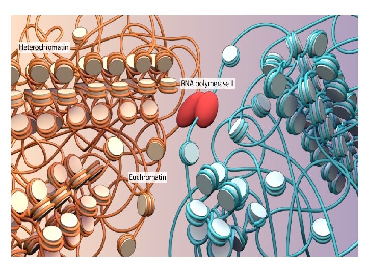
• Structure of chromatin in non-dividing cells • Two types of chromatin material have been recognized in a non-dividing cell: • (i) Euchromatin and (ii) Heterochromatin • Euchromatin is rich in gene concentration, and is often (but not always) under active transcription however the Heterochromatin contain very few structural genes i. e. gene that encode protein and remain condensed during interphase and early prophase, so called chromocentre.
• The major difference between heterochromatin and euchromatin is that heterochromatin is such part of the chromosomes, which is a firmly packed form and are genetically inactive, • while euchromatin is an uncoiled (loosely) packed form of chromatin and are genetically active.
Structure Euchromatin • The structure of euchromatin is reminiscent (suggestive) of an unfolded set of beads along a string, wherein those beads represent nucleosomes. • Nucleosomes consist of eight proteins known as histones, with approximately 147 base pairs of DNA wound around them.
• In euchromatin, the wrapping is loose so that the raw DNA may be accessed. • Each core histone possesses a `tail' structure, which can vary in several ways. • It is thought that these variations act as "master control switches, " which determine the overall arrangement of the chromatin. • In particular, it is believed that the presence of methylated lysine 4 on the histone tails acts as a general marker for euchromatin.
• Appearance • Euchromatin is a lightly packed form of chromatin (DNA, RNA and protein), generally appears as light-coloured bands and is rich in gene concentration, often (but not always) under active transcription. • This light staining/colour is due to the less compact structure of euchromatin.
• Unlike heterochromatin, it is found in both type of cells with nuclei i. e. eukaryotes and prokaryotes. • This indicates that the heterochromatin structure evolved later along with the nucleus, possibly as a mechanism to handle increasing genome size. • Euchromatin comprises the most active portion of the genome within the cell nucleus.
• Function • Euchromatin participates in the active transcription of DNA to m. RNA products. • The unfolded structure allows gene regulatory proteins and RNA polymerase complexes to bind to the DNA sequence, which can then initiate the transcription process. • Not all euchromatin is necessarily transcribed e. g. those euchromatin which transformed to heterochromatin are not transcribed because of to protect the genes.
• It is thought that the cell uses transformation from euchromatin into heterochromatin as a method of controlling gene expression and replication. • One example of constitutive euchromatin that is 'always turned on' is housekeeping genes, which code for the proteins needed for basic functions of cell survival.
Heterochromatin • Heterochromatin is a tightly packed form of DNA, which comes in different varieties. • Its major characteristic is that transcription is limited. • As such it is a means to control gene expression, through regulation of the transcription initiation.
• Structure • The two forms of chromatin were distinguished on the basis of staining i. e. the euchromatin is light stained, while heterochromatin stains intensely, indicating tighter packing. • Heterochromatin is usually localized to the periphery of the nucleus.
• Heterochromatin mainly consists of genetically inactive satellite sequences, and many genes are repressed to various extents, although some cannot be expressed in euchromatin at all. • Heterochromatin also replicate later in Sphase of the cell cycle and is found only in eukaryotes. • Both centromeres heterochromatic. and telomeres are
• There are two types of Heterochromatin: • (I) Constitutive heterochromatin • (II) Facultative heterochromatin
• Constitutive Heterochromatin • Constitutive heterochromatin is fixed and irreversible in form and function. • This type of heterochromatin is located around the centromeres and usually contains repetitive sequence.
• All cells of a given species will package the same regions of DNA in constitutive heterochromatin. • It is never expressed, thus in all cells any genes contained within the constitutive heterochromatin will be poorly expressed. • For example, all human chromosomes 1, 9, 16, and the Y chromosome contain large regions of constitutive heterochromatin.
• Facultative Heterochromatin • Facultative heterochromatin has the faculty to return to the normal euchromatic state. • The regions of DNA packaged in facultative heterochromatin will not be consistent between the cell types within a species. • Thus a sequence in one cell that is packaged in facultative heterochromatin (and the genes within poorly expressed) may be packaged in euchromatin in another cell (and the genes within no longer silenced).
• However, the formation of facultative heterochromatin is regulated, and is often associated with morphogenesis or differentiation. • An example of facultative heterochromatin is X-chromosome inactivation in female mammals: one X chromosome is packaged as facultative heterochromatin and silenced, while the other X chromosome is packaged as euchromatin and expressed.
• Function • Heterochromatin has been associated with several functions, from gene regulation to the protection of the integrity of chromosomes; • Some of these roles can be attributed to the dense packing of DNA, which makes it less accessible to protein factors that usually bind DNA or its associated factors. • For example, naked double-stranded DNA ends would usually be interpreted by the cell as damaged or viral DNA, triggering cell cycle arrest, DNA repair, or destruction of the DNA fragment such as by endonucleases in bacteria.
• Some regions of chromatin are very densely packed with fibers displaying a condition comparable to that of the chromosome at mitosis. • Heterochromatin is generally clonally inherited; when a cell divides the two daughter cells will typically contain heterochromatin within the same regions of DNA, resulting in epigenetic inheritance. • Variations cause heterochromatin to encroach on adjacent genes or recede from genes at the extremes of domains.
• Transcribable material may be repressed by being positioned (in cis) at these boundary domains. • This gives rise to different levels of expression from cell to cell, which may be demonstrated by positioneffect variegation. • Insulator sequences may act as a barrier in rare cases where constitutive heterochromatin and highly active genes are juxtaposed (e. g. the 5'HS 4 insulator upstream of the chicken β-globin locus, and loci in two Saccharomyces spp). • Chromosomal material that consists of uncoiled dispersed threads during interphase, is genetically active, and stains lightly with basic dyes.
• Structure of chromatin during cell division: • During interphase: During interphase the structure of chromatin is optimised to allow easy access for transcription and DNA repair. • The structural changes includes: • (i) Histone proteins which are the foundation blocks of chromatin modified by various post-translational modification to alter DNA packing. • (ii) Acetylation results in the loosening of chromatin and lends itself to replication and transcription. • (iii) When methylated they hold DNA together strongly and restrict access to various enzymes.
• During Metaphase. • The metaphase structure of chromatin differs vastly to that of interphase. • It is optimized for physical strength and manageability, forming the classic chromosome structure. • The physical strength of chromatin is vital for this stage of division to prevent sheer damage to the DNA as the daughter chromosomes are separated. • To maximize strength the composition of the chromatin changes as it approaches the centromere, primarily through alternative histon H 1 analogues.
• The primary functions of chromatin are: • 1) to package DNA into a smaller volume to fit in the cell. • 2) to reinforce the DNA macromolecule to allow mitosis • 3) to prevent DNA damage • 4) to control gene expression and DNA replication.