Standard S 7 L 2 a Students will


")


 • Mitosis begins (cell begins")


 • Chromatids (or pairs of")


 • Chromatids (or pairs of")


 • Two new nuclei form")


- Slides: 47
Standard S 7 L 2 a. Students will explain that cells take in nutrients in order to grow and divide and to make needed materials. S 7 L 2 b. Students will relate cell structures to basic cell functions.
Learning Target I can explain how cells grow and divide?
Mitosis & Cell Division
There are over 100 trillion cells in the body! 1. We all started out as a single cell, so how did we get so many? ? v Through a process of cell division.
2. Why is this important? A. This is important for growth. B. This is important for repair. C. This is important for replacement of cells. D. Some one-celled organisms reproduce by cell division.
3. How do cells divide? • By going through the cell cycle. • The cell’s life cycle is called the cell cycle. • The amount of time for this is different for different types of cells.
Cell Cycle
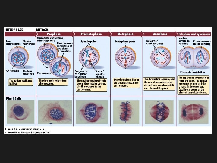
4. First Phase of the Cell Cycle: Interphase • This is the longest phase of the cell cycle. • This is a period of growth and development of the cell.
1 st: Interphase Draw and label this picture in the circle on your page.
1 st: Interphase • Chromosomes are copied (which means the # of chromosomes doubles) • Chromosomes appear as threadlike coils (called: chromatin) at the start, but each chromosome and its copy (called: sister chromosome) change to sister chromatids at the end of this phase
Label the picture on your page: label
Interphase under a microscope
5. Chromosomes • are structures in the nucleus that contains DNA, the hereditary material of life. • During interphase each chromosome is copied.
The duplicated strands • are identical. • are called “sister chromatids. ”
The center of a chromosome is called the centromere, it joins the two chromatids together to form a chromosome.
6. The nucleus divides in two ways, Mitosis & Meiosis (we will talk about this later)
7. Mitosis happens when body or somatic cells make exact copies of each other with the same number of chromosomes.
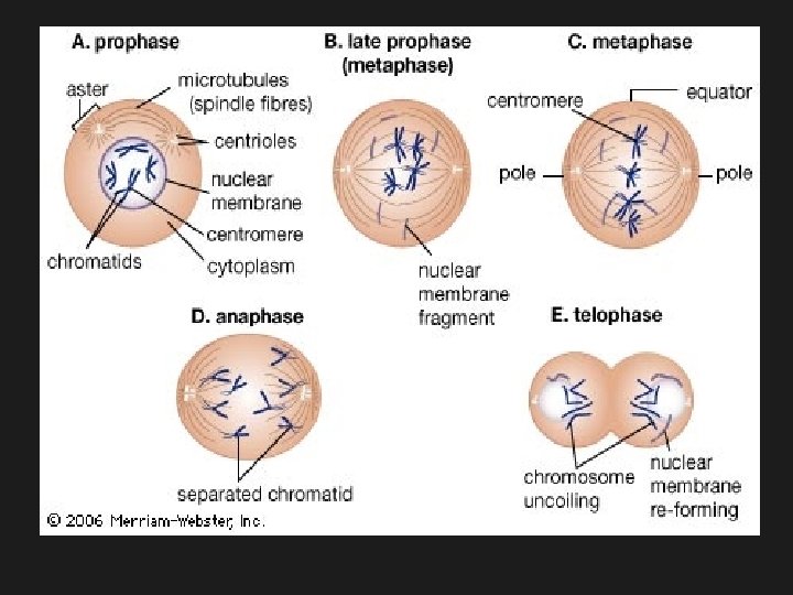
nd 2 : Prophase (1 st step of Mitosis) • Mitosis begins (cell begins to divide) • Centrioles (or poles) appear and begin to move to the opposite ends of the cell. • Spindle fibers form between the poles. • Nuclear membrane begins to disassemble (or come apart)
Draw and label this picture in the circle on your page centrioles Spindle Fibers Nuclear membrane dissolving
Picture of Prophase
rd 3 phase: Metaphase (2 nd phase of Mitosis) • Chromatids (or pairs of chromosomes) attach to the spindle fibers • The chromosomes line up at the equator.
3 rd: Metaphase Draw and label this picture in the circle on your page Chromatids Spindle Fibers
Metaphase under the Microscope
th 4 phase: Anaphase (3 rd phase of Mitosis) • Chromatids (or pairs of chromosomes) separate and begin to move to opposite ends of the cell. DNA
th 4 : Anaphase Draw and label this picture in the circle on your page Chromatids
Anaphase under the microscope
th 5 phase: Telophase (4 th phase of Mitosis) • Two new nuclei form • Chromosomes appear as chromatin (threads rather than rods) • Mitosis ends • Nuclear membrane begins to reform.
th 5 : Telophase Draw and label this picture in the circle on your page New nuclei Nuclear membrane reforming
Telophase under the microscope
Review • The phases of mitosis are prophase, metaphase, and telophase. • Prophase---spindle fibers form • Metaphase---Spindle Fibers Attached, Middle of Cell • Anaphase---Chromatids Separate; Away or Apart • Telophase---Two Nuclei
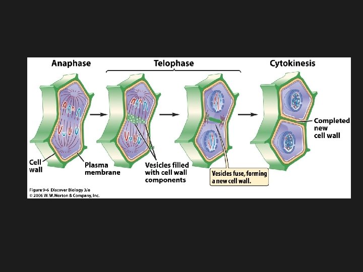
th 6 : Cytokinesis • Cell membrane moves inward to create two daughter cells. • Each with it own nucleus with identical chromosomes. add this to your notes: • The parent cell no longer exists. It became 2 daughter cells.
th 6 : Cytokinesis Draw and label this picture in the circle on your page
Trick for Remembering Phases 1. Interphase: Inner Growth Mitosis begins: PMAT 2. Prophase: Poles/pairing 3. Metaphase: Middle of Cell 4. Anaphase: Apart or Away 5. Telophase: Two Nuclei 6. Finally, Cytokinesis
How do plant cells divide after mitosis? • In plant cells, a cell plate forms in the middle of the cell. • The cell plate divides the cytoplasm into two parts. • New cell walls form along the cell plate. • New cell membranes form inside the cell walls.
Cell Plate
Results of Mitosis • Each cell in body has same number of chromosomes • Allows growth and replacement of damaged or worn out cells. • Asexual Reproduction: • Binary Fission-mitosis Budding---growing from side of parent Regeneration---regrow parts
3 important things about mitosis 1. 4 phases of mitosis – PMAT 2. Each new nucleus formed has the same number and type of chromosomes as the parent cell. 3. It allows for growth and replacement of cells.
2 types of Reproduction • Sexual and Asexual • Sexual reproduction is the process by which 2 sex cells, usually an egg and a sperm, join to form a zygote. This zygote will develop into a new organism with a unique identity.
Asexual Reproduciton • Asexual reproduction is the process by which an organism produces others of its same kind. The new organism is produced from ONE parent organism. • The hereditary information is identical to the parent.
Asexual Reproduction Regeneration • Uses mitosis and cell division to regrow body parts. • If the organism breaks into pieces, a whole new organism can grow. • Ex: sponges, sea stars, flat worms.
Budding • A new organism is growing from the body of the parent organism. • It is possible because of mitosis. • When the bud gets large enough it breaks away and lives on its own. • Ex. Hydra, potatoes, branches on trees.
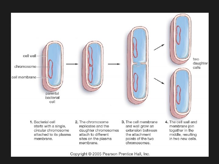
Binary Fission • It is the most common form of reproduction in prokaryotes. • It is like mitosis but since prokaryotes do not have a nucleus, the DNA is copied and then the single cell splits. • It occurs in some singlecelled eukaryotes.
Just some pictures you can look at on the next few slides