Spleen Dr Mahmoud AlAwaysheh MRCSI Muta University Anatomy


.")


")


- Slides: 47
Spleen Dr. Mahmoud Al-Awaysheh MRCSI Mu’ta University
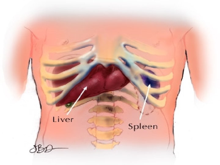
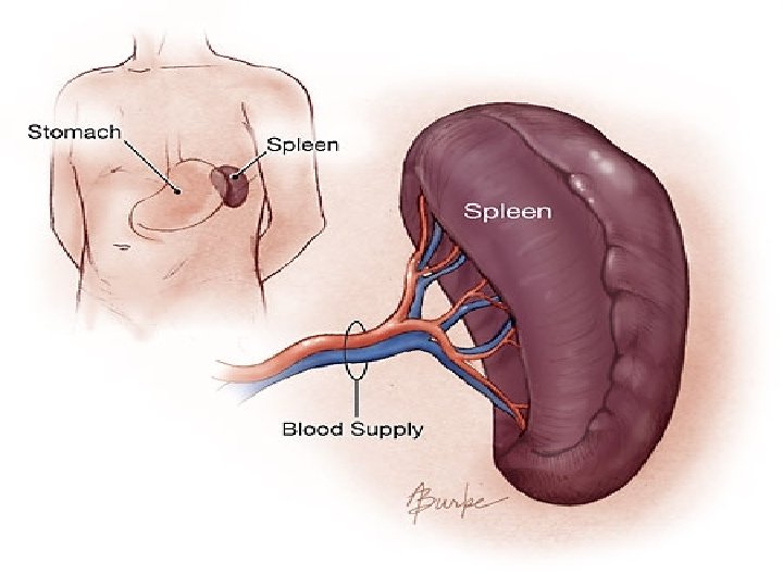
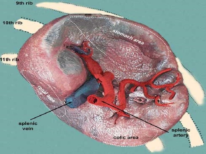
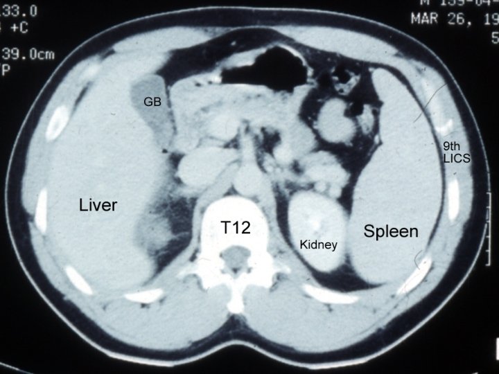
Anatomy: Spleen -Wt 75 -250 gm, LUQ, along 10 th rib, between gastric fundus &L hemidiaphragm. -Hilum: In the angle between stomach & L kidney, I n contact with tail of pancreas. -Concave Visceral surface: impressions. -Notch: infero lateral border, palpate in splenomegaly.
- Arterial: Splenic A. along upper border of tail of pancrease. - Vein: Splenic V. at hilum behind pancrease. join SMV to form portal V. - Lymphatics: efferent vessels from white pulp to L N in hilum to retropancrearic to coeliac nodes.
Physiology: Spleen
Functions: Spleen 1. Response to antigenic challenge 2. Destruction of abnormally shaped or rigid red cells 3. Phagocytosis of foreign substance 4. Platelet reservoir 5. Erythrocyte production
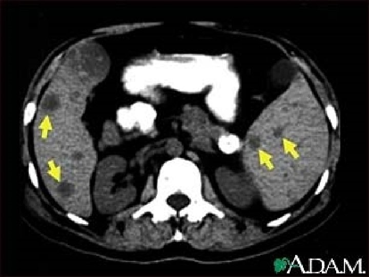
Investigation of the Spleen. FBC, Reticulocytes, tests for haemolysis. LFT & OGD in splenomegaly+portal hypertension in liver cirrhosis. Investigations for causes of splenomegaly including LN biopsy. Radiology: 1 -Calcification: splenic infarct, splenic a. aneurysm, hydatid cyst, TB. 2 -US, CT with contrast, MRI. 3 -Tc 99: is spleen site of RBC destruction? .
Congenital Abn of Spleen 1 -Agenesis: rare. 2 -Spleniculi: 10 -30% of population. Hilum (50%), splenic vessels & tail of pancrease (30%), mesocolon & splenic ligaments (20%). **Failure to remove spleniculi during splenectomy…. . persistant disease. 3 -Hamartomas: rare 4 -Non parasitic splenic cyst: 1 -True, dermoid, mesenchymal. 2 -False, trauma 3 - Pseudocyst after pancreatitis.
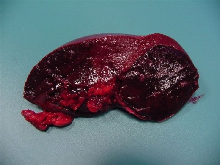
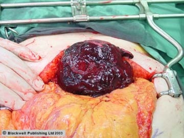
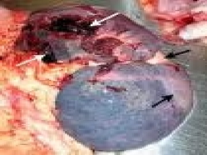
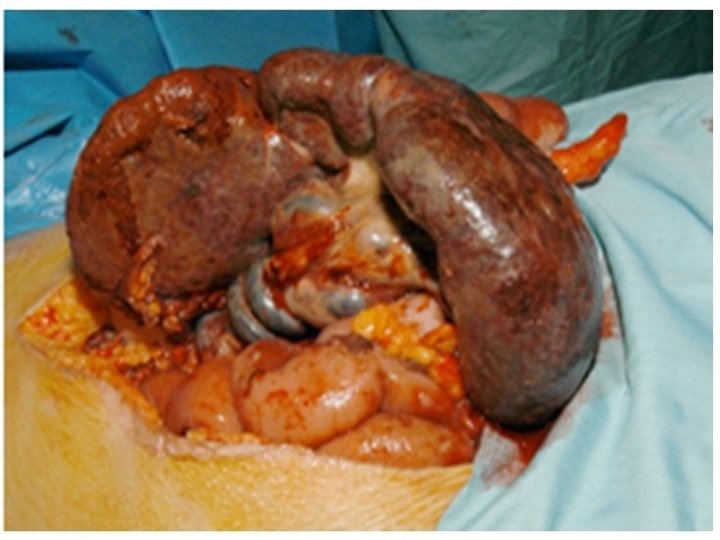
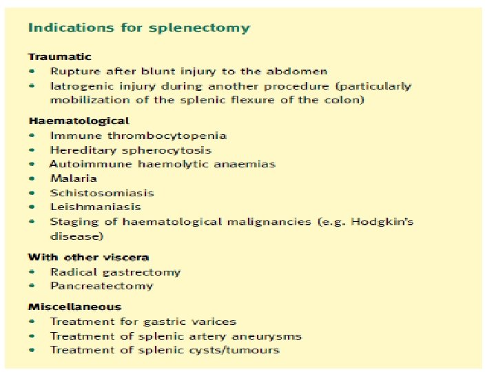
Consider it : Splenic Rupture 1 -Bunt abdominal trauma LUQ (RTA, Fall, . . . ). 2 -High Risk: diseased or enlarged spleen(rupture of a malarial spleen in trivial injury). 3 -Fractures of 9 th, 10 th, 11 th left ribs. 4 -Iatrogenic. Presentation: 1 -Succumbs rapidly from massive haemorrhage 2 -Initial shock, recovery, signs of late bleeding: Blood loss+tamponade+further bleeding. Reminder are, general signs of internal haemorrhage, local LUQ peritonitis signs, Kehrs sign+FAST US & CT. 3 -Delayed rupture: uncommon with these days scan use In ER.
Splenic rupture Management: 1 -Conservative: 1 -minimal or no abdominal findings+stable haemodynamically. 2 -CT, isolated injury, no hliar injury, no massive distruption of spleen. 2 -Immediate Laparotmy: 1 -continuingblood loss despite adequate resuscitation. 2 -associated abdominal organ injuries with blunt splenic trauma is up to 25 -50%. ******Consider splenic preservation******
Splenic Trauma Spleen Is one of most vascular organs , pass through it 350 liter of blood / day , contain 1 unit of blood at any moment ! Types of trauma : 1 - Blunt 2 - penetrating : Easily diagnosed , because patient almost always referred to Surgery.
Blunt Trauma of Spleen 25% of all blunt trauma of abdominal viscera More in male 3: 2 Most common cause is RTA Presentation : Asymptomatic abdominal pain (50%) , abdominal Distention , Hypotension (25%)
Diagnosis For stable patient : 1. Ultra. Sound 2. CT 3. Angiography
4. Plain radiography / chest and abdomen - The radiography signs of rupture are: 1. Obliteration of the splenic outline 2. Obliteration of the psoas shadow 3. Indentation of the left side of the gastric air bubble 4. Fracture of one or more lower ribs on the left side ( present 27 % of cases 5. Elevation of the left side of the diaphram 6. Free fluid between gas filled intestinal coils.
5. CT Modality of choice Used with contrast. Findings : 1. 2. 3. 4. Lacerations : irregular hypodense area with no enhancement. Sub-capsular hematoma : regular shape , cresentric. Intraparenchymal hematoma. Fragmentation with autosplenictomy.
Diagnosis Unstable patient : Open and See ! Peritoneal lavage FAST
Spleen injury grading scale : Stage 1 : Subcapsular Hematoma < 10 % of surface area. Capsular tear depth < 1 cm.
Spleen injury grading scale : Stage 2 : Subcapsular hematoma of 10 – 50 % of surface area. Laceration depth : 1 -3 cm Not involving trabecular vessels Intraparenchymal Hematoma < 5 cm in Diameter.
Spleen injury grading scale : Stage 3 : Sub-capsular Hematoma > 50 % , or Ruptured spleen. Laceration depth : > 3 cm Involving the trabecular Vessels. Intraparenchymal hematoma > 5 cm in diameter.
Spleen injury grading scale : Stage 4 : Laceration involving hilar or segmental vessels with devascularization of > 25 % of speen
Spleen injury grading scale : Stage 5 : Shattered spleen
Treatment 1. Conservative: Admit patient to ICU Repeated ultrasound For those with stage 1 or 2.
2 - splenectomy 3 - coservative splenoraphy. suturing of spleen to prevent further bleeding. 4 - splenic artery Embolization. - less mortality and morbidity than splenictomy - mortality is related to % of splenic tissue Embolized. - Complications : 1. 2. 3. Pancreatitis Splenic Abscess Pleural Effusion ( most common )
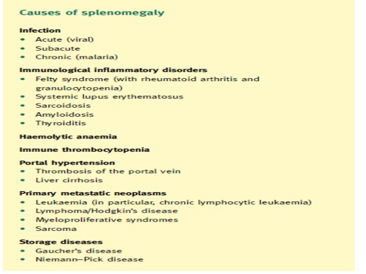
Splenomegaly & Hypersplenism: Clinical syndrome 1 -Splenic enlargement. 2 -Any combination of anaemie, leucopenia or thrombocytopenia. 3 -Compensatory bone marrow hyperplasia. 4 -Improvement after splenectomy.
Splenectomy for Blood ds 1 -ITP: -15 -50 y female. -CP: Ecchymoses purpuric patches of skin & MM. Post traumatic skin petechial haemorrhage. Epistaxis, Menorrhgia. 10% palpable spleen. - Investigations: B. T increased. C. T & P. T normal. Thrombocytopenia.
Treatment 1 -75% regress after 1 st attack in paediatrics. 2 -Steroid for short course in adult & children…recovery 3 - Surgery: Refractory (more than 9 months+low platelet+ 2 relapses). Two thirds cure.
Splenectomy for Blood ds 2 -Haemolytic Anaemias: 1 -Hereditary Spherocytosis. 2 -Sickel cell anaemia. 3 -Thalassaemia. 4 -Acquired autoimmune haemolytic anaemia
Heridatary spherocytosis -Autosomal dominant. spherocytic RBC. -Present in childhood. haemolytic jaundice+splenomegaly+pigment gall stones. -Fragility test RBC haemolyse in stronger saline solution 0. 6%(normally in 0. 47% saline solution). -Reticulocytes increase. -Radioactive Cr 51 labelling RBC. . RBC destruction.
Splenectomy for neoplasms 1 -Haemangioma : Most common benign tumour of spleen Haemangiosarcoma (rare). 2 -Lymphoma: most common cause of neoplastic enlargement. ---Splenectomy is for. . 1 -management. 2 -diagnosis& staging. (CT is alternative for staging). 3 -Myelofibrosis: Abnormal proliferation of mesenchymal elements in BM, spleen, liver & LN. -Over 50, gross splenomegaly with LUQ pain. -Splenectomy reduces the need transfusion & may relieve the pain.
Splenectomy Preop. Investigations: 1 -Blood, FFP, Cryoprecipitate, platelets. 2 -Coagulation profile. 3 -Antibiotic prophylaxis
Splenectomy Post operative complications: 1 -Slipped ligature from splenic a…Haemorrhage. 2 -Haematemesis(gastric mucosal damage) 3 -Gastric dilatation. 4 -Left basal atelectasis & Pleural effusion. 5 -Injury to tail of pancrease. pancreatitis, abscess, fistula. 6 -injury to greater curvature of stomach during ligation short gastric vessels…fistula.
Splenectomy 7 -Venous Thrombosis. *Prophylactic aspirin if platelets more than 1 million* 8 -Post splenectomy septicaemia: 1 -Strep pneumonia. 2 -N meningitides. 3 -H influenzae 4 -E. coli.
Higher Risk Groups: 1 -Young. 2 -Chemoradiotherapy. 3 -Splenectomy for blood dss. 9 - OPSI (overwhelming post splenectomy infection) is a real clinical danger.
Splenectomy OPSI: 1 -Prophylactic daily penicillin if under 5 y until they are 10. 2 -prophylaxis for 2 -3 years if older tha 5 y. Oral Penicillin, Erythromycin, Aomxicillin, Co-Amoxiclav. I. V same A. B above or Cefotaxime, Ceftriaxone or chloramphenicol if allergic to Penicillin or Cephalosporine
Splenectomy * Post operative vaccination give less than 50% antibody levels of those with preoprative vaccination.
Thank You Dr. Mahmoud Al. Awaysheh Mu’ta University