Soft tissue tumors Lecture 4 Soft tissue tumors


: Lipoma: Benign tumors of fat. Most common soft tissue")


, with abundant")


 penetrating the")


")
")


- Slides: 40
Soft tissue tumors Lecture 4
Soft tissue tumors Classified according to tissue type that they arise from: Fat. Fibrous tissue. Muscle Neurovascular tissue. Some of these tumors arise from: Pluripotent mesenchymal stem cells.
Soft Tissue Tumors
Causes: Most arise without any cause. 1 - Enviromental factores: Radiaion exposure. Burn injury. Toxin exposure. Associated with human herpes virus 8: (Kaposi sarcoma). 2 - Genetics: Chromosomal abnormalities & genetic derangements.
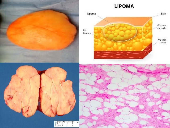
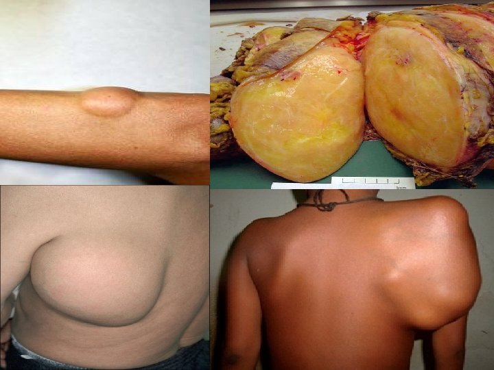
Tumors of adipose tissue (Fat): Lipoma: Benign tumors of fat. Most common soft tissue tumors in adults. Most lipomas are solitary lesions. Multiple lipomas suggest presence of rare hereditary syndromes. Gross appearance: Mobile, slowly enlarging, painless, soft, yellow, well-encapsulated masses of mature adipocytes. Histology: Mature fat cells with No pleomorphism.
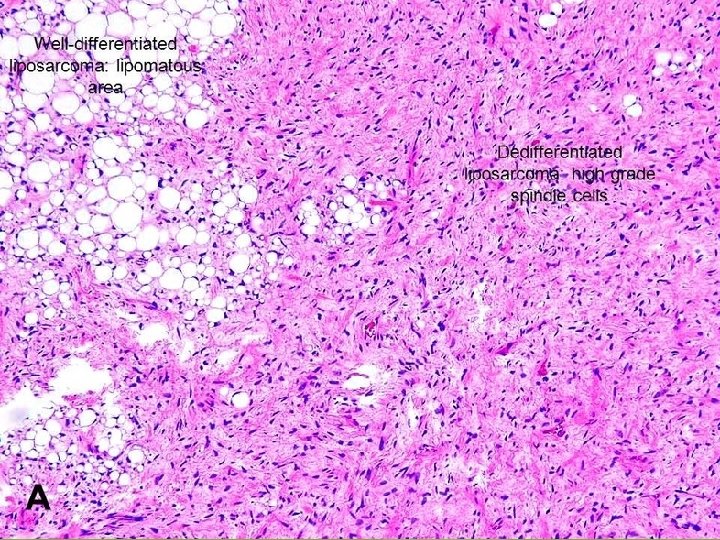
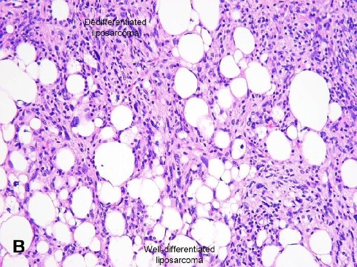
Liposarcoma Malignant neoplasms of adipocyte. Most common in fifth & sixth decades of life. Arise in deep soft tissues or in retroperitoneum. Well-circumscribed lesions. Morphology: Several different histologic subtypes: 1 - Well-differentiated liposarcoma. 2 - Myxoid/round cell liposarcoma: Characterized by abundant mucoid extracellular matrix. 3 - Poorly differentiated tumors.
Prognosis is influenced by the Histologic subtype: Well-differentiated tumors: q Grow slowly. q Associated with more favorable outlook. q Have Good prognosis. Myxoid/round cell & poorly differentiated tumors q. Aggressive tumors. q. Recur after excision. q. Have Poor prognosis.
Lipoblasts : Signet ring Appearance Cells that are indicative of fatty differentiation. Present in most cases. Have cytoplasmic lipid vacuoles depressing nucleu
Well-differentiated liposarcoma.
Myxoid liposarcoma: Fat cells & more primitive cells with lipid vacuoles (lipoblasts), with abundant myxoid matrix.
Fibrous tumors & tumor-like lesions It is fibrous tissue proliferations include heterogeneous group of lesions: 1. Nodular fasciitis: Ø Not a true tumor. Ø Reactive lesion. Ø Self-limited proliferation. 2. Fibromatosis: Ø Benign lesions but locally infilterative so it is difficult to be removed completely during surgical excision. 3. Fibrosarcomas: Ø Highly malignant neoplasm. Ø Recur locally. Ø Can metastasize.
Reactive Proliferations Nodular Fasciitis: q. Self-limited fibroblastic proliferation. q. Occurs in adults on volar aspect of forearm, chest or back. q. Solitary, rapidly growing & occasionally painful mass of few weeks duration. q. Preceding trauma is noted in 10 -15% of the cases. q. Rarely recurs after excision.
Nodular fasciitis: Highly cellular lesion composed of randomly oriented spindle cells with prominent mitotic activity surrounded by myxoid stroma.
Fibromatosis Benign lesion. Composed of fibroblastic proliferations. Can grow in infiltrative way. Have tendency to recur after surgical removal. Some lesions are locally aggressive but do not metastasize. Types of fibromatosis: 1 - Superficial fibromatosis. 2 - Deep fibromatosis.
Superficial fibromatosis: Arise in superficial fascia & include: 1 - Palmar fibromatosis (Dupuytren contracture) 2 - Plantar fibromatosis. 3 - Penile fibromatosis (Peyronie disease). They are more innocuous(Harmless). Come to clinical attention earlier, because they cause deformity of involved structure.
Plantar fibromatosis Dupuytren contracture
Deep fibromatosis: Desmoid tumors: Arise in abdominal wall & muscles of trunk, extremities, within abdomen (mesentery & pelvic walls). Could be: 1. Isolated lesions. 2. Multiple as component of Gardner syndrome: Autosomal dominant disorder including: q. Colonic adenomatous polyps & osteomas. q. Grow in locally aggressive manner. q. Recur after excision.
Morphology Gray-white, firm to rubbery, poorly demarcated, infiltrative masses, 1 -15 cm in greatest dimension.
Histologic examination: q Spindle cells arranged in broad large fascicles (clusterts) penetrating the adjacent tissue. q Few mitoses. q Tumor cells composed of: q 1. Fibroblast. q 2. Myofibroblast: Cell in between fibroblast & smooth muscle cell in phenotype. q Some lesions are cellular. q Others contain abundant dense collagen. q Lesion are disfiguring or disabling & occasionally painful q Recur if incompletely removed due to infiltrative nature.
Desmoid tumor
A- Excised tumor mass with its cut surface. B- Histopathology section showing spindle cells & abundant extracellular collagen in the stroma.
Fibrosarcoma Malignant neoplasms of fibroblasts. Most occur in adults. In deep tissues of thigh, knee, retroperitoneal area Grow slowly & present for several years at time of diagnosis. Recur locally after excision in more than 50% of cases. Can metastasize hematogenously to lungs.
Morphology Soft unencapsulated, infiltrative masses contain areas of hemorrhage & necrosis. Histologic types: 1 -Tumors resemble fibromatosis. 2 -Densely packed lesions with spindle cells growing in herringbone fashion: V-shaped weaving pattern (Type of fish). 3 -Pleomorphic tumors with myxoid stroma (myxofibrosarcoma). 4 -Highly cellular neoplasms exhibiting architectural disarray, pleomorphism, frequent mitoses & necrosis.
Fibrosarcoma: Malignant spindle cells arranged in a herringbone pattern. (Zigzag)
Pleomorphic fibroblastic sarcoma: There are fascicles of plump spindle cells in a swirling (storiform) pattern.
Skeletal muscle tumors are almost all malignant. Rhabdomyoma: Rare benign tumor, most often found in heart. (Spider cells)
Rhabdomyosarcoma Most common soft tissue sarcoma of childhood & adolescence, appearing before age 20. Most common in: Head & neck or genitourinary tract at sites where there is little or no normal skeletal muscle. Morphology: Three different histologic types: 1. Embryonal. 2. Alveolar. 3. Pleomorphic variants.
Embryonal: Soft, gelatinous, grapelike masses arising near mucosal surfaces of bladder or vagina In other cases they are poorly defined, infiltrating storiform masses. Rhabdomyoblast: Diagnostic cell in all types. 1 - Round cell. 2 - Elongated cell called tadpole or strap cells. Granular eosinophilic cytoplasm rich in thick & thin filaments. Contain cross-striations visible by light microscop
Rhabdomyosarcoma A- Embyonal. B- Alveolar. In both: Rhabdomyoblast is diagnostic cell with granular eosinophilic cytoplasm.
Rhabdomyosarcoma: Rhabdomyoblasts: Large round with abundant eosinophilic cytoplasm.
Elongated Rhabdomyoblast: Tadpole or strap cells. Contain cross-striations
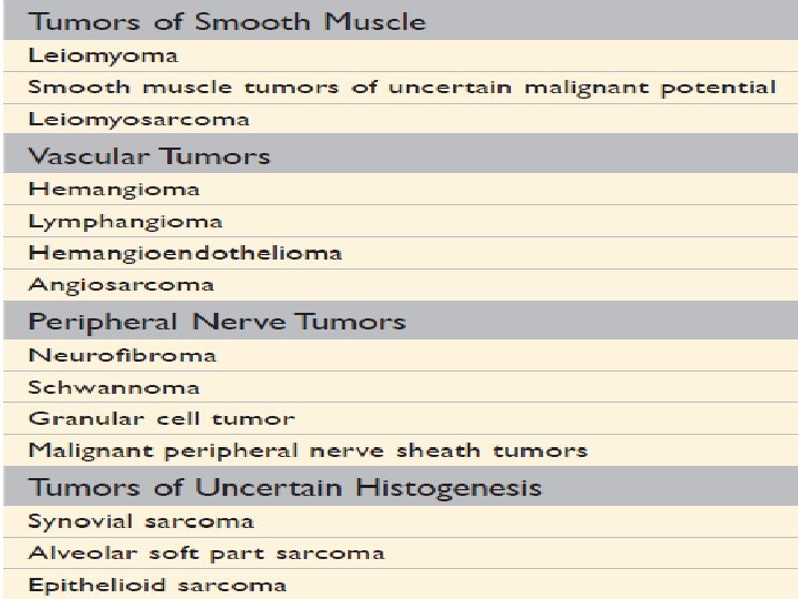
Smooth muscle tumors Leiomyoma: Benign well-circumscribed neoplasms arise from smooth muscle cells anywhere in body. Most commonly in uterus & skin.
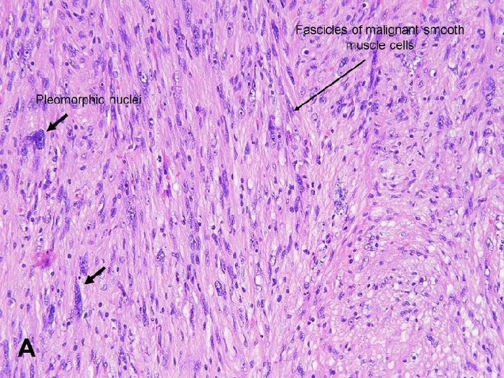
Leiomyosarcoma Occur in adults. Common sites: 1 - Skin & deep soft tissues of extremities. (Firm painless masses) 2 - Retroperitoneum. Large, bulky & cause abdominal symptoms. Histologic examination: Spindle cells with cigar-shaped nuclei arranged in interlacing fascicles.
Thank you Spindle cells with cigar-shaped nuclei arranged in interlacing fascicles.