RESPIRATORY SYSTEM Respiration Facts Your lungs contain almost


- Slides: 29

RESPIRATORY SYSTEM

Respiration Facts : Your lungs contain almost 1500 miles of airways and over 300 million alveoli. If the surface of all the alveoli were spread out flat it would cover a tennis court. Every minute you breathe in 13 pints of air.

More Facts Plants are our partners in breathing. We breathe in air, use the oxygen in it, and release carbon dioxide. Plants take in carbon dioxide and release oxygen. Thank goodness! People tend to get more colds in the winter because we're indoors more often and in close proximity to other people. When people sneeze, cough and even breathe -- germs go flying!

Respiration: Exchanging gases between the atmosphere and body cells Ventilation Gas exchange between the blood and lungs Gas transport in the bloodstream Gas exchange between the blood and body cells Cellular respiration

4 Distinct Processes of respiration • Pulmonary ventilation- air movement in and out of the lungs. • External Respiration- gas exchange between the lungs and the blood of capillaries. • Transport of gases- oxygen and carbon dioxide must travel to tissue cells of the body. • Internal Respiration- gas exchange from blood of the capillaries to and from the cells of the body.

Inhalation/Exhalation Diaphragm – muscle that dictates air pressure within the thoracic cavity to manipulate air to go in or out of the lungs

Inhalation Process • 1. ) Contraction of the diaphragm flattens the floor of the thoracic cavity, increasing the volume of the cavity and drawing air into the lungs. (75% of the process) • 2. ) Contraction of the external intercostal muscles assist in inhalation by elevating the ribs. (25% of the process) • 3. ) Accessory muscles like the sternocleidomastoid, serratus anterior, pectoralis minor and scalene muscles can assist in elevating the ribs increase inhalation speeds.

Exhalation Process • 1. ) Intercostal and transverse thoracic muscles depress the ribs and reduce the width and depth of the thoracic cavity. • 2. ) The abdominal muscles, including the external and internal oblique, transversus abdominis and rectus abdominis can assist to in exhalation by compressing the abdomen and forcing the diaphragm upward.

Inhalation/Exhalation • Decreasing volume increases pressure which forces air out. • Volume decreases= P outside < P inside • Increasing volume decreases pressure which forces air in. • Volume increases= P outside > P inside

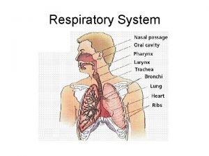
What is done with the Air we breathe? • Air we breathe enters the nasal cavity or mouth. • Your job is to explain what air passes through as it travels to the lungs and eventually the alveoli of the lungs, in addition what is done to air to prepare it for diffusion into the bloodstream. Clean it, cool it, warm it etc.

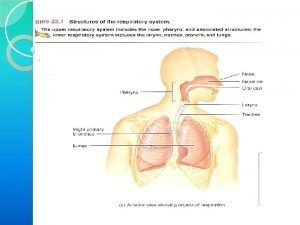
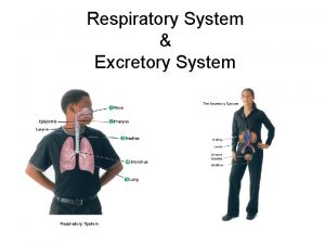
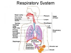
Upper Respiratory System • Nose • Nasal Cavity • Sinuses • Pharynx

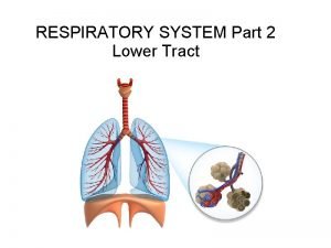
Lower respiratory System • Larynx • Trachea • Bronchioles • Alveoli

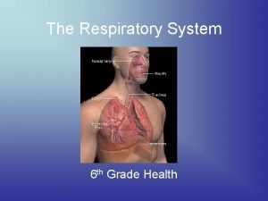
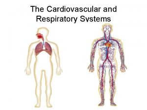
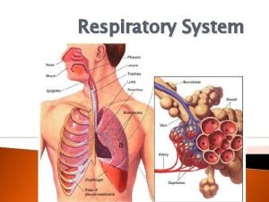
Major Organs Of the Respiratory System

Nasal Sinuses • Air-filled spaces in the bones of the skull –Maxillary, frontal, sphenoid, ethmoid • Open to the nasal cavity and are lined with mucus membrane that is continuous with that lining the nasal cavity • Reduce the weight of the skull, serve as resonant chamber for sound, warm and moisten air Red: Maxillary Checkered: Frontal Green: Ethmoid Yellow: Sphenoid

Nasal Cavity • Nostril- opening for air • Nasal Cavity- superior to the hard palate • Nasal Conchae-make the air turbulent to have it touch olfactory epithelium for smell, clean it with cilia and warm and moisten air for preparation to the trachea. (Superior, middle and inferior)

Nasal Cavity Cont. • Paranasal cavities- opening within the skull to lighten it. They also secrete mucus into the cavity for moistening and warming air. • Nasal Septum- extension of the skull that separates the nostrils. • Auditory tube (eustachian)opening to the tympanic membrane of the ear. Allows pressure to increase or decrease in the middle ear. Elevation

Pharynx • Naso- meaning air from the nasal cavity travels through to the larynx • Oro- meaning it directs air or food to be directed to the esophagus or larynx. Swallowing has the right of way so food and liquid goes to the esophagus. • Laryngo- posterior to the epiglottis so air only enters this portion. Epiglottis pushes down during swallowing.

Epiglottis • Elastic cartilage that will cover the larynx when muscles that control swallowing push it down to cover the opening at the laryngopharynx.

Larynx Vocal Folds Epiglottis • This structure is the combination of nine cartilages connected by membranes and ligaments. • Thyroid cartilage • Cricoid cartilage laryngeal prominence/adam’s apple Arytenoid cartilageattach to vocal folds to rotate and produce sound

Vocal Folds/Cords Arytenoid Cartilage Glottis- opening to trachea • So air vibrates across the vocal cords as the arytenoid cartilage rotates to manipulate how far the cords are stretched to produce different sounds or pitch. Epiglottis remains open!! Epiglottis Vocal cords

Here’s how it works!! https: //www. youtube. com/watch ? feature=player_detailpage&v=XGds 2 GAv. GQ

Trachea • Also known as the windpipe. • 3 layers- mucosa, submucosa, and adventitia. • mucosa- psuedostratified ciliated epithelium that traps dirt and pathogens. Goblet cells • submucosa- help produce mucous sheets within connective tissue. • adventitia- connective tissue supported by hyaline cartilage

Trachea Cont. • Tracheal cartilage- (hyaline cartilage) C shaped cartilage that allows for trachea to expand or constrict. • Trachealis muscle- pulls the cartilage ring in for constriction or relaxes for dilation. • Constriction = more air power. Ex. Coughing • Dilation = more air flow in • Also allows food to pass by without damage or discomfort.

Cavities of the lungs • Thoracic Cavity- cavity of the chest, diaphragm separates it from the abdominal cavity. • Pleural Cavity- space between the parietal pleura and visceral pleura which set cavity pressure to keep lungs inflated. Collapsing pressure is 4 Hg • Damage to this cavity and the lung will collapse

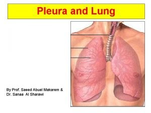
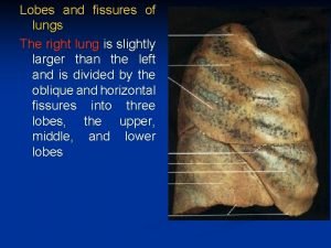
Lungs Facts • Lungs are not symmetrical, need to fit the heart in there. • Lungs don’t have the same amount of lobes, 3 on the right and 2 on the left. 3 Lobes 2 Lobes • Lungs are controlled by atmospheric pressure dictated by the diaphragm. • Lungs stay inflated by the pleural cavity maintaining a 4 mm Hg less than that of the alveoli.

Bronchi Subdivisions • Left and Right Primary • Secondary to Tertiary • Then Bronchioles!!!!!! • Bronchi tubes mimic that of the trachea but as they branch the following changes occur. • Decrease in cartilage, now elastic fibers • No more cilia or columnar epithelium. Need macrophages now. • Smooth muscle increases as passageway gets smaller for constriction to certain conditions

Alveoli • Simple squamous epithelium. Great for diffusion!!!!! • Close proximity to capillaries of pulmonary circulation. • Elastic fibers hold alveoli together and allow them to expand contract for inhalation and exhalation. • Smoking causes macrophages to hang in alveoli and eat all the junk!!! • Macrophages create an enzyme they release called elastase • Elastase breaks down the elastic fibers causing the alveoli from exhaling. (Emphysema)

Respiratory Disorders • Emphysema • Lung Cancer • Cystic Fibrosis • Asthma • Bronchitis • Pneumonia • Tuberculosis

Disease White Boards • How do you get the disease? • What are the symptoms? • What are the treatments? • Is it terminal? Explain why or why not, if not can it be in certain circumstances? • Are new strands existent on Earth today compared to the past? • If terminal, are there advancements to make us see the light?
 Stimulus for breathing
 Conducting zone and respiratory zone
 Interesting facts about respiratory system
 Monika shpokayte
 Digestive system circulatory system and respiratory system
 Internal respiration vs external respiration
 The nose serves all the following functions except
 Smoking damages your lungs
 Taking care of your respiratory system
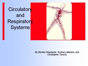
 How respiratory system work with circulatory system
 Circulatory system and respiratory system work together
 Give us your hungry your tired your poor
 64:8+9:9-63:7
 Decreased tactile fremitus
 Fremitus test
 Sce respiratory
 Physiology of lungs
 Lung 3 lobes
 Air path through the respiratory system
 J receptors
 Right lung lobes
 Conducting zone lungs
 Chemoreceptors in brain
 Mediastinal surface
 Lung fissures surface anatomy
 Bronchopulmonary segment
 Pulmonary ligament
 Bronchophont
 Barotrauma lungs
 Right lung lateral view
