RENAL PATHOLOGY INFECTION OF THE UPPER LOWER URINARY


- Slides: 42

RENAL PATHOLOGY INFECTION OF THE UPPER & LOWER URINARY TRACT Reference: Robbins & Cotran Pathology and Rubin’s Pathology

Objectives: At the end of the two lectures the students will be able to: Recognize the predisposing factors for infections of the kidney and urinary tract. Describe the different types of infections in the kidney and urinary tract. Recognize acute and chronic pyelonephritis. Describe the causes of urinary tract obstruction. Recognize drug induced nephritis. Key Outlines: Urinary Tract Obstruction: causes and clinical manifestations in children and adults. Infections of the Urinary Tract: Predisposing Factors and Clinical Manifestations. Pathology of Acute and Chronic Pyelonephritis including causes and complications of urolithiasis. Drug induced interstitial nephritis and renal necrosis.

Lecture outline for 2 lectures • Upper urinary tract infection or inflammation • Tubulointerstitial nephritis Ø Acute pyelonephritis Ø Chronic pyelonephritis Ø Drug induced tubulointerstitial nephritis • Urinary tract outflow obstruction Causes • Urolithiasis • Hydronephrosis • • Lower urinary tract infection Ureteritis • Cystitis •

ACUTE PYELONEPHRITIS

Acute Pyelonephritis • Tubulointerstitial nephritis consists of inflammatory disease primarily involving the renal tubules and interstitium. • Acute pyelonephritis is an acute suppurative inflammation of the upper urinary tract (the kidney and the renal pelvis) caused by bacterial infection. It typically follows infection of the lower urinary tract. • The most common causative organism is enteric gram-negative rods Escherichia coli. Other organisms are species of Proteus, Klebsiella, Enterobacter, and Pseudomonas. There are two routes by which bacteria can reach the kidneys: 1. Ascending infection from the lower urinary tract is the most important and common route by which the bacteria reach the kidney. The bacteria ascends from the urethra, into the urinary bladder and up the ureters to the kidneys. 2. Through the bloodstream/hematogenous (less common) e. g. from an infected heart valve in endocarditis, miliary tuberculosis etc.

Acute Pyelonephritis: predisposing factors 1. Urethral instrumentation (catheterization and cystoscopy). 2. Obstruction: obstruction at the level of the urinary bladder (e. g. stones and benign prostatic hypertrophy) incomplete emptying and increased residual urine stasis which allows bacteria to multiply bacteria ascend along the ureters infects kidneys 3. Incompetence of the vesicoureteral orifice increased residual urine favor bacterial growth and allows reflux of bladder urine with bacteria to ascend the ureter (normally ureteral insertion into the bladder is a competent one-way valve that prevents retrograde flow of urine). Vesicoureteral reflux is seen in 30% of young children with UTI and it is usually a due to a congenital defect of the valve. 4. Gender: UTI most commonly affects females (predisposed because of the close proximity of the urethra to the rectum and short urethra). 5. Pregnancy: Asymptomatic bacteriuria occurs in 10% of pregnant women, out of which some develop acute pyelonephritis. 6. Preexisting renal lesions 7. Diabetes mellitus: diabetic glycosuria predisposes to infection by providing a rich medium for bacterial growth. Diabetics also have increased risk of complications of pyelonephritis e. g. septicemia, necrotizing papillitis and recurrence of infection. 8. Immunosuppression & immunodeficiency

Robbins basic pathology, 9 th Ed Elsevier 2013

Acute Pyelonephritis: morphology Macroscopic • The kidney may be enlarged and swollen with a bulging cut surface. • Cut surface: small yellowish white subcapsular microabscesses (suppurative necrosis). • The renal papillae are diffusely blunted • Pelvi-calyceal mucosa may be hyperemic and covered by purulent exudate. • Rarely the kidney may be filled with large amounts of pus in the renal pelvis, calyces, and ureter, producing pyonephrosis.

Acute Pyelonephritis: morphology Microscopic • There is a dense acute tubulointerstitial inflammation (neutrophils) with tubular destruction. The neutrophils fill the tubules and collecting ducts. • The vessels and glomeruli often are preserved. • In severe cases of acute pyelonephritis inflammation extends beyond the renal capsule perinephric abscess. Pathology Education Informational Resource (PEIR) Digital Library http: //peir. path. uab. edu/library/_data/i/upload/2013/08/01/20130 801094757 -67538 f 5 c-me. jpg

Acute pyelonephritis: microscopy https: //web. duke. edu/pathology/Week 14/images/large/path 9 -06 and 9 -07. jpg

Acute Pyelonephritis: complications clinical features Ø Papillary necrosis • Fever, chills, sweats, malaise, flank pain with costovertebral angle tenderness. • Dysuria, frequency and urgency. Ø Perinephric abscess • Leukocytosis with neutrophilia is common. Ø Chronic pyelonephritis • Pyuria, hematuria and wbc casts in the urine. Ø Septicemia • Positive urine culture. • Differentiating upper from lower urinary tract infection is often clinically difficult. Ø Pyonephrosis

Papillary necrosis (necrotizing papillitis) • It is a type of pyelonephritis characterized by necrosis of the renal papillae (the apex of the renal pyramids). • It is seen in a) diabetics with acute pyelonephritis b) in acute pyelonephritis with significant urinary tract obstruction. c) interstitial nephritis associated with analgesic abuse. d) Chronic liver disease e) Renal transplant rejection f) Infection e. g. tuberculosis g) Sickle cell disease • Grossly: yellow white suppurative necrosis of the tips of the some or all renal papillae (renal pyramids). • Microscopically: coagulative necrosis at the tips of renal papillae with surrounding neutrophilic infiltrate Pathology Education Informational Resource (PEIR) Digital Library http: //peir. path. uab. edu/library/_data/i/upload/2013/08/01/20130801094931 -333 fc 99 e-me. jpg

CHRONIC PYELONEPHRITIS

Chronic Pyelonephritis • It is a chronic tubulointerstitial inflammation of the kidney caused repeated bouts of inflammation and healing. It results a scarred kidney with deformed renal pelvis and calyces. • It may caused either by Ø Chronic obstruction associated pyelonephritis e. g. obstruction of the ureter by calculi (stones), tumor within the ureter, or extrinsic compression etc. Ø Chronic reflux associated pyelonephritis. Chronic pyelonephritis usually is associated with urinary obstruction or reflux; results in scarring of the involved kidney and gradual renal insufficiency

Chronic Pyelonephritis: morphology Macroscopy • The kidneys are small and contracted. • The surface of the kidney is irregularly scarred with areas of depression. • The cortex in thinned out and there is deformity and blunting of the pelvicalyceal system. Robbins basic pathology, 9 th Ed Elsevier 2013

Chronic Pyelonephritis: morphology Microscopy • Tubules: tubular atrophy with thyroidization of tubules • Interstitium: interstitial fibrosis and chronic interstitial inflammation, • Glomeruli: periglomerular fibrosis and glomerulosclerosis.

Chronic pyelonephritis: microscopy

Chronic Pyelonephritis: clinical features • Most patients have episodic symptoms of urinary tract infection or acute pyelonephritis, such as recurrent fever and flank pain. • Slowly develops, patients may present with chronic renal failure. • Hypertension. • Some patients have a silent course until end-stage renal disease develops. • Imaging studies show abnormalities of the pelvicalyceal system and cortical scarring.

DRUG INDUCED TUBULOINTERSTITIAL NEPHRITIS

Drug induced tubulointerstitial nephritis • Drugs are an important cause of renal injury. • Drug-induced interstitial nephritis is an Ig. E (hypersensitivity) and T cell–mediated immune reaction to a drug; characterized by interstitial inflammation, often with abundant eosinophils, and edema • Acute drug-induced tubulointerstitial nephritis (TIN) occurs as an adverse reaction to various therapeutic drugs e. g. penicillins (methicillin, ampicillin), rifampin) diuretics (thiazides), nonsteroidal anti-inflammatory agents, etc. Morphology • Interstitium: shows edema and infiltration by chronic inflammatory cells (lymphocytes and macrophages). Eosinophils are usually present in large numbers. • Sometimes there are non-necrotizing granulomas and multinucleated giant cells in the interstitium. • The glomeruli are usually normal. Clinical features • Abnormal renal function test after few days or weeks after exposure to the drug. • Urine test: hematuria and eosinophils. • Can present as acute kidney injury with rising serum creatinine and oliguria. • It is important to diagnose because if remove the offending drug on time the injury maybe reversible.

URINARY TRACT OBSTRUCTION (URINARY OUTFLOW OBSTRUCTION)

Causes of urinary tract outflow obstruction • Congenital anomalies • Urolithiasis • Benign prostate hyperplasia • Tumors (prostate, cervix, urinary bladder etc. ) • Inflammations (prostatitis, urethritis etc. ) • Pregnancy • Others (paralysis of urinary bladder)

UROLITHIASIS

Urolithiasis/nephrolithiasis • Urolithiasis is the condition where urinary calculi are formed or located anywhere in the urinary system e. g. kidney, bladder, ureters etc. • Renal pelvis and calyces are common sites for calculi formation. Predisposing factors: • Metabolic factors: hypercalciuria, hyperphosphaturia, oxaluria, gout etc. • A persistently alkaline urine favors formation of calcium phosphate stones. • A persistently acidic urine favors formation of oxalate or uric acid stones. • Stasis facilitates precipitation of salts and stone formation. • Chronic dehydration: concentrates urine and favors stone formation • For unknown reasons, renal stones are more common in men than in women. • There may be a familial tendency toward stone formation.

Urolithiasis Stones vary in composition, depending on various factors. Types of stones seen are: 1. Calcium stones: 75% of kidney stones are calcium oxalate or phosphate (radio-opaque). 2. Infection stones: Some 10% of stones are caused by infection results in alkaline urine leads to formation of magnesium ammonium phosphate stones. Infection stones can occasionally fill the pelvis and calyces to form large staghorn calculi. 3. Uric acid stones: are formed in acidic p. H. Patients with hyperuricemia and gout are predisposed to uric acid stones, however it can be seen in people with no hyperuricemia and gout. Pure uric acid stones are radiolucent. 4. Cystine stones: are uncommon.

Urolithiasis: morphology • Majority (80%) of the stones are unilateral • Common sites of formation are renal pelvis and calyces and the bladder • Commonly multiple stones are found in one kidney • Stones vary in size from few mm to large stones that dilate the entire renal pelvis. • They range from hard to soft , from smooth to rough • The staghorn calculi take the shape of the pelvicalyceal system. They are large stones are usually composed of magnesium ammonium phosphate Sectioned Kidneys, Ureters, and Bladder showing location of Kidney Stones. SOURCE: pickup from 5 A 11872 Referenced from www@ http: //graphics 8. nytimes. com/images/2007/08/01/health/adam/17091. jpg Anatomy text: Clemente Anatomy. 4 th Ed. Carmine D. Clemente. Lippincott Williams & Wilkins. plate 233 figure 356

http: //peir. path. uab. edu/library/_data/i/upload/2013/08/01/20130801095842 -a 9530 c 10 -me. jpg

Staghorn calculi Qiao's Pathology: Kidney - Staghorn Calculus https: //www. flickr. com/photos/jianhua_qiao_md/5648360864/in/album 72157622371115615/ http: //peir. path. uab. edu/library/_data/i/upload/2013/08/01/20130801094321 -3468 c 066 -me. jpg

Urolithiasis: Clinical features • Stones may be asymptomatic esp. stones lodged in the renal pelvis • Stone can erode the mucosa hematuria. • Smaller stones can pass into the ureter, where they cause obstruction intense paroxysms of flank pain radiating toward the groin known as renal or ureteral colic. They may pass out in urine, painful. • Stones obstruct urine flow or to produce sufficient trauma to cause ulceration and bleeding predispose to bacterial infection. Complications • Recurrent infection • Hydronephrosis • Pyelonephritis • Pyoneohrosis • Acute urinary retention • Renal failure. Diagnosis and management In most cases the diagnosis is readily made radiologically In the past most kidney stones required surgical removal, but now ultrasonic disintegration (lithotripsy) and endoscopic removal are now effective.

HYDRONEPHROSIS (IS THE CONSEQUENCE OR COMPLICATION OF URINARY OUTFLOW OBSTRUCTION)

Hydronephrosis • It is the dilation of the renal pelvicalyceal system with resultant renal parenchymal atrophy. • It is caused mainly by obstruction to the outflow of urine. • The obstruction can be acute or chronic and be at any level of the urinary tract. Obstruction below the level of ureters causes bilateral hydronephrosis. • The obstruction can be: a) Congenital: e. g. atresia of urethra, abnormal compression of ureter by renal artery etc. b) Acquired: Ø Foreign bodies: Calculi Ø Tumors: Benign prostatic hyperplasia, prostate, bladder, cervix tumors etc. Ø Inflammation: Prostatitis, ureteritis, urethritis Ø Neurogenic: Spinal cord damage with bladder paralysis Ø Pregnancy

Hydronephrosis: pathogenesis • There is backflow of the urine into the kidney the calyces and pelvis become markedly dilated back flow in collecting ducts and ultimately renal cortex renal parenchyma atrophy. • The obstruction also triggers an interstitial inflammatory reaction leading eventually to tubular atrophy and interstitial fibrosis. • Initially the glomeruli are spared but eventually they become sclerotic. http: //www. medfriendly. com/images/hydronephrosis. jpg

Hydronephrosis: morphology Grossly: • Enlarged kidney due to dilation of the renal pelvis and calyces • Atrophy or compression of the renal parenchyma ensues. • Depending on the level of the obstruction, one or both ureters may also be dilated (hydroureter)

Hydronephrosis: gross Robbins basic pathology, 9 th Ed Elsevier 2013

Hydronephrosis: clinical features • Unilateral hydronephrosis may remain completely silent for long periods • Bilateral hydronephrosis can leads to oliguria to anuria and renal failure • With time the changes become irreversible • In the presence of hydronephrosis, the kidney is more susceptible to pyelonephritis, which causes additional injury • Early diagnosis and timely removal of obstruction within a few weeks usually permits full return of function

INFECTIONS OF THE LOWER URINARY TRACT

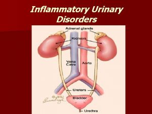
Lower urinary tract The ureters, urinary bladder, and the urethra also known as the lower urinary tract form the outflow part of the urinary system Transitional epithelium (urothelium) lines the ureters, bladder, and posterior urethra except the terminal urethra which is lined by squamous epithelium. Normal urothelium

Pathology Inflammation: • Ureteritis • Cystitis • Urethritis UTI (urinary tract infection) is the infection of the lower urinary tract (cystitis, prostatitis, urethritis). Ureteritis § Ureteritis is an inflammation of the ureters. It is a complication of descending infections of kidneys or ascending infections if there is vesicoureteric reflux. § Ureteritis is often associated with ureteral obstruction ( e. g. calculi, intraluminal blood clots or extrinsic compression of ureter by an adjacent tumors or lymph nodes and the pregnant uterus can compress the ureters).

CYSTITIS • It is inflammation of the urinary bladder. It may be acute or chronic. It is the most common urinary tract infection and is often seen as a nosocomial infection in hospitalized patients. • Predisposing factors include bladder calculi, bladder outlet obstruction (e. g. prostatic hyperplasia), diabetes mellitus, immunodeficiency, prior instrumentation or catheterization, radiation therapy, and chemotherapy. • The risk of cystitis is increased in females because of a short urethra, especially during pregnancy. • Common in patients in whom indwelling catheters remain for prolonged periods. • Bacterial cystitia is most common form of cytstitis. It is caused mainly by coliform bacteria e. g. E coli, Proteus vulgaris, Pseudomonas and Enterobacter spp.

Cystitis: pathology § Acute cystitis : edema, hemorrhage and a neutrophilic infiltrate. § Chronic cystitis: a predominance of lymphocytes and fibrosis. Occasionally there are numerous lymphoid follicles called follicular cystitis or dense infiltrates of eosinophils eosinophilic cystitis. Special forms of cystitis: • Tuberculous cystitis: there is granulomatous cystitis with or without necrosis. • Hemorrhagic cystitis: shows mucosal hemorrhages. Is often seen in acute bacterial infection, adenovirus infection, bleeding diatheses (e. g. , leukemia, treatment with cytotoxic drugs and disseminated intravascular coagulation). • Chronic interstitial cystitis (chronic pelvic pain syndrome): is a persistent, painful form of chronic cystitis typically affects middle-aged women. It is characterized by intermittent, suprapubic pain, urinary frequency, urgency, hematuria and dysuria without evidence of bacterial infection; cause is unknown; urine cultures are usually negative. The disease is refractory to all forms of therapy. Eentually bladder fibrosis contracted bladder.

Special forms of cystitis contd… › Malakoplakia: uncommon inflammatory disorder of unknown etiology. Seen in the bladder and other sites within and outside the urinary tract; characterized by plaques on the mucosal surface of the bladder. Histology shows a chronic inflammation with numerous macrophages. Some of these macrophages contain laminated, mineralized concretions of basophilic calcium called Michaelis-Gutmann bodies. › Polypoid cystitis: is an inflammatory condition resulting from irritation to the bladder mucosa in which there is marked submucosal edema resulting in polypoidal (polyp-like)projections. › Schistosomiasis: in it the S. hematobium eggs (picture) can be seen and they ellicit a granulomatous reaction with an eosinophilic infiltrate. The ova can calcify ova and appear as grains of sand in bladder wall. Occasionally the entire urinary bladder become calcified known as calcific cystitis.

Acute and chronic cystitis: clinical features § Excessive urinary frequency, painful burning urination (dysuria) and lower abdominal or pelvic discomfort. § Examination of urine usually reveals inflammatory cells and causative organism can be identified by urine culture. § Most cases of cystitis respond well to treatment with antimicrobial agents.
 Diagnostico etiologico
 Ira pré renal renal e pós renal
 Urised otc
 Sterile pyuria ppt
 Urine infection symptoms
 Complicated urinary tract infection
 Urinary tract infection in pregnancy ppt
 Vasa recta vs peritubular capillaries
 Ajkd atlas of renal pathology
 Ajkd atlas of renal pathology
 Classification of upper respiratory tract infection
 Acute upper respiratory infection unspecified คือ
 Nearest whole number upper and lower bounds
 Upper bound and lower bound in array
 Upper lower bounds
 Limites de control
 Lower bound of 5
 Roof plan parts
 Upper egypt and lower egypt
 Upper and lower control limit formula
 Larangoscopy
 Dante's inferno 7th level of hell
 Programme upper lower
 Greatest lower bound and least upper bound
 What divided upper and lower egypt
 Upper epidermis function in photosynthesis
 Well ordering principle
 Invertible upper triangular matrix
 Uti upper and lower
 How to find upper and lower bounds
 Upper and lower bounds 3 significant figures
 Upper airway anatomy
 Upper lower class
 Box-and-whisker plots worksheet
 Upper specification limit and lower specification limit
 2 significant figures upper and lower bounds
 Sample and population difference
 Upper and lower respiratory tract
 Fetal pig urinary system labeled
 Figure 15-3 the urinary system
 Uria medical term
 Frog spinal nerves
 Chapter 47 urinary and reproductive disorders


