Renal Cell Carcinoma Dr Amit Gupta Associate Professor

. • Most common in sixth")

 renal cell carcinoma extending into the collecting system")
, obesity, high dietary")

 flank pain (45%)")


 • High dose IL-2 only")
- Slides: 20
Renal Cell Carcinoma Dr. Amit Gupta Associate Professor Dept. of Surgery
Epidemiology • Male predominance (M: F 1. 5: 1). • Most common in sixth to eighth decades; peak incidence in sixth decade • Metastatic disease in 30% at diagnosis, and eventually in 50% (lung, liver, bone, distant LN, adrenal, brain, opposite kidney, soft tissue) • Most sporadic RCCs are unilateral and unifocal
• Stage at diagnosis is the most important prognostic factor • Predominant histologic type: adenocarcinoma arising from tubular epithelium • Adenocarcinoma subtypes: – clear cell (75– 85%) – chromophilic/ papillary (10– 15%) – chromophobe (5– 10%) – oncocytic (rare) – Sarcomatoid (1– 6%; poor prognosis)
Papillary (chromophilic) renal cell carcinoma extending into the collecting system
Risk factors • Tobacco , urban environmental toxins (cadmium/ asbestos/ petrols), obesity, high dietary fat intake, acquired cystic renal disease from renal failure • Association with von Hippel-Lindau disease: – autosomal dominant – loss of 3 p – >70% chance developing RCC (almost all clear cell histology) risk of developing multiple other benign and malignant tumors (retinal angiomas, CNS hemangioblastomas, pheochromocytoma , pancreatic cancer)
Pathology • Round to ovoid • Circumscribed by a pseudo capsule of compressed parenchyma and fibrous tissue • Nuclear features can be highly variable
Diagnosis • Common signs and symptoms: – – – hematuria (80%) flank pain (45%) flank mass (15%) classic triad of prior three only present in 10% normocytic/normochromic anemia, fever, weight loss • Less common signs and symptoms: – hepatic dysfunction without mets – Polycythemia – hypercalcemia (occurs in 25% of patients with RCC mets)
Paraneoplastic syndromes in 20% of patients with RCC
Diagnosis • Labs: CBC, LFT, BUN/Cr, LDH, urinalysis • Imaging: – CT abdomen – MRI abdomen if CT suggests IVC involvement • Metastatic evaluation: – Chest X ray – Bone scan or MRI brain only if clinically indicated
CT scan shows right renal tumor with perinephric stranding suggesting invasion of the perinephric fat
Contrast inferior venacavogram in patient with a right renal tumor shows involvement of the subdiaphragmatic vena cava
• PET: equivocal findings on conventional imaging • Percutaneous renal biopsy or aspiration: limited role
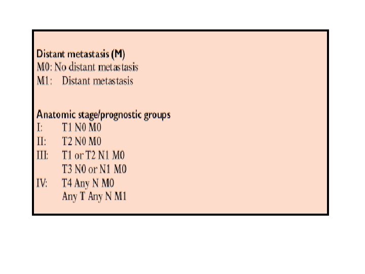
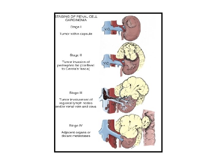
Staging AJCC 7 th Edition
Prognostic Factors For RCC
Management Stage I-III Nephrectomy • Open radical nephrectomy, but laparoscopic gaining popularity • Nephron sparing surgery via partial nephrectomy, if possible (open or laparoscopic) • Possible to spare adrenal gland in ~75% cases No role for adjuvant chemo/immunotherapy
No widely accepted role for neoadjuvant or adjuvant radiotherapy. Retrospective data suggest possible utility in select cases: – Positive surgical margins – Locally advanced disease with perinephric fat invasion and adrenal invasion (IVC/renal vein extension alone does not increase local recurrence significantly) – LN+ – Unresectable (pre-op RT)
Stage IV Cytoreductive nephrectomy: improved survival with nephrectomy followed by interferon alpha vs. interferon alpha alone
Systemic therapy • Immunotherapy (IL-2, interferon alpha, or combination) • High dose IL-2 only FDA approved treatment for • Biologic agents show promise in recent trials • Bevacizumab • Sorafenib or sunitinib • Temsirolimus Consider chemo (gemcitabine ± 5 -FU or capecitabine) Focal palliation of metastases • RT alone • Metastasectomy • Combination of both