Differential diagnosis Papillary carcinoma Poorly differentiated carcinoma Medullary

- Slides: 22
Differential diagnosis • Papillary carcinoma • Poorly differentiated carcinoma • Medullary carcinoma • Anaplastic carcinoma • Squamous cell carcinoma, poorly differentiated • Basal cell carcinoma • Lymphocytic thyroiditis
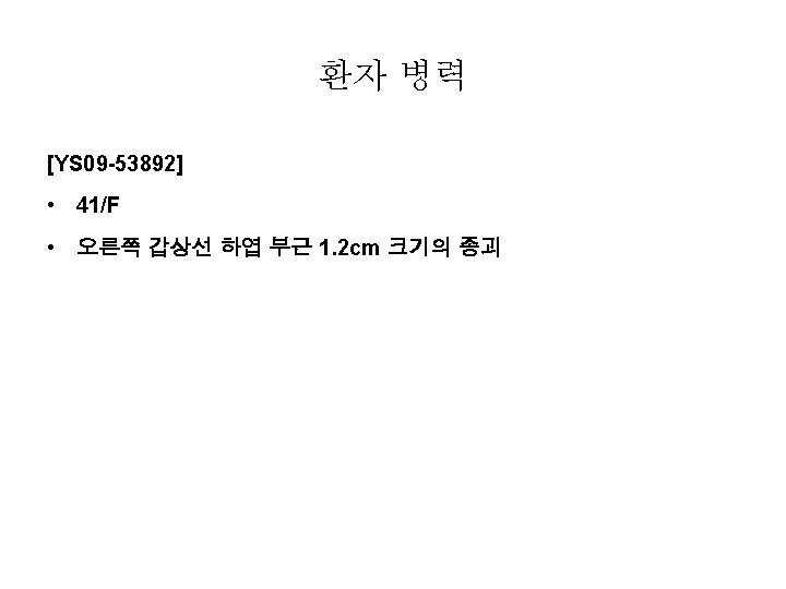
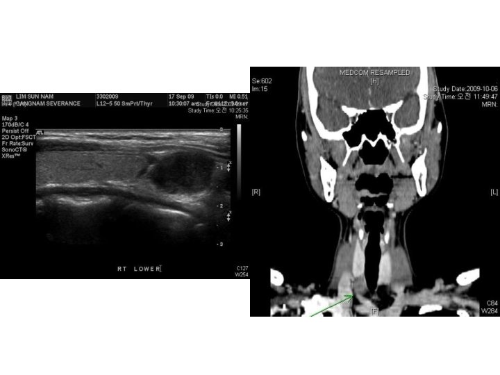
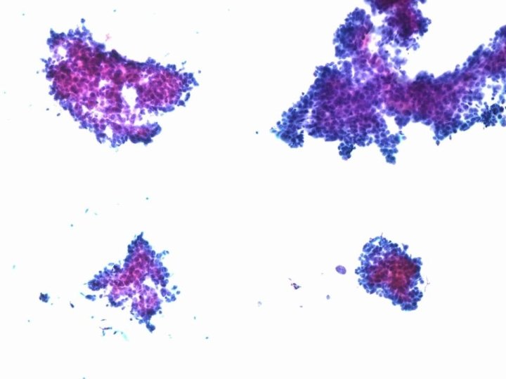
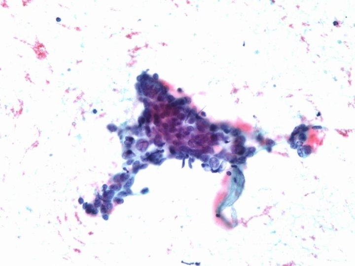
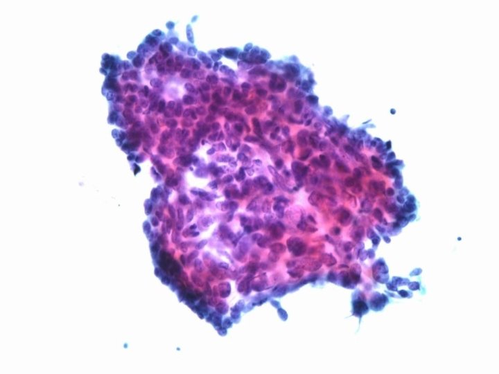
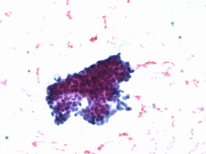
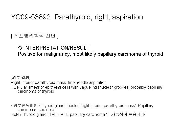
YC 09 -53892 Parathyroid, right, aspiration [ 세포병리학적 진단 ] ◇ INTERPRETATION/RESULT Positive for malignancy, most likely papillary carcinoma of thyroid [외부 결과] Right inferior parathyroid mass, fine needle aspiration - Cellular smear of epithelial cells with vague intranuclear grooves, probably papillary carcinoma of thyroid <외부판독의뢰>Thyroid gland, labeled 'right inferior parathyroid mass': Papillary carcinoma, see note. Note) Thyroid gland 에서 기원한 papillary carcinoma 의 가능성이 높습니다.
Differential diagnosis • Undifferentiated thyroid carcinoma • Squamous cell carcinoma of thyroid • Thymic carcinoma
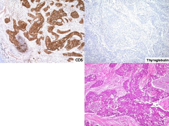
CD 5 Thyroglobulin
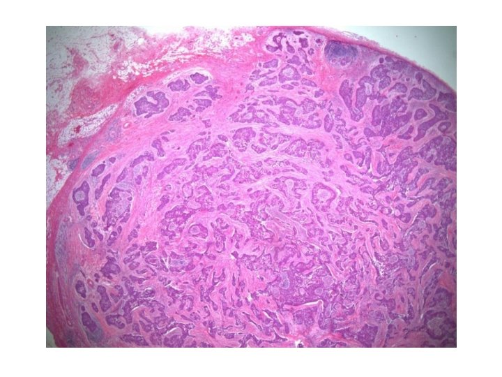
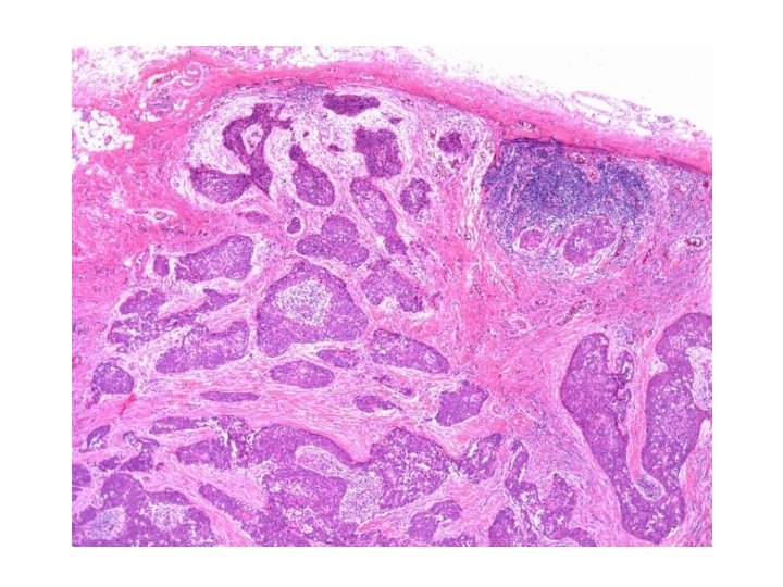
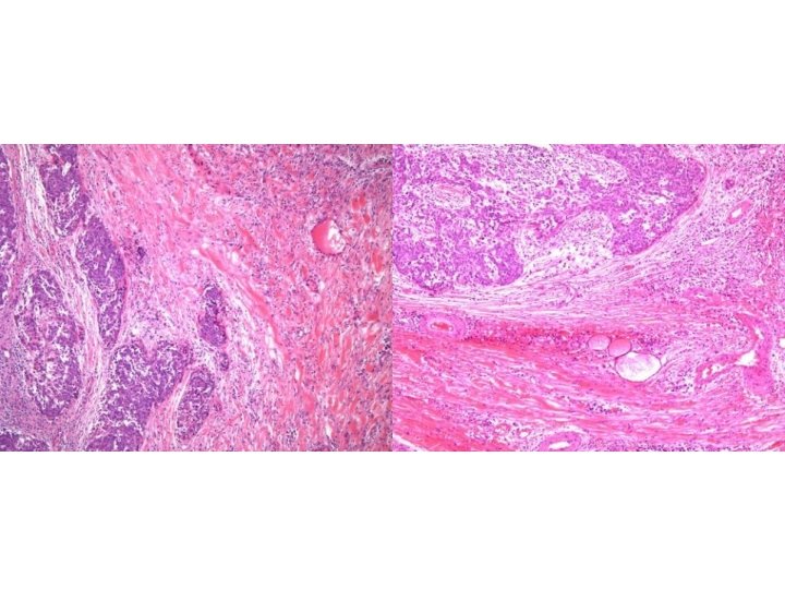
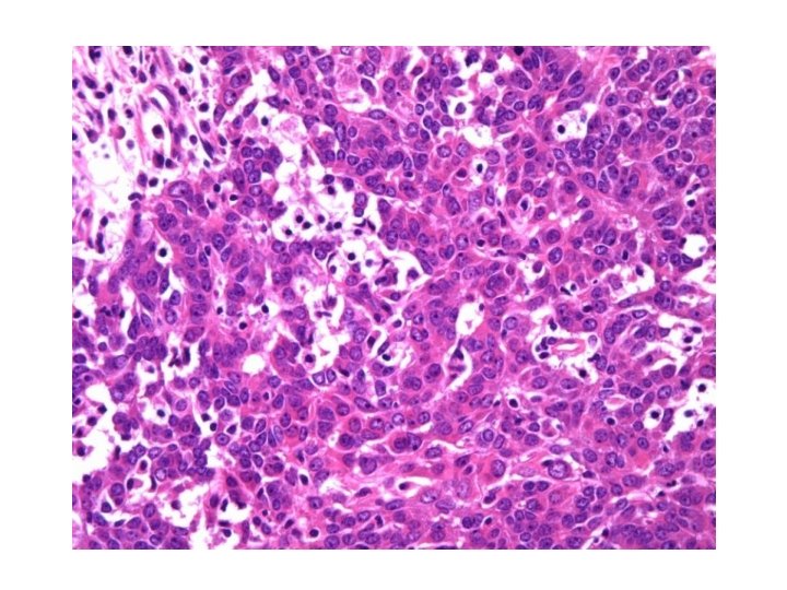
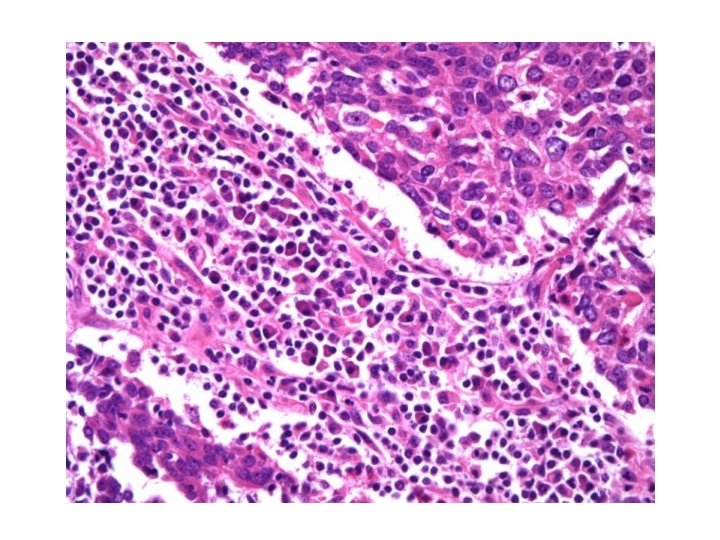
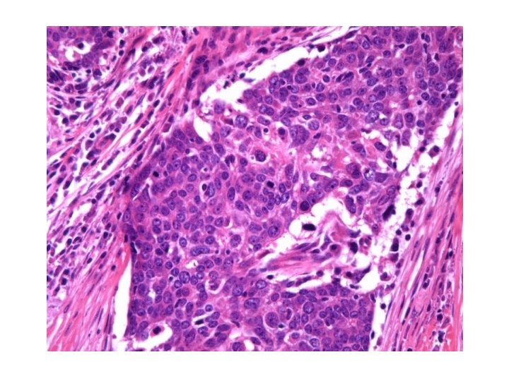
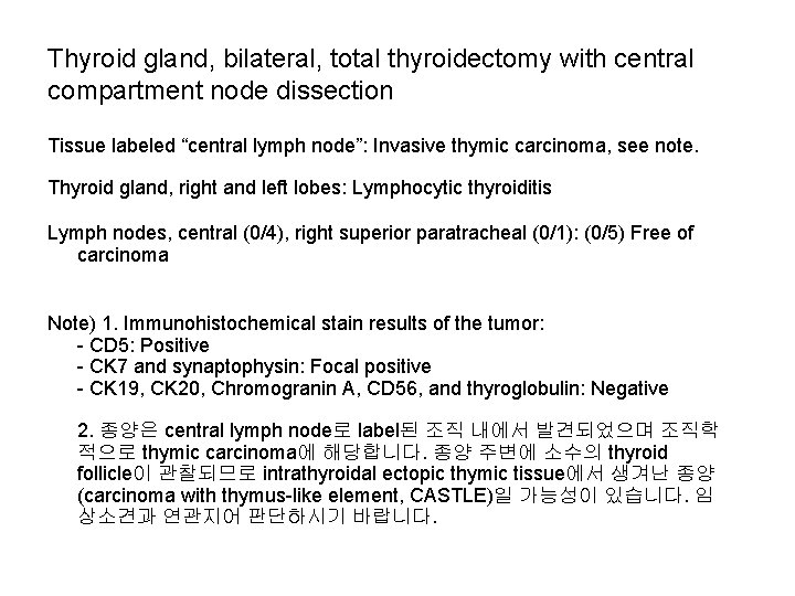
Thyroid gland, bilateral, total thyroidectomy with central compartment node dissection Tissue labeled “central lymph node”: Invasive thymic carcinoma, see note. Thyroid gland, right and left lobes: Lymphocytic thyroiditis Lymph nodes, central (0/4), right superior paratracheal (0/1): (0/5) Free of carcinoma Note) 1. Immunohistochemical stain results of the tumor: - CD 5: Positive - CK 7 and synaptophysin: Focal positive - CK 19, CK 20, Chromogranin A, CD 56, and thyroglobulin: Negative 2. 종양은 central lymph node로 label된 조직 내에서 발견되었으며 조직학 적으로 thymic carcinoma에 해당합니다. 종양 주변에 소수의 thyroid follicle이 관찰되므로 intrathyroidal ectopic thymic tissue에서 생겨난 종양 (carcinoma with thymus-like element, CASTLE)일 가능성이 있습니다. 임 상소견과 연관지어 판단하시기 바랍니다.
Carcinoma showing thymus-like differentiation (Kaohsiung J Med Sci 2008; 24: 591– 7) • Carcinoma showing thymus-like differentiation (CASTLE) is a rare malignant neoplasm that was originally described by Miyauchi et al in 1985. Since then, only 25 cases of this unusual tumor have been reported. • Microscopic examination revealed confluent nests and lobules of epithelioid and spindle-shaped cells with pleomorphic vesicular nuclei, distinct nucleoli with small lymphocytes and plasma cell infiltration, and focal squamous differentiation. • Immunohistochemical tests showed that the tumor was positive for CD 5, weakly positive for cytokeratin, and negative for thyroglobulin and vimentin.
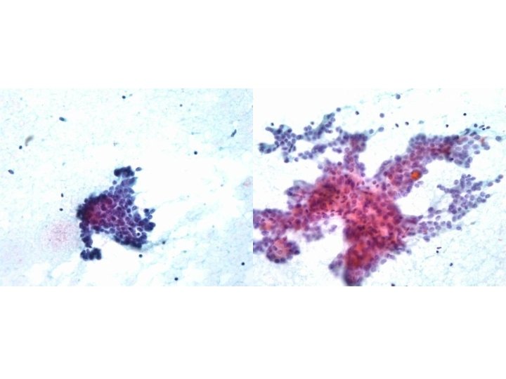
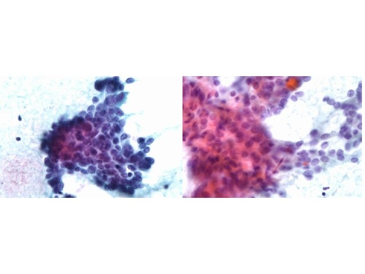
A case of malignant thymoma mimicking thyroid carcinoma: a pitfall in fine-needle aspiration. Endocr J. 2004 Apr; 51(2): 237 -41. Matsuura B et al. A case of malignant thymoma presenting as an anterior neck mass A fine-needle aspiration cytology from the tumor showed tight clusters of epithelial cells with crowded ovoid nuclei. The tumor was initially diagnosed as thyroid carcinoma, clinically and cytologically. A thymoma with a dominant epithelial component has to be considered in the differential diagnosis of a suspected papillary carcinoma of the thyroid.