Lymphangitis Lymphadenitis Dr Amit Gupta Associate Professor Dept


 (MC) Staphylococcus aureus Pseudomonas")


 count • Blood culture • Leading -edge culture")
 imaging was very useful in determining")


")


, syphilis, Castleman dis.")


 Presenting Signs and Symptoms Cervical nodes most commonly involved Stages of")
")


- Slides: 35
Lymphangitis Lymphadenitis Dr Amit Gupta Associate Professor Dept Of Surgery
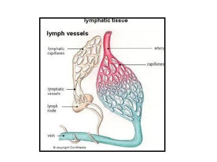
Introduction • Lymphatic system encompasses a network of vessels, glands, and organs located throughout the body. • Transports fluids, fats, proteins, and other substances in the body. • Lymph nodes filter the lymph fluid. • Foreign bodies such as bacteria and viruses are processed in the lymph nodes to generate an immune response to fight infection.
When pathogenic organisms enter the lymphatic channels local inflammation and subsequent infection ensue, manifesting as red streaks on the skin. The inflammation or infection then extends proximally toward regional lymph nodes. Bacteria can grow rapidly in the lymphatic system
Definition Inflammation of the lymphatic channels that occurs as a result of infection at a site distal to the channel
Etiology • • Species of group A beta-hemolytic streptococci (GABHS) (MC) Staphylococcus aureus Pseudomonas species Streptococcus pneumoniae Pasteurella multocida Gram-negative rods, gram-negative bacilli, and fungi Aeromonas hydrophila Wuchereria bancrofti Diabetes, immunodeficiency, varicella, chronic steroid use, or other systemic illnesses have increased risk of developing serious or rapidly spreading lymphangitis.
Nodular lymphangitis Superficial inoculation with one of the following organisms: – – – Sporothrix schenckii Nocardia brasiliensis Mycobacterium marinum Leishmania panamensis L guyanensis Francisella tularensis
Prognosis • With uncomplicated lymphangitis is good. • Antimicrobial regimens are effective in more than 90% of cases. • Without appropriate antimicrobial therapy cellulitis may extend along the channels; necrosis and ulceration may occur. • Morbidity and mortality is related to the underlying infection. • Mortality associated with lymphangitis alone, lymphangitis caused by GABHS can lead to bacteremia, sepsis, and death.
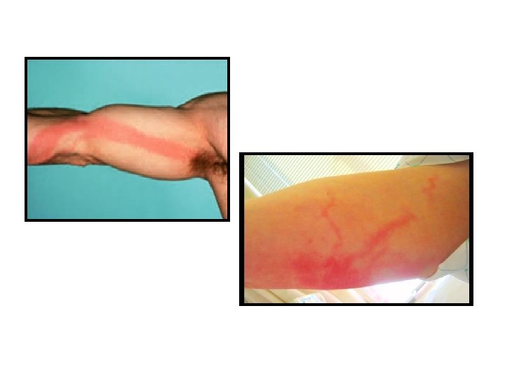
Clinical presentation • H/o minor trauma to an area of skin distal to the site of infection • Children with lymphangitis: fever, chills, and malaise, headache, loss of appetite, muscle aches. • H/o recent cut or abrasion or of an area of skin • Can progress rapidly to bacteremia and disseminated infection and sepsis
Physical examination • Erythematous and irregular linear streaks extend from the primary infection site toward draining regional nodes. • Primary site may be an abscess, an infected wound or an area of cellulitis • Blistering of the affected skin may occur • Lymph nodes associated with the infected lymphatic channels are often swollen and tender • Patients may be febrile and tachycardic
Differential diagnosis • Contact dermatitis • Cellulitis • Septic thrombophlebitis • Superficial thrombophlebitis • Necrotizing fasciitis • Myositis • Sporotrichosis
Investigations • Complete blood cell (CBC) count • Blood culture • Leading -edge culture or aspiration of pus • Cultures and Gram staining of fluid. Threshold sensitivity of Gram staining: 100, 000 microorganisms per milliliter, concentration rarely found in cellulitis or lymphangitis.
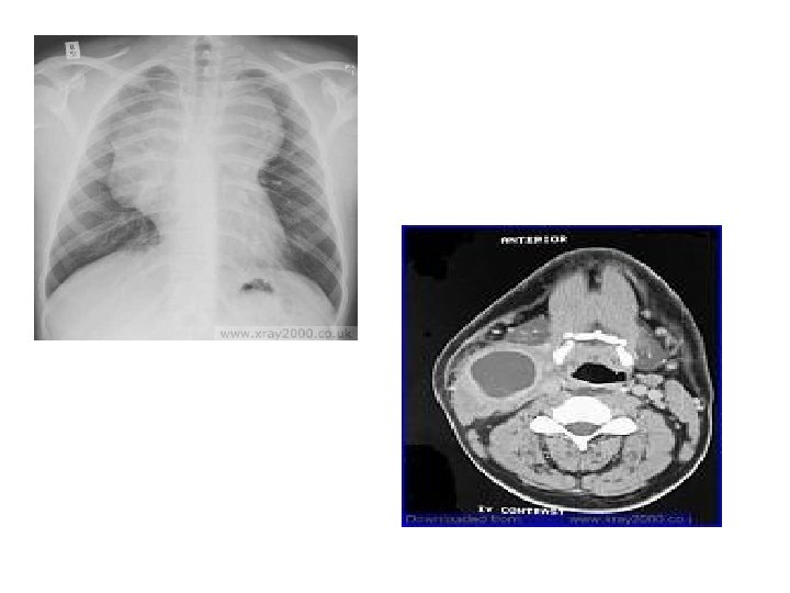
A study found that multidetector computed tomography (MDCT) imaging was very useful in determining the morphology (cellulitis with a few small subcutaneous nodules and channels) and the extension of the lesion in a case of nodular lymphangitis caused by Mycobacterium marinum
Management • Antibiotics • Oral • Parenteral (with signs of systemic illness (eg, fever, chills and myalgia, lymphangitis). • Analgesics • Anti -inflammatory • Elevation and immobilization of affected areas reduces swelling, pain, and the spread of infection. An abscess may require surgical drainage.
Nodular lymphangitis • Treatment of nodular lymphangitis is determined by identifying the underlying cause. • Sporotrichosis is most often identified in this disease and is commonly found among gardeners.
Antibiotics • • Dicloxacillin Cephalexin Cefazolin Cefuroxime Ceftriaxone Clindamycin Nafcillin Trimethoprim and sulfamethoxazole (TMP/SMZ)
Lymphadenitis
Lymphadenitis acute or chronic Rarely biopsied Classification pattern etiology predominant histologic
Lymphadenitis - etiology Microbial • viral • bacterial, mycobacterial • fungal • Protozoal Nonmicrobial Autoimmune storage disease
Lymph node inflammation Lymph node hyperplasia: 1. follicular 2. paracortical 3. sinuses 4. mixed patterns
Lymph node inflammation - patterns • Follicular hyperplasia bacteria, RA, HIV(early), syphilis, Castleman dis. • Paracortical hyperplasia viruses, dermatopathies, vaccination, drug hypersensitivity, Kikuchi, SLE, draining pus or carcinoma
Suppurative Bacterial Lymphadenitis • Staphylococcus aureus and Group A Streptococcus • Anaerobes • Usually acute onset, fever, CBC • Management: antibiotics • If not resolving or getting worse – Ultrasound and/or CT with contrast to evaluate for phlegmon/abscess/infiltrate • FNA vs Surgical I&D vs Surgical Excision if abscess is identified
Suppurative Lymphadenitis with Overlying cellulitis
Subacute Lymphadenitis • 2 -6 weeks • Usually no improvement with antibiotics • DD: • Atypical Mycobacteria • Cat Scratch disease • Toxoplasmosis • TB
Atypical Mycobacteria • Leading cause of sub-acute disease • Species involved: • Mycobacterium avium-intrucellulare • Mycobacterium scrofulaceum • Develops over weeks to months • Lymph nodes may have violaceous skin over the node • No fever, normal behavior, no pain • Diagnosis: acid fast stain and culture, can take weeks. PCR. • Treatment: surgical excision of involved lymph nodes, some offer antibiotics (Clarithromycin plus Rifabutin)
Tuberculosis lymphadenitis (Scrofula) Presenting Signs and Symptoms Cervical nodes most commonly involved Stages of Tubercular Lymphadenitis • Periadenitis • Cold abscess • 'Collar stud' abscess • Sinus
Tuberculosis (Scrofula)
Tuberculous lymphadenitis is popularly known as Collar stud abscess, due to its proximity to the collar bone and superficial resemblance to a collar stud
History • Fever, malaise, anorexia, myalgias • Pain or tenderness of node • Sore Throat / URI / Toothache / Ear pain • Insect Bites • Exposure to animals • History of travel or exposure to TB • Immunizations • Medications
Physical Exam • • • General – Febrile or toxic appearing Skin – Cellulitis, impetigo, rash ENT – Otitis, pharyngitis, teeth, and nasal cavity Neck – Size – Unilateral vs Bilateral – Tender vs Nontender – Mobile vs Fixed – Hard vs Soft Lungs – Consolidations suggesting TB Abdomen – Hepatosplenomegaly
Laboratory Workup • CBC with Differential • ESR • Throat culture • Serology – EBV, CMV, Toxoplasmosis, Bartonella, Syphilis, HIV • LDH, uric acid
Imaging Workup • CXR if malignancy sus. – To look for mediastinal lymphadenopathy • Ultrasound – Abscess? – Benign vs. malignant • Sometimes CT/MRI – To evaluate for abscess • EKG/ECHO – If suspect Kawasaki Disease • Biopsy – FNA or Excisional
Treatment Incision drainage with proper evacuation of the abscess, followed by anti-tubercular antibiotic treatment.