PROTISTS For more than half of the long


- Slides: 28

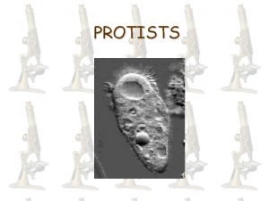
PROTISTS For more than half of the long history of life on earth, all life was microscopic. These prokaryotes lacked internal membranes, except for invagianations of surface membranes in photosynthetic bacteria. The first evidence of a different kind of organism is found in tiny fossils in rock 1. 5 bilion years old. These fossils cells are much larger than bacteria and contain internal membranes and what appear to be small, membrane-bounded structures. The complexity and diversity of form among these single cells is astonishing. The step from relatively simple to quite complex cells marks one of the most important events in the evolution of life, the appearence of a new kind of organism, the eukaryote. Eukaryotes that are clearly not animals, plants.

PROTISTS 2 • Eukaryotic cells are distinguished from prokaryotes by the presence of a cytoskeleton and compartmentalization that includes a nuclear envelope and organelles, exact sequence of events that led to large, complex eukaryotic cells is unknown but several key events are agreed upon, loss of a rigid cell wall allowed membranes to fold inward, increasing surface area. Membrane flexibility also made possible for one cell to engulf another. • FOSSIL EVIDENCE DATES THE ORIGINS OF EUKARYOTES • Indirect chemical traces hint that eukaryotes may go as far back as 2. 7 billion years, but no fossils as yet support such an early appearance, in the rocks about 1. 5 billion years old , we begin to see the first microfossils.

PROTISTS 3 • The Nucleus and ER arose from membrane infoldings • Many Prokaryotes have infolding of their outer membranes extending into the cytoplasmembranes in Eukariotes is called the endoplasmic reticulum (ER) and the nuclear envelope, AN extension of the ER network that isolates and protects the nucleus, is thought to have involved from such infolding. m that serve as passageways to the surface. The netework of internale

THE TEORY OF ENDOSYMBIOSIS Endosymbiosis means living toghether in close association. Endosymbiosis, a concept that is now widely accepted, saggests that a critical stage in the evolution of eukaryotic cells involved endosymbiotic relationships with prokaryotic organisms. According to this theory, energy producing bacteria may have come to reside within larger bacteria, eventually evolving into what we now know as mitochondria. Possibly the original host cell was anaerobic with hydrogen-dependent metabolic pathways. The symbiont had a form of respiration that produced H 2. The host depended on the symbiont for H 2 under anaerobic conditions and was able later to adapt to an O 2 -rich atmosphere using the symbiont’s respiratory pathway.

CHLOROPLASTS EVOLVED FROM ENGULFED PHOTOSYNTETIC BACTERIA • Photosyntetic bacyteria may have come to live within other larger bacteria, leading to the evolution of chloroplasts, the photosyntetic organelles of plants an algae. The history of chloroplast evolution is an example of the care that must be he taken in phylogenetic studies. All chloroplasts are likely derived from a single line of cyanobacteria, but the organisms that host these chloroplasts are not monophyletic. This apparent paradox is resolved by considering the possibility of secondary, and even tertiary endosymbiosis. This figure explains how red and green algae both obtained their chloroplasts by engulfing photosyntetic cyanobacteria. The brown algae most likely obtained their chloroplasts by engulfing one or more red algae, a process called secondary endosymbiosis

DEFINING PROTISTS • Protist are the most diverse of the four kingdoms in the domain Eukarya. Protists are united on the basis of a saingle negative characteristic. They are eukaryotes They are not fungi, plants or animals. Many are unicellular, but numerous colonial and multicellar groups also exist. Most are microscopic, but some are as large as trees. They represent all symmetries and exhibit all types of nutrition. The origin of etukaryotes, which began with ancestral protists, is among the most significant events in the evolution of life.

PROTISTA IS NOT MONOPHYLETIC One of the most important statements we can make about the kingdom Protista is that it is paraphyletic and not a kingdom at all; as a matter of convenience, single-called eukaryotic organisms typically been grouped together and called protists. This lumps 200000 different and only distantly related forms together. The “single-kingdom” classification of the Protista is artificial and not representative of any evolutionary relationship.

PROTIST CELL SURFACES VERY WIDELY Protist posses a vary array of cell surfaces. Some protists, such as amoebas, are surrounded only by their plasma membrane. All others protists have a plasma membrane with an extracellular matrix (ECM) deposited on outside of the membrane. Some ECMs form strong cell walls for istance, diatoms and foraminifera secrete glassy shells of silica. Many protists with delicate surfaces are capable of surviving unfavorable environmental conditions. How do they manage to survive so well? They form cysts, which are dormant forms with restants outer coverings in which cell metabolism is more or less competely shut down. Not all cysts are so sturdy, however. Vertebrate parastic amoebas, for examble, form cysts that are quite resistant to gastric acididity, but will not tolerate dessication or high temperature.

PROTISTS HAVE SEVERAL MEANS OF LOCOMOTION • Movement in protists is also accompilshed by diverse mechanisms. Protists move chiefly by either flagellar rotation or pseudopodial movement. Many protists wave one or more flagella to propel themselves through the water, and others use banks of short, flagella-like structures called cilia to create water currents for their feeling or propulsion. Pseupodos are the chief means of locomotion antong amoebas, whose pseudopods are large blunt extensions of the cell body called lobopodia. Axopodia can be extended or retracted. Because the tips can adhere to adjacent surfaces, the cell can move by a rolling motion, shortening the axopodia in front and extending those in the rear.

PROTISTS HAVE A RANGE OF NUTRITIONAL STRATEGIES Protists can be heterotrophic and autotrophic. Some autotrophic protists are photosinthetic and are called phototrophs. Others are heterotrophs that obtain energy from organic molecules synthesized by other organisms. Among the heterotrophic protists are some called phagotrophs, which ingest visible particles of food by pulling them into intracellular vesicles called vacuoles phagosomes. Lysosomes fase with the food vacoule, introducing enzymes that digest the food particles within. Digested molecules are absorbed across the vacuolar membrane. Protists that ingest the food in soluble form are called osmotrophs. Another example of the protists’tremendous nutritional flexibility is seen in mixotrophs, protists that are both phototrophic and heterotrophic.

THE PROTISTS’ REPRODUCTION The protists have two types of reproduction: ASEXUAL REPRODUCTION: Asexual reproduction involves mitosis, but the process often differs from the mitosis in multicellular animals. For example, the nuclear membrane often persists throughout mitosis, with the microtubolar spindle forming within it. In some species, a cell simply splits into nearly equal halves after mitosis. Sometimes the daughter cell is considerably smaller than its parent and then grows to adult size-a type of cell division called budding. SEXUAL REPRODUCTION: Most eukaryotic cells also possess the ability to reproduce sexually. Meiosis is a major evolutionary innovation that arose in ancestral protists and allows for the production of haploid cells from diploid cells. Sexual reproduction is the process of producing offspring by fertilization, the union of two haploid cells. The great advantage of sexual reproduction is that it allows for frequent genetic recombination, which generates the variation that is the starting point of evolution.

DIPLOMONADS, A TYPE OF PROTIST. • DIPLOMONADS HAVE TWO NUCLEI: They are unicellars and move with flagella. This groups lacks mitochondria, but ahs two nuclei. One of them is Giardia intestinalis. Is a parasite that passfrom human to human contaminating the water causing diarrhea. Electron micrographs of Giardia cells stained with mitochondrial specific antibodies reveal degenerate mitochondria. • PARABASALIDS HAVE UNDULATING MEMBRANES: Parabasalids contain an intriguing array of species. Some live in the gut of termites and digest cellulose, the main component of the termite’s wood-based diet. The symbiotic relationship is on layer more complex because these parabasalids have a symbiotic relationship with baacteria that also aid in the digestion of cellulose. The persintent activity of these three symbiotic organisms from three different kingdoms can lead to the collapse of a home built of wood of recycle tons of fallen trees in a forest.

EUGLENOZOA • Euglenoids are the only eukaryotes to possess mitochondria. They have also chloroplasts and are autotrophic, but the others lack chloroplasts, ingest their food, and are heterotrophic. Some euglenoids with chloroplasts may become heterotrophic in the dark; the chloroplasts become small and nonfunctinal. Photosynthetic euglenoids may sometimes feed on dissolved or particulate food. Interlocking proteinaceus strips arranged in a helical pattern form a flexible structure called the pellicle, wwhich lies within the plasma membrane of the euglenoids. Because its pellicle is flexible, a euglenoid is able to change its shape. Reproduction in this phylum occurs by mitotic cell division. The nuclear envelope remains intact throughout the process of mitosis. No sexual reproduction is known to occur in this group.

EUGLENA • In Euglena the genus for which the phylum is named, two flagella are attached at the base of a flask-shaped opening called reservoir, which is located at the anterior end of the cell. One of flagella is long and has a row of very fine, short, hairlike projections along one side. A second, shorter flagellum is located within the reservoir but does not emerge from it. Contractile vacoules collect excess water from all parts of the organism and empty it into the reservoir, which apparently helps regulate the osmotic pressure with the organism. The stigma, which also occurs in the green algae, helps these photosynthetic organisms move toward light. Cells of Euglena contain numerous small chloroplasts. These chloroplasts, like those of the green algae and plants, contain chlorophylls a and b, thogether with carotenoids. Although the chloroplasts of euglenoids differ somewhat in strueture from those green algae, they probably had a common origin. Euglena’s photosynthetic pigments are light-sensitive. It seems likely that euglenoid chloroplasts ultimately evolved from a symbiotic relaytionship through ingestion of green algae. Recent phylogenetic evidence indicates that Euglena had a multiple origins within the Euglenoids, and the concept of a single Euglena genus is now being debated.

KINETOPLASTS A second major group within the Euglenozoa is the kinetoplastids. The name kinetoplastid refers to a unique, single mitochondrion in each cell. The mitochondria have two types of DNA: minicircles and maxicircles. This mitochondrial DNA is responsible for very rapid glycolysis and also for an unusual kind of editing of the RNA by guide RNAs encoded in the minicircles.

TRYPANOSOMES • Parasitism has evolved multiple times within the kinetoplasts. Trypanosomes are a group of kinetoplastids that cause many serious human diseas, the most familiar being trypanosomiasis, also known as African sleeping siekness, which causes extreme lethargy and fatigae. • Leishmaniasis, which is transmitted by sand flies, is a trypanosomic disease that causes skin sores and in some cases can affect internal organs, leading to death. About 1. 5 milion new cases are reported each year. The rise in leishmaniasis in South America correlates with the move of infected individuals from rural to urban environments, where there is a greather chance of spreading the parasite.

ALVEOLATA • Dinoflagellates: Most are photosyntetic anicells with two flagella. They live in both marine and freshwater environments. Some dinoflagellates are luminous and contribute to the twinking or flashing effects seen in the sea at night, especially in the tropics. The flagella, protective coats, and biochemistry of dinoflagellates are distinctive, and the dinoflagellates do not appear to be directly related to any other phylum. Most dinoflagellates have chlorophylls a and b, in addition to carotenoids, so that in the biochemistry of their chloroplasts, they resemble the diatoms and the brown algae. Possibly this lineage acquired such chloroplasts by forming endosymbiotic.

RED TIDE OF ALVEOLATA • The poisonous and destractive “red tides” that occur frequently in coastal areas are ofen associated with great population explosions, or blooms of dinoflagellates, whose pigments color the water. Red tides have a profund, detrimental effect on the fishing industry worldwide. Some 20 species of dinoflagellates produce powerful toxins that inhibit the diaphragm and cause respiratory failure in many vertebrates. When the toxic dinoflagellates are abundant, many fishes, birds and marine animals may die. • Although sexual reproduction does occur under starvation conditions, dinoflagellates reproduce primarily by asexual cell division. Asexual cell divions relies on a unique form of mitosis in which the permanently condensed chromosomes divide within a permanent nuclear envelope. After the numerous chromosomes duplicate, the nucleus divides into two daughter nuclei. • Also, the dinoflagellate chromosome is unique among eukaryotes in that the DNA is not generally complexed with histone proteins. In all other eukaryotes, the chromosomal DNA is complexed with histones to form nucleosomes, structures that represent the firsr order of DNA packaging in the nucleous. How dinoflagellates maintain distinct chromosomomes with a small amount of histones remains a mystery.

APICOMPLEXANS • Apicomplex are spore-forming parasites of animals. They are called apicomplexans because of a unique arrangement of fibrils, microtuboles, vacuoles, and other cell organelles at one end of the cell, termed an apiral complex. The best known apicomplexan is the malarial parasite Plasmodium. • They can also take a lot of virus: • PLASMODIUM AND MALARIA: Plasmodium glides inside the red blood cells of its host with amoeboid-like contractily. Like other apicomplexans, Plasmodium has a coplex life circle involving sexual and asexual phases and alternation beetween different hosts, in this case mosquitoes (Anopheles gambiae) and humans. Even though Plasmodium has mitocondria, it grows best in a low O 2, high-CO 2 enviroment. Effort to eradicate malaria have focused on eliminating the mosquito vectors, developing drugs to poison the parasites that have entered the human body; and developing vaccines.

OTHER APICOMPLEXANS • GREGARINES: Gregarines are another group of apicomplexans that use their distinctive apical complex to attach themselves in the intestinal epithelium of arthropods, annclids and mollusks. Most of the gregarine body, aside from the apical complex, is in the intestinal cavity, and nutrients appear to be obtained through the apicomplex attachment to the cell. • TOXOPLASMA: Using its apical complex, Toxoplasma gondii invades the epithelial cells of the human gut. Most individuals infected with the parasite mount an immune response, preventing any permanent damge. In the absence of a fully functional immune system, however, Toxoplasma can damage brain, heart, and skeletal tissues, during extended infections. Individuals with AIDS are particularly susceptible to Taxoplasma infections.

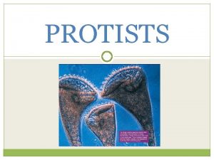
CILIATES • MODE OF LOCOMOTION: As the name indicates, most ciliates feature large numbers of cilia (tiny beating hairs). Their cilia are usually arranged either in longitudinal rows or in spirals around the cell. Cilia are anchored to microtubules beneath the plasma membrane, and they beat in coordinated fashion. In some groups, the cilia have specialized functions, becoming fused into sheets, spikes and rods that may then function as months, paddles, teeth, or feet. The ciliates have a pellicle, a tough but flexible outer covering, that enables them to squeeze through or move around obstacles.

STRAMENOPILA (BROWN ALGAE) • • The name Stramenopila refers to unique, fine hairs found on the flagella of members of this group, although a few species have lost their hairs during evolution. BROWN ALGAE: Brown algae are the most conspicuous seaweeds in many northern regions. The life cycle vof the brown algae is marked by an alternation of generations between a multicellular sporophyte and a multicellular gametophyte. Some sporophyte cells go to through meiosis and produce spores. These spores germinate and undergo mitosis to produce the large individuals we recognize, such as the kelps. The gametophytes are often much smaller, filamentous individuals, perhaps a few centimetres in width. Even in acquatic environment, trensport can be a challenge for the very large brown algal species. Distinctive transport cells that stack one upon the other enhance transport within some species. However, even though the large look loke plants, it is important to realize thet they do not contain the complex tissues such as xylem thet are found in plants.

DIATOMS • Diatoms, members of the phylum Chrysophuta, are photosyntetic, unicellular organisms with unique double shells made of opaline silica, which are often strikingly marked. The shells of diatoms are like small boxes with lids, one half of the shell fitting inside the other. Their chloroplasts, containing chlorophylls a and c, as well as carotenoids, resemble those of the brown algae and dinoflagellates. Diatoms produce a unique carbohydrate called chrysolaminarin.

OOMYCETES • All oomycetes are either parasites or saprobes (organisms that live by feeding on dead organic matter). They are distingued from other protists by the structure of their motile spores, or zoospores, which hear two unequal flagella, one pointed forward and the other backward. Zoospores are produced asexually in a sporangium. Sexual reproduction involves the formation of male and female reproductive organs that produce gametes. Most oomycetes ae found in water, but their terrestrial relatives are plant pathogens. Two important oomycetes are: Phytophtora infastans that attaches the potatoes and the Saprolegaia that is a fish that can cause serious losses in fish hatcheries.

RHODOPHYTA • Rhodophyta are type of protists that are red algae. This lineage lacks flagella and centrioles and has the accessory photosyntetic pigments, which are arranged within structures called phycobilisomes. • The origin of the over 7000 species of Rhodophyta has been a source of controversy. Evidence supporting very early eukaryotic origins and common anchestry with green algae has been considered. Molecular comparisons of the chloroplasts in red and green algae support a single endosymbiotc origin for both. • Comparisons of the nuclear DNA coding for the large subunit of RNA polymerase II from two red algae, a green alga, and another protist support the conclusion that the Rhodophyta emerged before the evolutionary lineage that led to plants, animals and fungi.

CHOANOFLAGELLIDA • Choanoflagellates are most like the common ancestor of the sponges and indeed all animals. Choanoflagellates have a single emergent flagellum surrounded by a funnel-shaped, contractile collar composed of closely placed filaments, a structure that is exactly matched in the sponges, which are animals. These protists feed on bacteria strained out of the water by their collar. Colonial forms resemble freshwater sponges. The close relationships of Choanoflagellates to animals was further demonstrate by the strong homology between a surface receptor found in Choanoflagellates and sponges. This surface receptor initiates a signaling pathway involving phosphorylation.

Amoebas • So far, we have organized the protists based on their closest relatives. Some lineages vary tremendously if you consider just a single trait. For example, the stramenopiles include autotrophic, marine algae, and terrestrial plant pathogens. It is also possible for unrelated organisms to acquire similar traits. That is the case with amoebas, which have similar cell morphology, but are not monophyletic.

SLIME MOLDS • Slime molds originated at least three distinct times, and the three lineages are very distantly related. Like water molds, these organisms were once considered fungi. We will explore two lineages: the plasmodial slime molds, which are uge, single celled, multinucleate, oozing masses, and the cellular slime molds in which single cells combine and differentiate creating an early model of multicellularity.
 Lirik lagu more more more we praise you
 More more more i want more more more more we praise you
 5730x5
 Most millionaires attended private schools
 5-1 changing households
 Er than more than
 Greater than god and more evil
 Tall+short h
 Once upon a time there lived a wise old teacher
 Từ ngữ thể hiện lòng nhân hậu
 The joy luck club scar
 Ring clasp rpd
 Hybrid mythological creatures
 Half man half horse name
 Half woman half snake greek mythology
 λερναια υδρα
 Half man half horse name
 Akers clasp vs c clasp
 Top half bottom half
 Narnia half mens half geit
 Half empty or half full
 Gerald croft
 Percent greater than 100 and less than 1
 Greater than less than fractions
 Keywords for greater than
 Greater than less than fractions
 Your love is deeper than the ocean higher than the heavens
 Dot
Dot Compound inequality examples


