Porphyrins Synthesis of Porphobilinogen and Heme Porphyrins are

Porphyrins Synthesis of Porphobilinogen and Heme

Porphyrins are large heterocyclic organic ring structures. The naturally occurring porphyrins of biological significance are the hemes. Hemes in biological systems consist of ferrous iron (Fe 2+) complexed with four nitrogens of the specific porphyrin molecule identified as protoporphyrin IX. The major function of heme in humans is its role in the coordination of O 2 molecules in hemoglobin.

Synthesis of heme

1 -The first reaction in heme • biosynthesis takes place in the mitochondria and involves the condensation of one glycine and one succinyl-Co. A by the pyridoxal phosphate-requiring (vitamin B 6) enzyme, δ-aminolevulinic acid synthase (ALAS) forming the compound δaminolevulinic acid.

2 -the mitochondrial ALA is transported to • the cytosol, where ALA dehydratase (also called porphobilinogen synthase) dimerizes two molecules of ALA to produce the pyrrole ring compound porphobilinogen. 3 -The next step in the pathway involves • the head-to-tail condensation of four molecules of porphobilinogen to produce the linear tetrapyrrole intermediate, hydroxymethylbilane. The enzyme for this condensation is porphobilinogen deaminase

4 -The most important fate of • hydroxymethylbilane is converted to uroporphyrinogen III by the enzyme, uroporphyrinogen-III synthase. 5 -uroporphyrinogen III is • decarboxylated by the enzyme uroporphyrinogen decarboxylase to coproporphyrinogen III

6 -Following its synthesis, • coproporphyrinogen III is transported to the interior of the mitochondrion, where two propionate residues are decarboxylated, yielding vinyl substituents on the two pyrrole rings. The colorless product is protoporphyrinogen IX. These reactions are catalyzed by oxygen-dependent coproporphyrinogen-III oxidase

7 -In the mitochondrion, protoporphyrinogen • IX is converted to protoporphyrin IX (structure shown below) by protoporphyrinogen IX oxidase. The final reaction in heme synthesis also – takes place in the mitochondrion and involves the insertion of the ferrous iron (Fe 2+) atom into the ring system generating heme b. The enzyme catalyzing this reaction is known as ferrochelatase.

Protoporphyrin IX

Heme Metabolism and Disposition of Bilirubin • The largest repository of heme in the human body is in red blood cells, which have a life span of about 120 days. There is thus a turnover of about 6 g/day of hemoglobin, which presents 2 problems. First, the porphyrin ring is hydrophobic and must be solubilized to be excreted. Second, iron must be conserved for new heme synthesis.
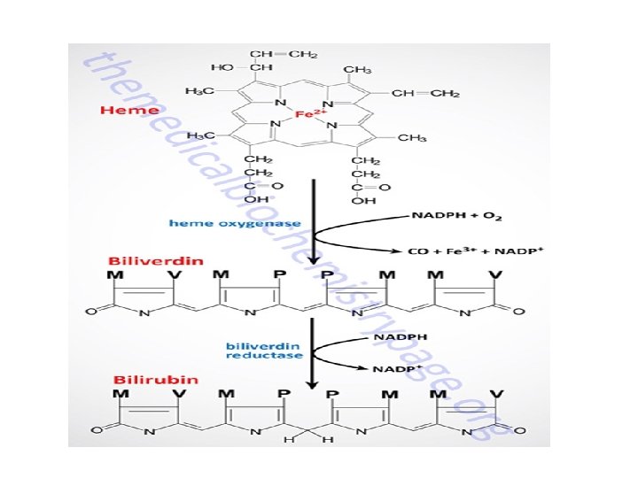

Pathway for the degradation of heme to bilirubin The ring of heme is opened through the • action of heme oxygenase which also results in the relase of the iron as the ferric form (Fe 3+) and also releases carbon monoxide, CO. The product of the heme oxygenase reaction is biluverdin. Biliverdin is converted to bilirubin via the action of biliverdin reductase. The various substituents on the pentameric rings of biliverdin and bilirubins are M: methyl, P: propyl, V: vinyl

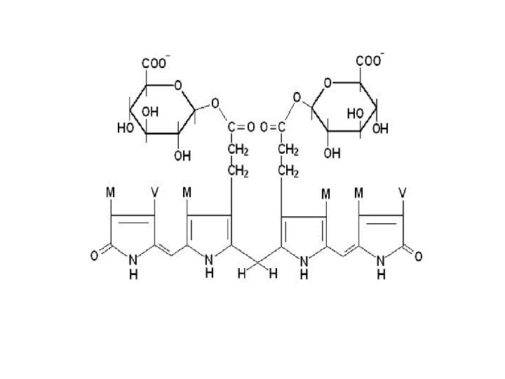
In hepatocytes, bilirubin-UDP- • glucuronosyltransferase adds two equivalents of glucuronic acid to bilirubin to produce the more water soluble, bilirubin diglucuronide derivative. The increased water solubility of the tetrapyrrole facilitates its excretion with the remainder of the bile as the bile pigments

In newborn infants, or in individuals with • abnormally high red cell lysis, or liver damage with obstruction of the bile duct, the bilirubin and its precursors accumulate in the circulation; the result is hyperbilirubinemia, the cause of the abnormal yellowish pigmentation of the eyes and tissues known as jaundice. All newborn infants undergo turnover of the red blood cells that contain fetal hemoglobin (Hb. F) so that new red blood cells containing adult hemoglobin (Hb. A) can be produced.

In some cases the activation of the UGT 1 A gene at • birth is insufficient to handle all the red cell turnover resulting in neonatal jaundice, apparent around day 2 or 3. If the blood levels of bilirubin do not decline in a short period of time these infants will need to be treated by phototherapy. The blue-green wavelength light (460 -490 nm) used in biliblankets is sufficient to induce breakdown of bilirubin in the skin so that it can be cleared from the blood. In rare cases the phototherapy does not work fast enough and in these cases it is appropriate to treat the infants with phenobarbital which enhances the induction of the UGT 1 A gene

In normal individuals, intestinal bilirubin is acted on by • bacteria to produce the final porphyrin products, urobilinogens and stercobilins, that are found in the feces. The stercobilins oxidize to brownish pigments which impart the brown to brown-black color to normal feces. Indeed, the color of the stool can be quite diagnostic since chalky clay colored feces are indicative of a defect in the hepato-biliary circulation, such as in bile obstruction. Some of the urobilinogen produced by intestinal bacteria is reabsorbed from the gut and enter the circulation. These urobilinogens are converted to the urobilins which are then excreted in the urine. Oxidation of the urobilins imparts the yellowish coloration to urine.

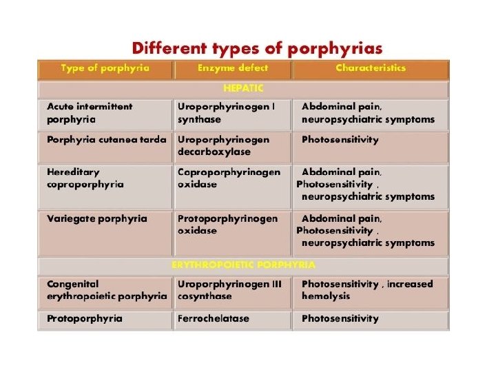
Clinical Aspect of Heme Metabolism and Bilirubin Clinical problems associated with heme • metabolism are of two types. Disorders that arise from defects in the enzymes of heme biosynthesis are termed the porphyrias) and cause elevations in the serum and urine content of intermediates in heme synthesis. Inherited disorders in bilirubin metabolism lead to hyperbilirubinemia

Hyperbilirubinemias Bilirubin levels are measured in the serum by • an assay utilizing Ehrlich diazo reagent and results in the formation of an azobilirubin product. Conjugated bilirubin does not require addition of alcohol to promote the azotization reaction and thus, this is referred to as measurement of direct bilirubin. The reaction with unconjugated bilirubin requires the addition of alcohol and thus is referred to as the measurement of indirect bilirubin. Normal bilirubin measurements are 0. 3– 1. 2 md/d. L for total (indirect + direct).
 results in a yellow-orange discoloration of")
Excess circulation and accumulation of • bilirubin (hyperbilirubinemia) results in a yellow-orange discoloration of the tissues and is most easily visible as icteric (yellowish) discoloration in the sclera of the eyes. Bilirubin toxicity (bilirubin encephalopathy) can be life threatening in neonates. Bilirubin encephalopathy is characterized by yellow discoloration of the basal ganglia in babies with intense jaundice and was first described over a century ago and the term "kernicterus" was coined to describe these physical changes. Any increase in plasma bilirubin above 20 mg/d. L is considered dangerous in neonates.

Porphyrias The porphyrias are both inherited and • acquired disorders in heme synthesis. These disorders are classified as either erythroid or hepatic, depending upon the principal site of expression of the enzyme defect. Eight different porphyrias have been classified encompassing defects in each of the enzymes of heme synthesis.
- Slides: 22