PLANT PATHOGENESI S PLANT PATHOGENESIS STEPS 1 Prepenetration


 Directly")


 Examples: Hail, Frost, Insect feeding, Cultivation heat scorching, topdressing,")


 The nature of cell")
 Primary cell wall (cellulose, pectates) Secondary cell wall (almost entirely cellulose)")


. Produced")


- Slides: 35
PLANT PATHOGENESI S
PLANT PATHOGENESIS STEPS: 1. Pre-penetration 2. Penetration 3. Post Penetration PRE-PENETRATION Attachment / Colonization of the Pathogen with the Host Sources of inoculums: • Soil: Bacteria, Rhizoctonia, Sclerotinia, • Infected plant parts: Seed, cuttings, bulbs, corms, tubers etc. • Diseased debris: Alternaria, Phytophthora • Alternate hosts: Rusts- wheat rust: Barberis vulgaris • Collateral hosts: Viruses: BCMV, PVY, Powdery mildews etc.
ARRIVAL OR LANDING OF INOCULUMs Inoculums is carried by different agencies like, • Wind: powdery & Downy mildew, rusts • Water: bacteria, Colletotrichum spp. Fusarium • Soil: Rhizoctonia, bacteria, Sclerotinia • Insect vectors: viruses- Potato virus Y by aphid: Myzus persicae • Planting material, : viruses: BCMV, TMV • Tools: TMV, bacteria, fungi, –Some pathogens may also use their own force to reach host e. g. nematode larvae, zoospores
Differs in different host pathogen combinations • Attachment of the pathogen to the host • Spore germination in fungi, multiplication in bacteria and hatching of nematode eggs • Perception of the host surface Attachment / Contact 1. Electro taxis 2. Chemo taxis 3. Rheo taxis Recognition of the Pathogen by host or vice versa
PENETRATION • Direct penetration • Indirect penetration –Fungi may penetrate in either way –Bacteria mostly enter through wounds and some time by natural openings –Viruses, viroids, phytoplasma, RLOs etc. by mechanical means (wounds) and by vectors
Direct penetration • Through Appressorium (e. g. Involved in direct penetration (eg. Colletotrichum spp. and M. grisea) • Germtube e. g. (Apple scab V. inequalis) • Appressorium give rise to penetration peg • Haustoria e. g. powdery mildews • Infection cushion e. g. Fusarium, Sclerotinia –Includes fungi imperfecti & ascomyctes
Leaf structure
METHODS FOR FUNGAL DIRECT PENETRATION
DIRECT PENETRATION BY • Nematodes : larvae • Higher parasitic plants: haustori
INDIRECT PENETRATION Penetration through natural openings a. Stomata b. Hydathodes c. Nectorthodes d. Lenticels
STOMATAL PENETRATION Appressorium Penetration peg Sub stomatal cavity Infection hyphe (penetrate diff cells) Directly haustoria (Biotrophs)
THROUGH STOMATA • Some fungal and bacterial pathogens • Penetration depends upon –Size of stomata –No. of stomata –Location –Time of opening & closing of stomata • Majority of the fungi produces appressorium • Stomatal invaders includes: ustilaginales, uredinales & Peronosporales
PENETRATION OF FUNGI
ENTRY THROUGH HYDATHODES Bactera e. g. Xanthomonas campestris pv. Oryzae NECTORTHODES Erwinia amylovora causing fire blight of apple & pear E. amylovora LENTICELS
PENETRATION THROUGH WOUNDS (TRAUMA INFECTION) Examples: Hail, Frost, Insect feeding, Cultivation heat scorching, topdressing, mowing traffic damage Pathogen 1 st multiply on wounds in the presence of moisture and then penetrate host cell by • Directly • Haustoria • Enzymes or toxins
MECHANICAL OR CHEMICAL BACK GROUND OF PENETRATION 1. 2. 3. 4. ENZYMES TOXINS GROWTH REGULATORS SUPRESSORS ENZYMES 1. CUTINASE 2. PECTOLYTIC ENZYMES 3. CELLULASES 4. HEMICELLULASES 5. LIGNINASES
TOXINS 1. HOST SPECIFIC TOXIN 2. NON-HOST SPECIFIC TOXIN GROWTH REGULATORS 1. AUXIN 2. GIBBERELLINS 3. CYTOKININ 4. ETHYLENE 5. ABA FEW POLYSACCHARIDES
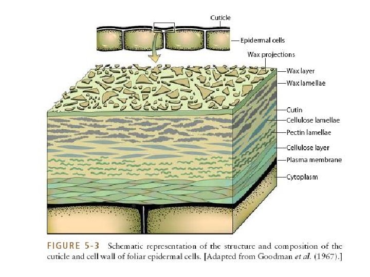
THE ENTIRE PROCESS OF DIRECT PENETRATION, HOWEVER, DEPENDS ON (a) The nature of cell wall layers, (b) The potentialities of the enzyme system of the pathogen, and (c) The potential force the pathogen can exert. The main components of the host ‘cell wall • Pectin • Cellulose • Hemicellulose • Lignin • Small quantity of protein
Middle lamella (pectates) Primary cell wall (cellulose, pectates) Secondary cell wall (almost entirely cellulose) Intercellular spaces Cell lumen Plasmodesmata
The three main softening or solubilizing enzymes found in fungi v Cellulolytic v. Pectolytic v. Lignolytic • The outer layer of host plant cell may have a layer of wax which is followed by cuticle impregnated with wax. • The cutin gradually decreases with the depth of the epidermis and is replaced by pectin which occurs as a homogeneous layer in some host plant. • Subsequently in the secondary wall of the cell pectin is replaced by cellulose. • Cellulose layers contain appreciable amounts of protein.
q No pathogen secretes enzymes that would degrade wax. Only mechanical entry can enable a pathogen to breach this layer. q. Cutin layer is penetrated either by pres sure (mechanically exerting force) or by the action of degrading enzymes: cutinase, cutin esterase, and carboxyl cutin esterase. Cutinase breaks cutin into fatty acid and hydroxyfatty acid.
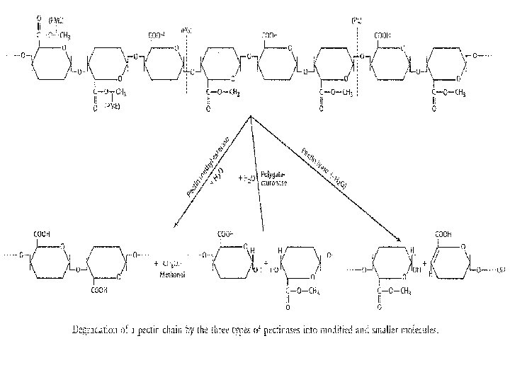
q. Pectic substances which form basic material of middle lamella, primary and secondary walls are degraded by pectinolytic enzymes. The Pectinolytic enzymes act against pectin and pectic substances in many steps: (a)The enzymes pectin methyl esterases (PME) hydrolyse the pectic substances into methanol and pectic acid. (b) The enzymes pectin glycosidases, polygalacturonases (PG) and polymethyl galacturo nases (PMG) degrade the pectic acid and methylated chains of pectin. Besides these, wall modifying enzyme (WME) modifies the pectic material for subsequent degrada tion.
The degradation of pectic substances provides nutrients for many fungal pathogens and due to weakening of the cell wall facilitates inter and intra cellular invasion by hyphae. Pectic enzymes are produced both constitutively and adaptively. High C/N ratio of the substrate favors increased mycelial growth and low enzyme synthesis whereas lower ratios give poorer growth but increased enzyme synthesis. Pectic enzymes have no effect on protoplast and thus cannot do much Killing of host cells. They may be secreted by some of the pathogens involved in spotting and blotching of leaves, al though the macerating effect may be masked by necrosis caused by toxins.
Cellulose forms the structural framework of cell walls. Cellulolytic enzymes act upon the cellulose and break it to simple compounds and make the way for the easy penetration of the pathogen into the host cell. There are two theories to explain the mechanism of cellulose degradation and maceration by cellulolytic enzymes which are adaptive enzymes: the unienzyme theory and the multienzyme theory. Ø According to uni enzyme theory, complete degradation of cllulose into glucose units is by a single enzyme: Cellulose Cellulase Cellobiose Cellobiase & Glucose. Ø Whereas, the multi enzyme theory explains cellulose degradation in a series of steps through two groups of enzymes.
§ One group of enzymes loosens the cellulose fibrils of the crystalline area by hydrolytic mechanism. § The other group of enzymes penetrates the cellulose lattice and causes hydrolytic cleavage of hydrogen bondage. §In the spaces between the cellulose microfibrils of the cell wall, hemicelluloses are present which are degraded by hydrolytic cleavage by enzymes hemicellulases. §The degradation of hemicelluloses by the enzymes hemicellulases produces simpler sugars. In vascular wilt diseases the molecules released by cellulose degradation cause plugging of the vessels.
The most complex chemical compound in the plant cell wall is lignin which occurs chiefly in the matrix surrounding the cellulose fibrils. It is one of the most struc turally complex biopolymers whose degradation is caused by polyphenol oxidases pro duced by wood rotting fungi. But fungi cannot utilize the degradation products. The attack of nematodes is both internal as well as external. Both endophytic and ectophytic nematodes use force as well as enzymes to dissolve or break the cell wall to come in contact with cell protoplasm through their stylet. But endophytic nematodes enter bodily in the host cell, whereas the ectophytic ones remain outside the host.
TOXINS THAT AFFECT A WIDE RANGE OF HOST PLANTS Several toxic substances produced by phytopathogenic microorganisms have been shown to produce all or part of the disease syndrome not only on the host plant, but also on other species of plants that are not normally attacked by the pathogen in nature. Such toxins, callednonhost specific or nonhost selective toxins. These toxins increase the severity of disease caused by a pathogen, i. e. , they affect the virulence of the pathogen, but are not essential for the pathogen to cause disease, i. e. , they do not determine the pathogenicity of the pathogen. Several of these toxins, e. g. , tabtoxin and phaseolotoxin, inhibit normal host enzymes, thereby leading to increases in toxic substrates or to depletion of needed compounds. Several toxins affect the cellular transport system, especially H+/K+ exchange at the cell membrane. Some, e. g. , tagetitoxin, act as inhibitors of transcription in cell organelles, such as the chloroplasts. Others, e. g. , cercosporin, act as photosensitizing agents, causing the peroxidation of membrane lipids.
GROWTH REGULATORS IN PLANT DISEASE Plant growth is regulated by a small number of groups of naturally occurring compounds that act as hormones and are generally called growth regulators. The most important growth regulators are auxins, gibberellins, and cytokinins, but other compounds, such as ethylene and growth inhibitors, play important regulatory roles in the life of the plant. Growth regulators act in very small concentrations and even slight deviations from the normal concentration may bring about strikingly different plant growth patterns. The concentration of a specific growth regulator in the plant is not constant, but it usually rises quickly to a peak and then declines quickly as a result of the action of hormone inhibitory systems present in the plant. Growth regulators appear to act, at least in some cases, by promoting the synthesis of messenger RNA molecules. This leads to the formation of specific enzymes, which in turn control the biochemistry and the physiology of the plant.
Auxins The auxin occurring naturally in plants is indole 3 acetic acid (IAA). Produced continually in growing plant tissues, IAA moves rapidly from the young green tissues to older tissues, but is destroyed constantly by the enzyme indole 3 acetic acid oxidase, which explains the low concentration of the auxin. Increased auxin (IAA) levels occur in many plants infected by fungi, bacteria, viruses, mollicutes, and nematodes, although some pathogens seem to lower the auxin level of the host. Thus, the basidiomycete Exobasidium azaleae causing azalea leaf and flower gall.
GIBBERELLINS Gibberellins are normal constituents of green plants and are also produced by several microorganisms. Gibberellins were first isolated from the fungus Gibberella fujikuroi, the cause of the foolish seedling disease of rice. The best known gibberellin is gibberellic acid 3. Compounds such as vitamin E and helminthosporol also have gibberellin like activity. Gibberellins have striking growth promoting effects. They speed the elongation of dwarf varieties to normal sizes and promote flowering, stem and root elongation, and growth of fruit. Such elongation resembles in some respects that caused by IAA, and gibberellin also induces IAA formation. Auxin and gibberellin may also act synergistically. Gibberellins seem to activate genes that have been previously “turned off. ” The foolish seedling disease of rice, in which rice seedlings infected with the fungus Gibberella fujikuroi grow rapidly and become much taller than healthy plants, is apparently the result, to a considerable extent at least, of the gibberellin secreted by the pathogen.
CYTOKININS Cytokinins act by preventing genes from being turned off and by activating genes that have been previously turned off. The role of cytokinins in plant disease has just begun to be studied. Cytokinin activity increases in clubroot galls, in crown galls, in smut and rust galls, and in rust infected bean leaves. In the latter, cytokinin activity seems to be related to both the juvenile feature of the green islands around the infection centers and the senescence
ETHYLENE Ethylene is produced by several plant pathogenic fungi and bacteria. In the fruit of banana infected with Ralstonia solanacearum, the ethylene content increases proportionately ith the (premature) yellowing of the fruit, whereas no ethylene can be detected from normal fruits.
POLYSACCHARIDES Fungi, bacteria, nematodes, and possibly other pathogens constantly release varying amounts of mucilaginous substances that coat their bodies and provide the interface between the outer surface of themicroorganism and its environment. Exopolysaccharides appear to be necessary for several pathogens to cause normal disease symptoms either by being directly responsible for inducing symptoms or by indirectly facilitating pathogenesis by promoting colonization or by enhancing survival of the pathogen. The role of slimy polysaccharides in plant disease apears to be particularly important in wilt diseases caused by pathogens that invade the vascular system of the plant. In vascular wilts, large polysaccharide molecules released by the pathogen in the xylem may be sufficient to cause a mechanical blockage of vascular bundles and thus initiate wilting. Although such an effect by the polysaccharides alone may occur rarely in nature, when it is considered together with the effect caused by the macromolecular substances released in the vessels through the breakdown of host substances by pathogen enzymes, the possibility of polysaccharide involvement in the blockage of vessels during vascular wilts becomes obvious.