HISTOPLASMOSIS HISTOPLASMOSIS Darlings disease Causative fungus Histoplasma capsulatum


agar with cycloheximide and chloramphenicol are inoculated At")


- Slides: 38
HISTOPLASMOSIS
HISTOPLASMOSIS Darlings disease Causative fungus: Histoplasma capsulatum Disease of reticuloendothelial system Intracellular parasite Dimorphic fungus World wide in distribution but is most common in America
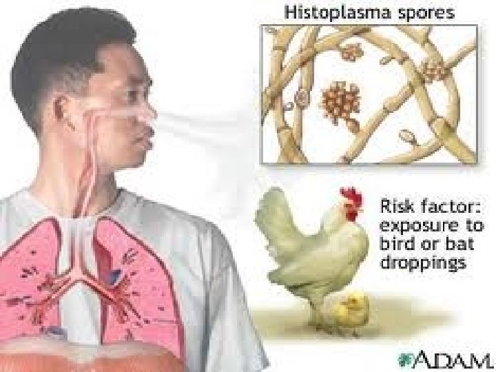
PATHOGENESIS Source of infection : soil enriched with excreta of birds or bats Route of infection : inhalation of spores
CLINICAL FEATURES 90 -95% are asymptomatic Acute pulmonary histoplasmosis : flu like symptoms malaise fever chills profuse sweating sore throat
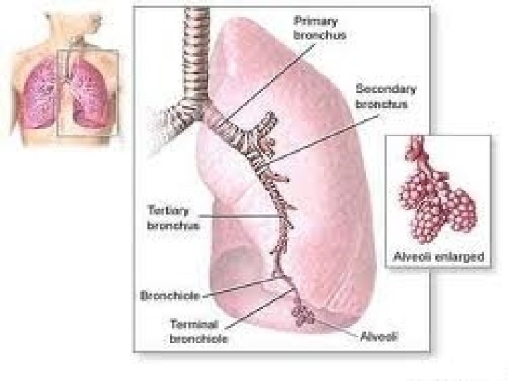
Continued……. . chest pain cough dyspnoea Chronic progressive pulmonary histoplasmosis: acute stage progresses leading to haemoptysis Apical and subapical cavities
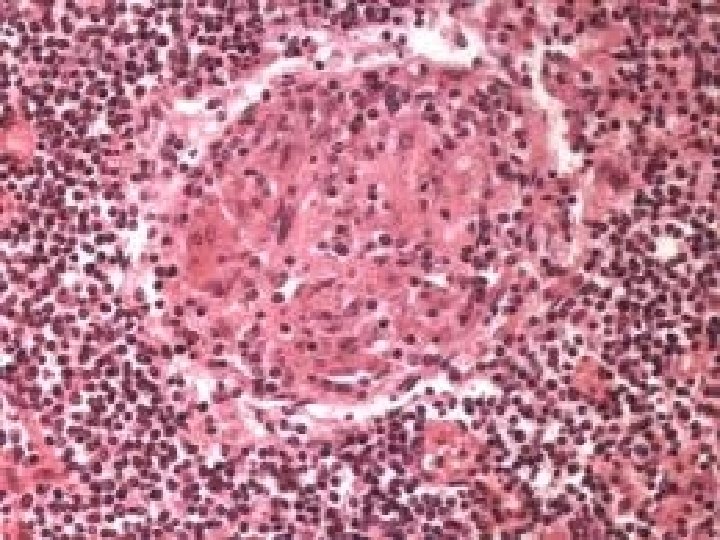
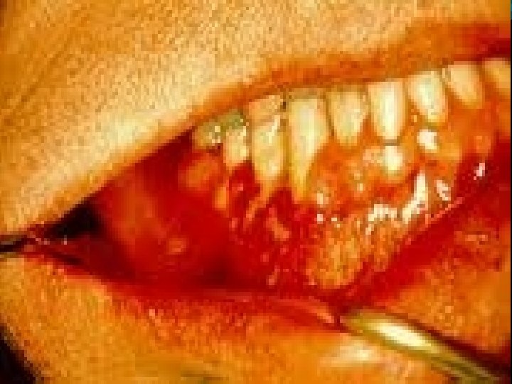
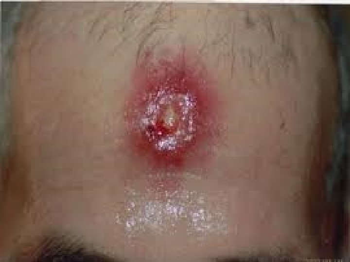
Disseminated histoplasmosis: develops in minority of infected individuals Involvement of RES leads to lymphadenopathy hepatosplenomegaly fever and anaemia Cutaneous and mucocutaneous: granulomatous ulcerative lesions
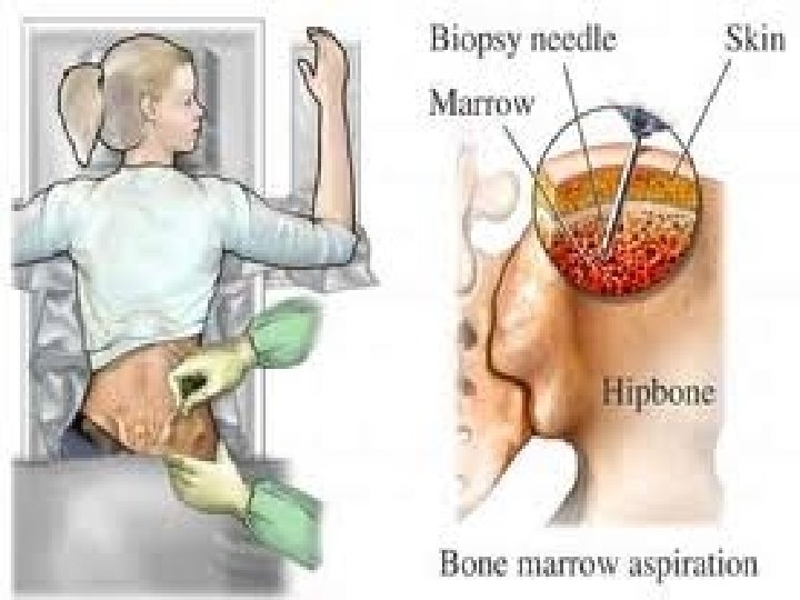
LAB DIAGNOSIS SPECIMENS organs sputum bone marrow aspirate peripheral blood scrapings from ulcers biopsies of lymph nodes and other
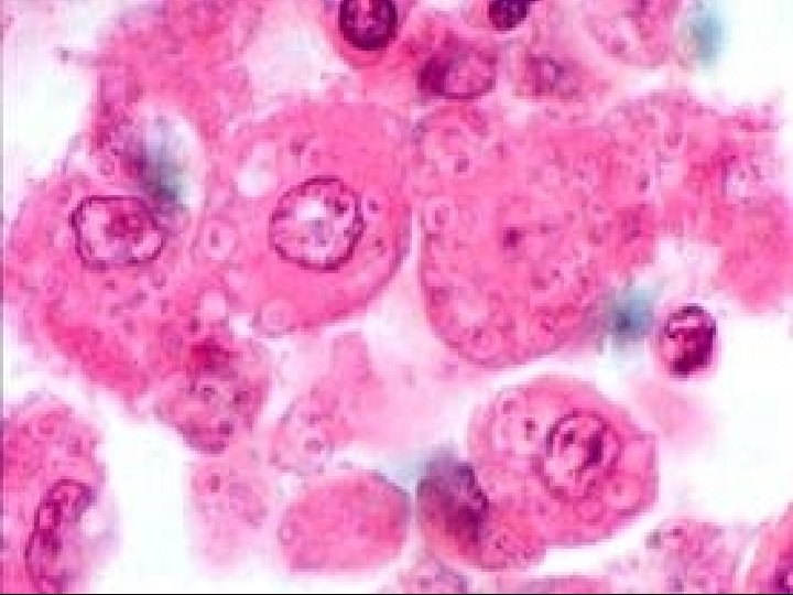
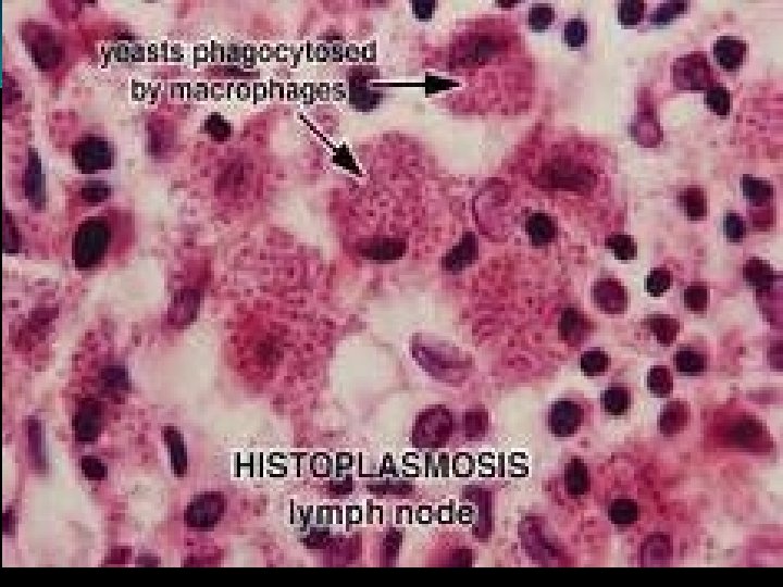
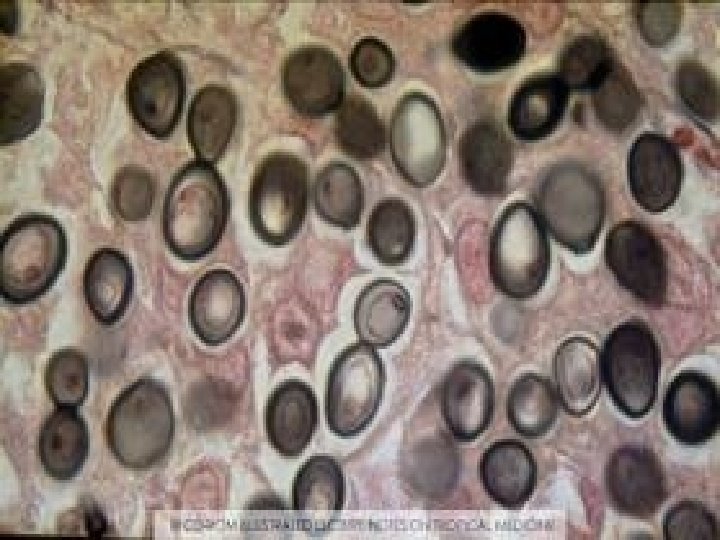
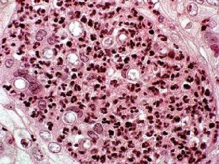
DIRECT EXAMINATION Smears of sputum or pus are stained with giemsa or wright stain On microscopic examination H. capsulatum appears as small, oval yeast cell (2 -4 micron) Packed within the cytoplasm of macrophages or monocytes
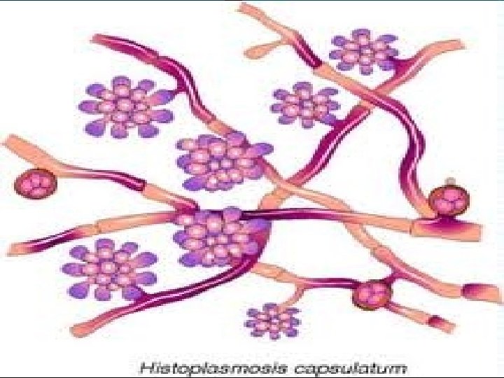
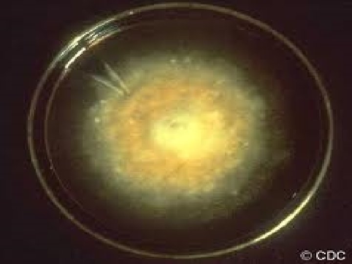
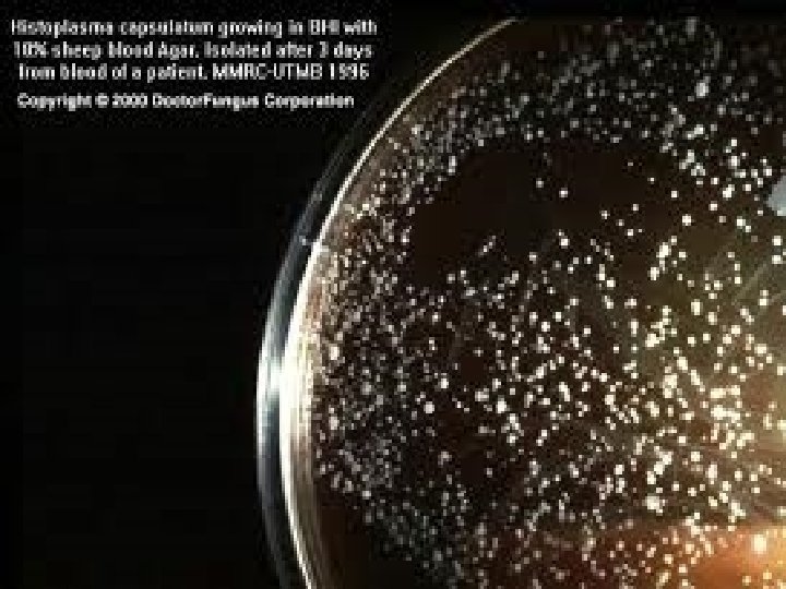
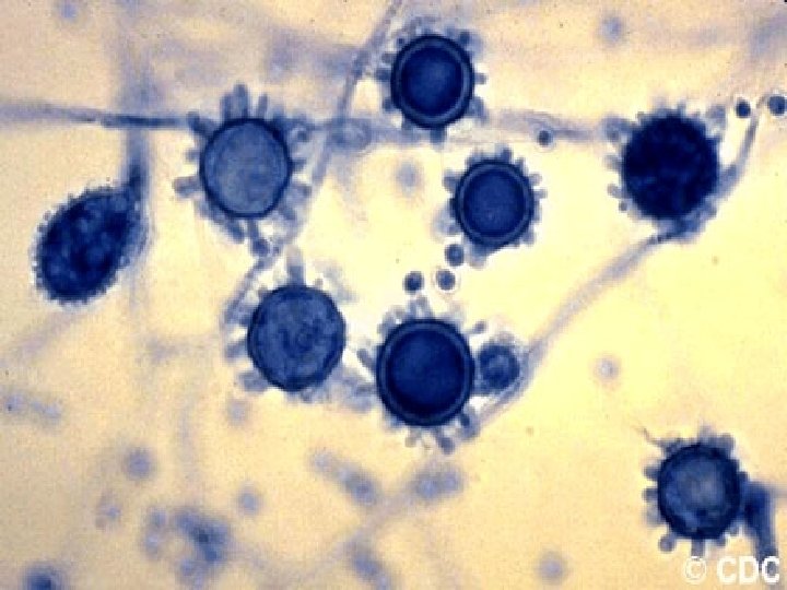
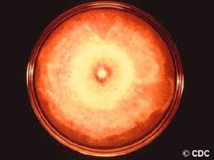
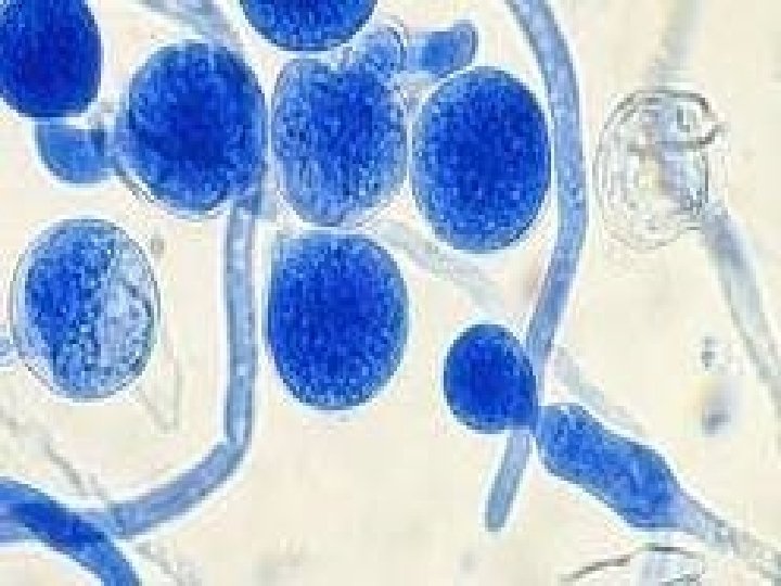
CULTURE SDA or brain heart infusion (BHI)agar with cycloheximide and chloramphenicol are inoculated At 37 c yeast phase is formed At 25 c appears as white cottony mycelial growth containing large(8 -20 microns)thick walled , spherical spores with tubercles or finger like projections
SEROLOGICAL TESTS latex agglutination precipitation complement fixation They become positive 2 weeks after infection Increase in titre of antibody indicates a progressive disease
HISTOPLASMIN SKIN TEST Delayed hypersensitivity test Similar to tuberculin test but antigen used is histoplasmin Positive reaction indicates past or present infection, but does not differentiate active and passive infections
AFRICAN HISTOPLASMOSIS Causative fungus: Histoplasma duboisii Mainly confined with in the continent of Africa Primarily involves skin and subcutaneous tissues It is morphologically identical to H. capsulatum in its mycelial phase but differs in yeast phase
O C Y S I S A L B M O T S
BLASTOMYCOSIS Causative fungus: Blastomyces dermatitidis Dimorphic fungus Characterised by suppurative and granulomatous lesions particularly in lungs Also effects skin, bone and genitourinary tract North american blastomycosis
PATHOGENESIS Route: inhalation Source : soil containing spores CLINICAL FEATURES: PULMONARY BLASTOMYCOSIS: Primary infection of lung may resemble TB or histoplasmosis May be asymptomatic or may leads to focal consolidations, miliary lesions, abscess
CUTANEOUS BLASTOMYCOSIS Primary lesion is papule secondary nodules ulcerative lesions DISSEMINATED Mainly seen in immunocompromised individuals including AIDS
LAB DIAGNOSIS • Specimens sputum pus scrapings from skin lesions
DIRECT MICROSCOPY 10%KOH mount thick walled yeast cells with a single broad based bud • H&E stain and PAS stains also show yeast cells in section
CULTURE SDA or blood agar At 25 c mycelial phase occurs slowly on incubation. filamentous with septate hyphae and many round or oval conidia • At 37 c yeast phase is seen-cells with thick, double contoured walls
Cultures should be incubated for atleast six weeks before discarding them as negative.
TREATMENT Not recommended in asymptomatic cases AMPHOTERICIN B KETOCONAZOLE ITRACONAZOLE