
RABIES Dr Himani Pandya Family Rhabdoviridae n Comprises
RABIES Dr Himani Pandya
Family Rhabdoviridae n Comprises of two genera: o Genus Lyssavirus- Contains Rabies virus and Rabies related viruses o Genus Vesiculovirus- Contains Vesicular stomatitis viruses
History n n n Disease of man and animals by bite of a infected (rabid, Mad) dog. Hydrophobia in man. Zinke in 1804 proved that causative agent of rabies is associated with saliva of rabid dogs. In 1881 Pasteur established that Rabies virus is present in the brain of infected animal. Pasteur also made the first Anti rabies vaccine.
History n n Pasteur established that the rabies virus was present in the brain of infected animal By serial intracerebral passage in rabbit, he obtained Fixed virus and demonstrated that dogs can be rendered immune by a series of injections of fixed virus of graded infectivity The vaccine was prepared by drying of pieces of spinal cord from rabbit infected with fixed virus In July 1885 - a 9 yrs old boy Joseph Mister was bitten by dog- 13 inoculations of cord vaccine- boy survived
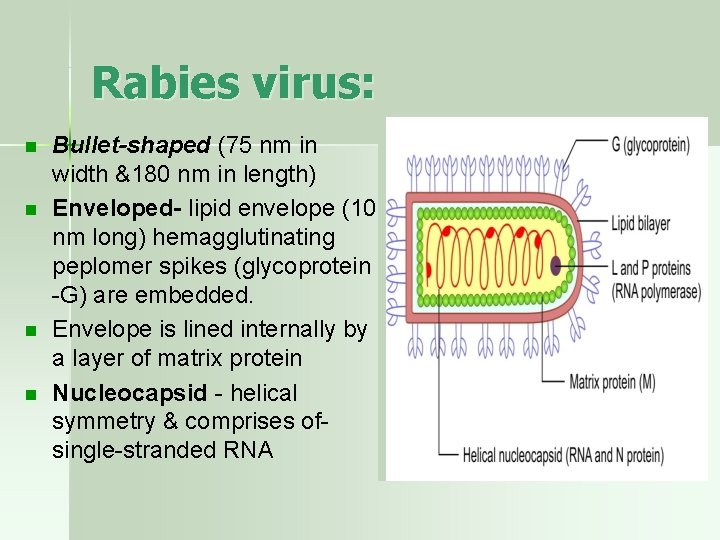
Rabies virus: n n Bullet-shaped (75 nm in width &180 nm in length) Enveloped- lipid envelope (10 nm long) hemagglutinating peplomer spikes (glycoprotein -G) are embedded. Envelope is lined internally by a layer of matrix protein Nucleocapsid - helical symmetry & comprises ofsingle-stranded RNA
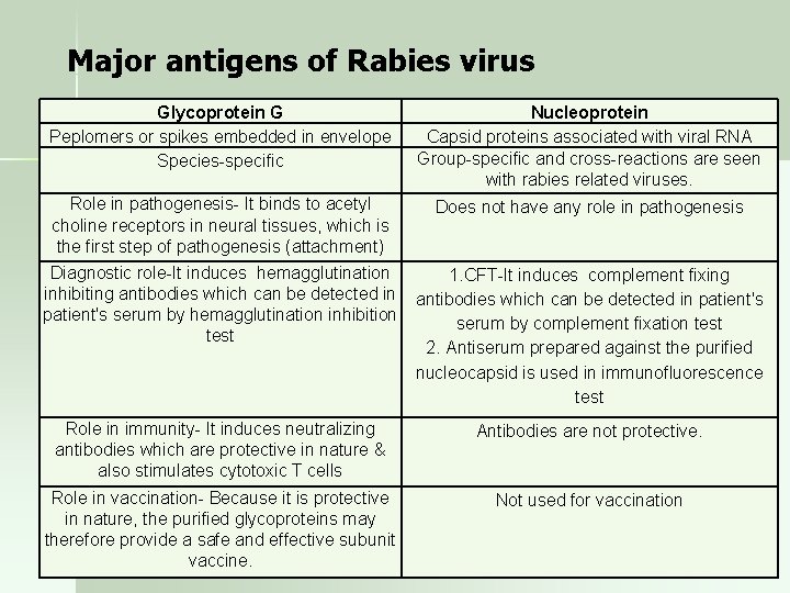
Major antigens of Rabies virus Glycoprotein G Peplomers or spikes embedded in envelope Species-specific Nucleoprotein Capsid proteins associated with viral RNA Group-specific and cross-reactions are seen with rabies related viruses. Role in pathogenesis- It binds to acetyl choline receptors in neural tissues, which is the first step of pathogenesis (attachment) Does not have any role in pathogenesis Diagnostic role-It induces hemagglutination inhibiting antibodies which can be detected in patient's serum by hemagglutination inhibition test 1. CFT-It induces complement fixing antibodies which can be detected in patient's serum by complement fixation test 2. Antiserum prepared against the purified nucleocapsid is used in immunofluorescence test Role in immunity- It induces neutralizing antibodies which are protective in nature & also stimulates cytotoxic T cells Antibodies are not protective. Role in vaccination- Because it is protective in nature, the purified glycoproteins may therefore provide a safe and effective subunit vaccine. Not used for vaccination
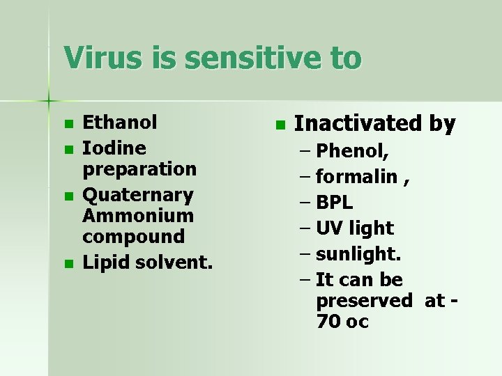
Virus is sensitive to n n Ethanol Iodine preparation Quaternary Ammonium compound Lipid solvent. n Inactivated by – Phenol, – formalin , – BPL – UV light – sunlight. – It can be preserved at 70 oc
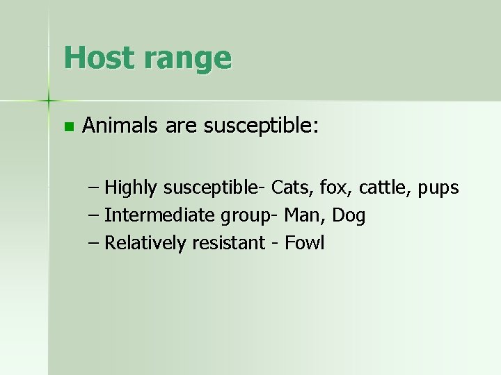
Host range n Animals are susceptible: – Highly susceptible- Cats, fox, cattle, pups – Intermediate group- Man, Dog – Relatively resistant - Fowl
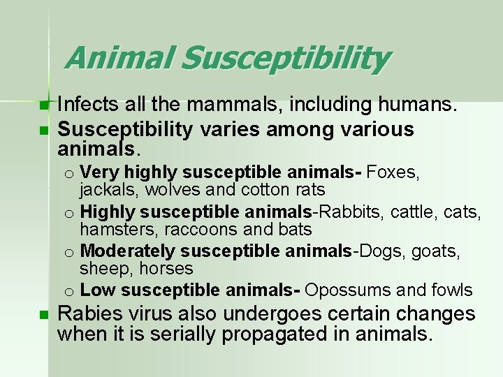
Animal Susceptibility n n Infects all the mammals, including humans. Susceptibility varies among various animals. o Very highly susceptible animals- Foxes, jackals, wolves and cotton rats o Highly susceptible animals-Rabbits, cattle, cats, hamsters, raccoons and bats o Moderately susceptible animals-Dogs, goats, sheep, horses o Low susceptible animals- Opossums and fowls n Rabies virus also undergoes certain changes when it is serially propagated in animals.
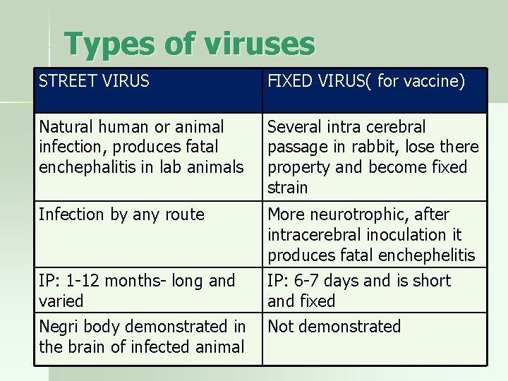
Types of viruses STREET VIRUS FIXED VIRUS( for vaccine) Natural human or animal infection, produces fatal enchephalitis in lab animals Several intra cerebral passage in rabbit, lose there property and become fixed strain Infection by any route More neurotrophic, after intracerebral inoculation it produces fatal enchephelitis IP: 1 -12 months- long and varied Negri body demonstrated in the brain of infected animal IP: 6 -7 days and is short and fixed Not demonstrated
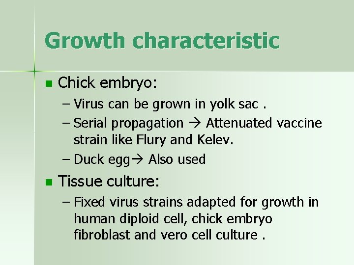
Growth characteristic n Chick embryo: – Virus can be grown in yolk sac. – Serial propagation Attenuated vaccine strain like Flury and Kelev. – Duck egg Also used n Tissue culture: – Fixed virus strains adapted for growth in human diploid cell, chick embryo fibroblast and vero cell culture.
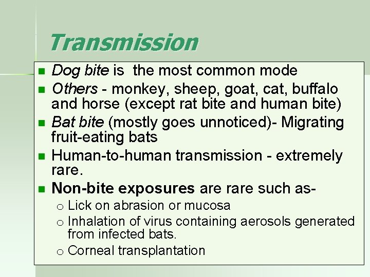
Transmission n n Dog bite is the most common mode Others - monkey, sheep, goat, cat, buffalo and horse (except rat bite and human bite) Bat bite (mostly goes unnoticed)- Migrating fruit-eating bats Human-to-human transmission - extremely rare. Non-bite exposures are rare such aso Lick on abrasion or mucosa o Inhalation of virus containing aerosols generated from infected bats. o Corneal transplantation
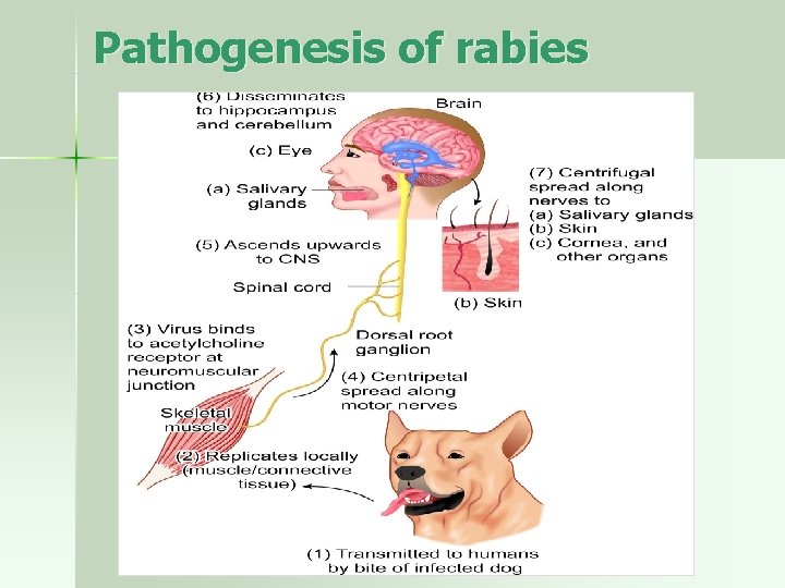
Pathogenesis of rabies
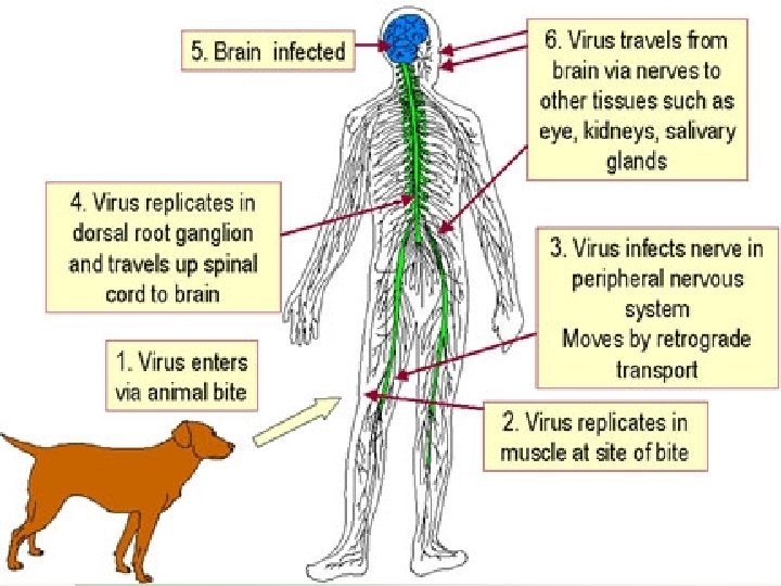
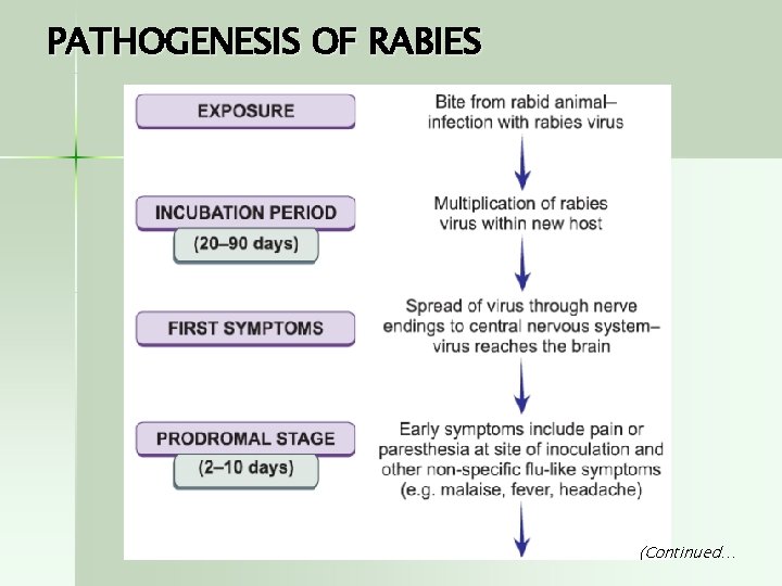
PATHOGENESIS OF RABIES (Continued…
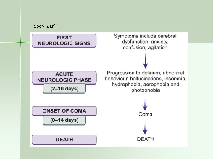
…Continues)
Clinical stages 1. Prodromal Stage: marked by Low grade fever, headache, malaise, fatigue , anorexia, apprehension , anxiety , agitation , irritability, nervous ness, insomnia or depression. Earliest symptoms are neuritic pain or parasthesia at the site of entry. Lasts 2 -4 days
Acute neurological phase: – Hyperexcitability: anxiety, agitation, hallucination, bizarre behavior. – Agitation or seizures with intermittent normal periods. – Autonomic dysfunction: inc in lacrimation and salivation – Pathognomonic features: HYDROPHOBIA Difficulty in drinking water. – Painful spasm of larynx or pharynx leads to choking or gagging, that patients develop a dread of even the sight or sound of water – coma
Generalized convulsions. Coma and death n Death occurs in 1 -6 days due to respiratory arrest during convulsions
In Animals: In dogs IP is 3 -6 wk. n n 1) Prodromal Stage: - 2 -3 days – Alert , troubled air , restlessness, change in disposition, snapping at imaginary objects, licking and neuronal sign develops. 2) Acute Stage : – Furious type of rabies: common, dog run amok and biting indiscriminate with out provocation. – Lower jaw droops. – Drooling of saliva from the mouth. – Progress to Paralysis convulsions death – Dumb type: Paralytic form, animal is unable to feed – As infective as furious type.
In cats : Similar to canine rabies. n In cattle and horses : Irritability , restless ness, and unusual aggressiveness. n In wild animals: the chief characteristic is loss of fear of man and other animals. n
Histopathological features Brain: n Characteristic features: Intracytoplasmic inclusion bodies ( Negri body) in neurons. n – Most abundant in cerebellum and hippocampus. – Composed of rabies virus protein and viral RNA
LABORATORY DIAGNOSIS • Samples • Corneal smears • Skin biopsy (face and neck) for antemortem diagnosis • Saliva • Brain postmortem 1. DIRECT MICROSCOPY a) ANTEMORTEM- IFT b) POST MORTEM- NEGRI BODY
Lab. diagnosis n n Human rabies: 1)Demonstration of rabies virus Ag by Direct Immunofluorescence. – Most common method. – Antirabies serum tagged with fluorescein isothiocynate + specimen. – Specimen : Corneal smear, facial skin biopsy and brain o High sensitivity and specificity o “Gold standard
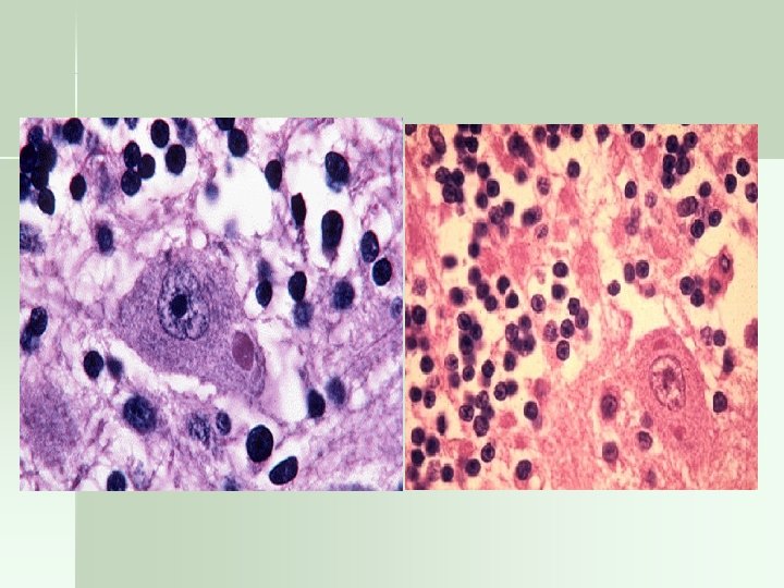
Negri body detection n n Useful for post mortem diagnosis of rabies. They are intracytoplasmic eosinophilic inclusions with characteristic basophilic inner granules. Sharply demarcated, spherical to oval, and about 210 µm in size. Most common sites of Negri bodies are neurons of cerebellum and hippocampus.
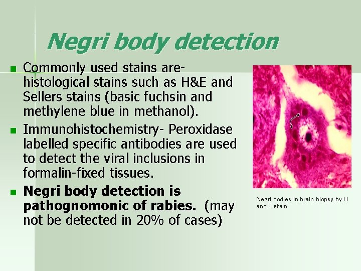
Negri body detection n Commonly used stains arehistological stains such as H&E and Sellers stains (basic fuchsin and methylene blue in methanol). Immunohistochemistry- Peroxidase labelled specific antibodies are used to detect the viral inclusions in formalin-fixed tissues. Negri body detection is pathognomonic of rabies. (may not be detected in 20% of cases) Negri bodies in brain biopsy by H and E stain
n 3) Virus isolation: – By mouse inoculation. – Chances of isolation are grater in early stages. – Samples: CSF, porcine tissue , saliva and urine. n 4) Demostration of high titer Ab in the CSF.
Lab. Diagnosis of Animal rabies n Aim: -To assess the risk of infection – To decide the post exposure treatment. n Specimen: The whole carcass, Severed head of animal or brain is removed and divided into two portion. – One portion is put in 50% glycerol. – Second portion is put into Zenkers fixative. n A portion of Hippocampus and cerebellum of the brain should be there as Negri bodies are abundant
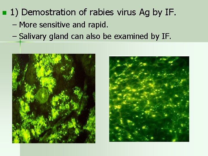
n 1) Demostration of rabies virus Ag by IF. – More sensitive and rapid. – Salivary gland can also be examined by IF.
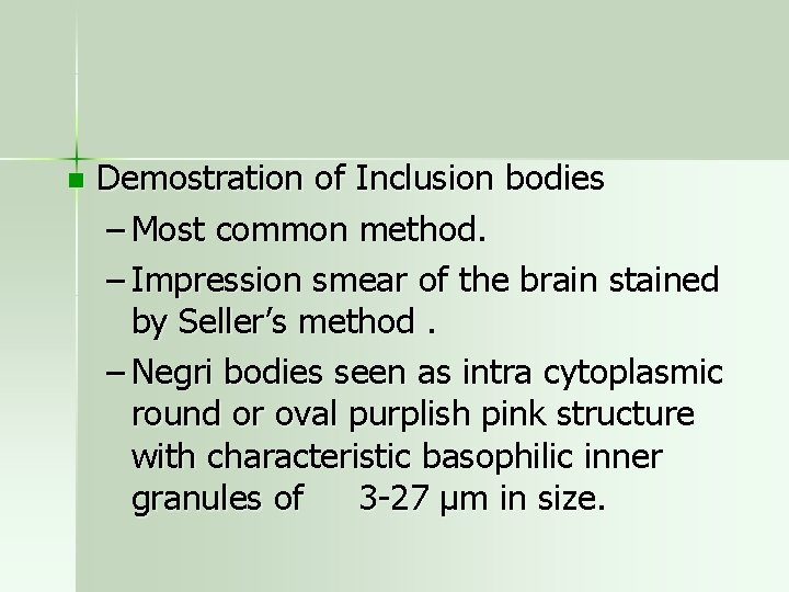
n Demostration of Inclusion bodies – Most common method. – Impression smear of the brain stained by Seller’s method. – Negri bodies seen as intra cytoplasmic round or oval purplish pink structure with characteristic basophilic inner granules of 3 -27 µm in size.
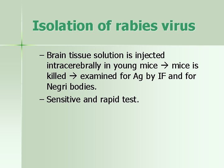
Isolation of rabies virus – Brain tissue solution is injected intracerebrally in young mice is killed examined for Ag by IF and for Negri bodies. – Sensitive and rapid test.
MCQ 1 n Shape of Rabies virus is a Round b Icosahedral c Bullet shaped (helical) d Square
MCQ 2 n Lab. diagnosis is done by demonstrating a Viral Antigen in facial tissue, Cornea b Postmortem demonstration of Negri bodies c Animal experiment d All of above
MCQ 3 n Rabies is a a Neurotropic virus b Hepatotropic virus c Enterovirus d None of above
MCQ 4 n Characteristics of Fixed virus are a It is more neurotropic b All are true c Obtained after several intra cerebral passages in rabbit d Negri bodies are not seen in the infection
MCQ 5 n Choose the correct statement a Man is intermediately susceptible animal b Fowl is most susceptible c Fox is less susceptible to rabies d Dog is highly susceptible to rabies
Rabies – Pre and Post exposure treatment and Vaccines
Rabies Prophylaxis n Specific prophylaxis given prior to exposure. In animal: Pre exposure n In man: Veterinary doctors and dog handlers and trainers n
Post exposure prophylaxis n Local treatment: Scrub immediately with soap and running water. – Treated with Cetavlon, tincture, or aqueous solution of iodine or alcohol. – In severely infected wounds antirabies serum is applied topically. – Anti tetanus serum and antibiotics are given to prevent sepsis.
Anti rabies vaccine Neural vaccine Suspension of nervous tissue of animals injected with fixed rabies virus. 1) Semple vaccine: Most widely used n n – 5% Suspension of sheep brain injected with fixed virus and inactivated with phenol at 37°C. – No residual live virus.
n n BPL vaccine: – BPL inactivating agent. – Modification of Semple vaccine. – Mostly used. Infant brain vaccine: – Encephalogenic factors present in brain tissue basic protein associated with myelin is responsible for neurological complications. – These reactions are scanty of absent in nonmyelinated neural tissue of newborn infant.
Non-neural vaccine n 1) Egg vaccine: – Duck egg vaccine: Fixed virus grown in duck egg and inactivated with BPL and is poorly immunogenic. – Live attenuated chick embryo vaccine a) LEP vaccine: 40 -50 egg passage level. Only for dogs aged 3 months or more. b) HEP vaccine: 180 passage level. Not recommended for man as it contain live virus.
n 2) Tissue culture vaccine: – A) HDCV Vaccine: First cell culture vaccine developed by Koprowsky, Wikkor & plotkin. Pitman Moore strain is grown on human diploid cells. Expensive – B) PCEC Vaccine: BPL inactivated Flury strain. – C) Vero cell vaccine: Non-tumerogenic continues cell lines.
n Sub Unit vaccine: – The glycoprotein subunit on viral surface (Protective Ag) clones and recombinant vaccine produces.
Vaccination Schedules n Indication for ARV: – When a person has been bitten , scratched or licked by an animal which can’t be apprehended. n Observe the animal for 10 days… – If the animal remain healthy for more than 10 days then discontinue the treatment. – If animal dies then full cores of treatment is required.
Dosage of vaccine n Depends on degree of risk to the patient: CLASSIFICAITON OF EXPOSURE n CLASS I : (Mild risk) – Licks on healthy unbroken skin. – Scratches without oozing of blood. – Consumption of unboiled milk of suspected animal.
n n CLASS II : ( Moderate risk) – Licks on fresh cuts. – Scratches with oozing of blood. – All bites except those on head, palm and finger. – Minor wound , 5 in No. n CLASS III: ( Severe risk) – All bites or scratches with oozing of blood on neck , head , face , palm and fingers. – Lacerated wound on any part of the body. – Multiple wound more than 5 in No. – Bites from wild animals.
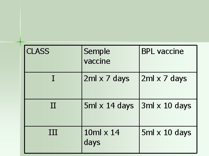
CLASS Semple vaccine BPL vaccine I 2 ml x 7 days II 5 ml x 14 days 3 ml x 10 days III 10 ml x 14 days 5 ml x 10 days
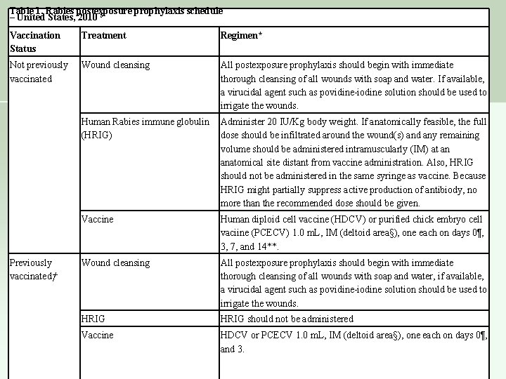
Table 1. Rabies postexposure prophylaxis schedule – United States, 2010 5 Vaccination Status Treatment Regimen* Not previously vaccinated Wound cleansing All postexposure prophylaxis should begin with immediate thorough cleansing of all wounds with soap and water. If available, a virucidal agent such as povidine-iodine solution should be used to irrigate the wounds. Human Rabies immune globulin (HRIG) Administer 20 IU/Kg body weight. If anatomically feasible, the full dose should be infiltrated around the wound(s) and any remaining volume should be administered intramuscularly (IM) at an anatomical site distant from vaccine administration. Also, HRIG should not be administered in the same syringe as vaccine. Because HRIG might partially suppress active production of antibiody, no more than the recommended dose should be given. Vaccine Human diploid cell vaccine (HDCV) or purified chick embryo cell vaciine (PCECV) 1. 0 m. L, IM (deltoid area§), one each on days 0¶, 3, 7, and 14**. Wound cleansing All postexposure prophylaxis should begin with immediate thorough cleansing of all wounds with soap and water, if available, a virucidal agent such as povidine-iodine solution should be used to irrigate the wounds. HRIG should not be administered Vaccine HDCV or PCECV 1. 0 m. L, IM (deltoid area§), one each on days 0¶, and 3. Previously vaccinated†
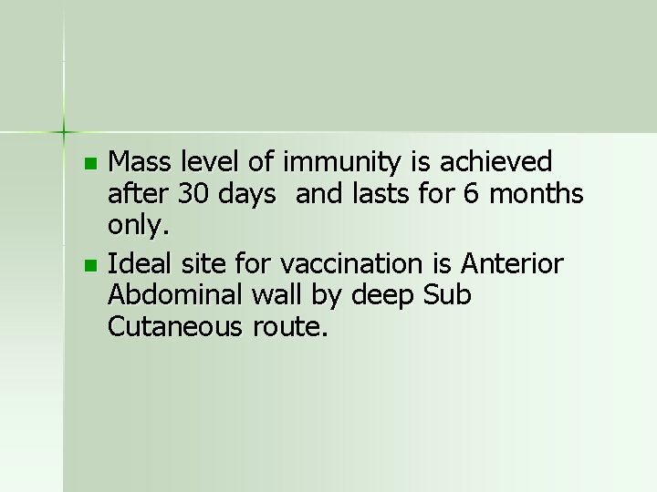
Mass level of immunity is achieved after 30 days and lasts for 6 months only. n Ideal site for vaccination is Anterior Abdominal wall by deep Sub Cutaneous route. n
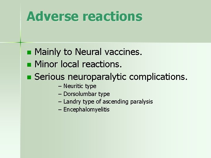
Adverse reactions Mainly to Neural vaccines. n Minor local reactions. n Serious neuroparalytic complications. n – Neuritic type – Dorsolumbar type – Landry type of ascending paralysis – Encephalomyelitis
For passive immunization Antirabies serum (ARS). – HORSE ARS: Given as early as possible , maximum in 5 days. 40 IU / Kg of body wt. – Human Rabies Ig ( HRIg) 10 -20 IU/Kg of body Wt. Half is given around wound locally and other half is given IM in gluteal region.
- Slides: 53