Pituitary tumors Classification Immunohistochemistry M Tohidi MD APCP


 pituitary adenoma (PAS x 100). The basophilic cells are also")


")

. In some GH omas, the")


 index ü Usually < 3% in noninvasive tumors ü Generally higher in")
 index ü There is statistically significant, but numerically minimal difference in MIB-1")


- Slides: 40
Pituitary tumors Classification & Immunohistochemistry M. Tohidi, MD, APCP Research Institute for Endocrine Sciences
History ü Over the years, several classification schemes have evolved. ü Simple tinctorial-based classification was accepted as dogma & dominated pituitary pathology for several decades. ü A classification based on tinctorial characteristics of tumor cell cytoplasm fails to provide information regarding cytogenesis, hormone content, cellular composition, endocrine activity & biologic behavior.
General classification of pituitary Tumors ü Benign adenoma ü Invasive adenoma ü Carcinoma ü Oncocytic tumors ü Metastatic tumors (breast, lung & GI) ü Others: craniopharyngiomas, meningiomas, germ cell tumors; rare granular cell tumors, pituicytomas, and gangliomas; and the even rarer gangliocytomas, lymphomas, astrocytomas, and ependymomas
General classification of pituitary Tumors Pituitary adenomas are the most common lesions in the sella region; ü they represent approximately 10– 15% of intracranial neoplasms in most neurosurgery series. ü depending on the sections examined they are noted in 3– 24% of unselected autopsies.
Classification of pituitary adenomas according to different parameters Pituitary adenomas can be classified according to: ü Biologic behavior ü staining affinities of the cell cytoplasm ü size ü endocrine activity ü histologic characteristics ü hormone production and contents ü ultrastructural features ü granularity of the cell cytoplasm ü cellular composition ü cytogenesis ü growth pattern
Classification of pituitary adenomas by an anatomical approach Principle: size based on radiological findings ü Microadenomas (the greatest diameter is <10 mm) ü Macroadenomas (the greatest diameter is ≥I 0 mm) Four grades of this radio-anatomical: Ø Stage I: microadenomas (<1 cm) without sella expansion Ø Stage II : macroadenomas (≥ 1 cm) and may extend above the sella Ø Stage III: macroadenomas with enlargement and invasion of the floor or suprasellar extension Ø Stage IV: destruction of the sella
Size of adenomas by immunophenotype Microadenoma 85% Macroadenoma Nearly 100% Macroademoa Prolactinomas in females Densely granulated GH cell adenoma FSH/LH adenomas ACTH-Secreting adenomas (80% microadenoma) Sparsely granulated GH cell adenoma TSH adenomas Plurihormonal adenoma Acidophil stem cell adenomas Mixed GH/PRL tumors Null cell adenoma Prolactinomas in males Silent type 3 adenoma
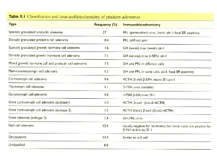
Classification of pituitary adenomas by histological criteria ü Immunohistological characterization of the tumors in terms of hormone production. IHC staining for pituitary hormones generally correlates with hormone serum levels. Some pituitary adenomas have no readily identifiable hormone production. ü Ultrastructural criteria which is especially useful for nonfunctional lesions : - Confirm the pituitary origin - Characterize the cytological differentiation of tumor cells in terms of anterior pituitary cell types.
Classification of pituitary adenomas by functional criteria ü Principle: endocrine activity of the tumor - PRL-producing (lactotroph ) adenomas ACTH-producing (corticotroph) adenomas GH-producing (somatotroph) adenomas TSH–producing (thyrotroph) tumors Mixed PRL- and GH-producing adenomas Clinically non-functioning (the endocrine-inactive) adenomas This group is comprised predominantly of gonadotroph adenomas which synthesize : • FSH • LH • The alpha or beta subunits
Silent versus null call adenoma Ø Silent adenomas * Positive immuno-staining for one or more pituitary hormones • Unassociated with clinical and biochemical evidence of hormone excess. - immuno-staining reactive but lacks biologic activity - a defect may exist in the secretory machinery. Ø Null cell adenoma * Immuno-negative for all adenohypophysial hormones. * Unaccompanied clinically and biochemically by hormone over secretion.
A five-tier classification, clinico-pathologic in nature, which focuses on endocrine activity, imaging, operative findings, histology, immunocytochemistry, and ultrastructure. The collected data provide valuable information to the clinical endocrinologist, neurosurgeon, and oncologist involved in the assessment of a tumor's biologic behavior, growth potential, therapeutic responsiveness, and prognosis.
Level 1: Functional classification of adenohypophysial tumors A. Endocrine hyperfunction 1. Acrornegalyig/gantism, elevated GH levels 2. Hyperprolactinemia and sequela 3. Cushing's disease, elevated ACTH, and cortisol levels 4. Hyperthyroidism with inappropriate hypersecretion of TSH 5. Significantly elevated FSH and LH and/or alpha subunit 6. Multiple hormonal overproduction B. Clinically nonfunctioning C. Functional status undetermined D. Endocrine hyperfunction due to ectopic sources 1. Clinical acromegaly secondary to ectopic GH-releasing hormone overproduction (hyperplasia/adenoma] 2. Cushing's disease secondary to ectopic corticotropin-releasing hormone overproduction (hyperplasia/adenoma)
Level 2: Imaging/Surgical Classification of Adenohypophysial Tumors Based on location 1. lntrasellar 2. Extrasellar extension (suprasellar, sphenoid sinus, nasopharynx, cavernous sinus, etc. ) 3. Ectopic (rare) B. Based on size 1. Microadenoma (< 10 mrn) 2. Macroadenoma ( 10 mml C. Based on growth pattern 1. Expansive 2. Grossly invasive of dura, bone, nerves, and brain 3. Metastasizing (craniospinal or systemic)
Level 3: Histologic Classification of Adenohypophysial Tumors Adenoma 1. Typical 2. Atypical (pleomorphism, enhanced mitotic activity, high MIB-1 labeling index) If growth pattern can be evaluated: 1. Expansive 2. Histologically invasive (bone, nerve, vessels, etc. ) B. Carcinoma (metastasis and/or brain invasion) C. Non-adenoma 1. Primary or secondary non-adenohypophysial tumors 2. Pituitary hyperplasia mimicking adenoma
Atypical adenoma Atypical morphologic features such as : ü Nuclear pleomorphism ü Elevated mitotic index ü Elevated a ki-67 (MIB-1) labeling index > 3% ü Extensive nuclear staining for P 53 Tumors with these features without documented metastases named as atypical adenoma.
Basophilic and atypical (anaplastic) pituitary adenoma (PAS x 100). The basophilic cells are also PAS-positive. Basophilic adenomas secrete ACTH, more rarely TSH or sometimes a gonadotropic hormone (FSH or LH). Note the presence of large or pleomorphic nuclei and especially the numerous mitoses indicative of rapid growth (anaplastic adenoma).
Level 4: Immunohistochemical Classification of Adenohypophysial Tumors Principle immunoreactivity • A. GH • B. PRL • C. GH and PRL, • D. ACTH • E. FSU/LH/alpha-SU • F. TSH • G. Rare hormone combinations • H. Immuno-negative Secondary immunoreactivity PRL, alpha-SU(f), TSH, FSH, LH Alpha- SU(i) Alpha- SU(f), TSH(i) LH, alpha-SU(i) PRL, GH, ACTH(i) Alpha-SU, GH(f), PRL(i)
Level 5: EM options based on ultra-structural features of tumor cells Mandatory for separation of 5 to 7 due to overlapping irnmunohistochemical profiles. Slow-growing 6 is similar to 1, whereas 5 and 7 may be aggressive. Mandatory for diagnosis if clinical presentation and TSH IR are not convincing. Mandatory for identification of tumor types but it is not essential for clinical management, since 11 to 14 have overlapping immunohistochemical profiles and similar biologic behavior.
Sample report format Doe, John 43 years, male Source: Sella, Cavernous sinus -------------------A. Acromegaly B. Macro-, invasive C. Pituitary adenoma, typical D. Growth hormone, prolactin, alpha-Subunit E. Densely granulated somatotroph adenoma
Immunohistochemistry
Solid sheets of uniform cells with round nuclei and neuro-endocrine nuclear features.
Pituitary adenoma H & E stain Reticulin stain
Normal pituitary versus pituitary adenoma. Note the delicate acinar pattern of a normal pituitary gland (left), in contrast with disruption of the normal reticulin network in adenoma (right) (Wilder's reticulin stain).
Development of immunohistochemical methods allowed the distinction of cell types based on their contents of specific hormones. The major cell types and their corresponding products include : Ø Somatotrophs (GH), 50% of the cells & predominantly in the lateral wings Ø Lactotrophs (PRL), 15 -25% & predominate at the postero-lateral edges Ø Mammosomatotrophs (GH, PRL) Ø Thyrotrophs (TSH) 5% of the cells and are located in the anteromedial regions of the gland. Ø Corticotrophs (adrenocorticotropin, betaendorphin, melanocytestimulating hormone), 15 -20% of the cells & primarily in the central mucoid wedge Ø Gonadotrophs (FSH, LH) approximately 5 -10 of the cell populations and are scattered throughout the anterior lobe
In addition to the hormone-producing cells, a second cell population is also present (folliculo-stellate cells) in the normal gland. The latter cells have a dendritic shape and typically encircle the hormone-positive cells. The folliculostellate cells are positive for : Ø S-100 protein Ø variably positive for GFAP. In contrast to their presence in the normal anterior pituitary, S-100 protein-positive folliculostellate cells are generally absent from pituitary adenomas.
Folliculostellate cells are stromal sustentacular cells that surround acini of the normal gland; they are immunoreactive for S 100 protein.
Neuroendocrine markers Pituitary adenomas are typically positive for neuroendocrine markers, including: ü Chromogranin (100%) ü Synaptophysin (92%) ü N 5 E (80%). The use of synaptophysin IHC has obviated the need for EM to document infrequent neurosecretory granules to verify adenoma diagnosis.
Cytokeratins v More than 90% of adenomas contain CK 8. v The use of CAM 5. 2 has the added advantages that highlights the difference between small fragments of normal compressed gland & the adenoma. v It also allow separation of densely from sparsely granulated GH adenomas predicting the response to somatostatin analogs. Ø In sparsely granulated GH cell adenomas, the staining is globular, corresponding to the presence of fibrous bodies. Ø Perinuclear staining is typical of densely granulated GH cell and mammo-somatotroph adenomas Ø Corticotroph cell adenomas exhibit more diffuse cytoplasmic staining for CK 8. v CK 20 is uniformly negative in adenomas, except for : Ø Corticotroph adenoma Ø Sparsely granulated GH adenomas Always CK 20 positive
he IHC of CAM 5. 2 keratin (CK 8). In some GH omas, the tumor cells shows globules of keratin, which are called as fibrous bodies, by IHC (a). b Shows magnified image of dot type fibrous bodies. In densely granulated adenomas, keratin is immunostained in cytoplasm (c). In electron microscopy, fibrous bodies shows smooth endoplasmic reticulum located in the Golgi region (d)
Pituitary adenoma Immunoperoxidase staining for GH
GH-producing adenoma he IHC of GH of densely and sparsely granulated adenomas. Densely granulated adenoma shows strong and diffuse cytoplasmic immunoreactivity for GH (a). Sparsely granulated adenomas show weak GH immunostaining (b)
sparsely granulated prolactinoma H&E stain immunoperoxidase stain for prolactin.
Pituitary carcinoma ü Pituitary carcinomas are rare. ü Diagnosis rests on the demonstration of metastases. ü These tumors are typically positive for generic neuroendocrine markers and for one or more anterior pituitary hormones. ü The most frequently synthesized hormones are prolactin and ACTH while the production of growth hormone, TSH, and FSH/LH is rare.
Ki-67 (MIB-1) index ü Usually < 3% in noninvasive tumors ü Generally higher in invasive adenoma but does not correlate perfectly. ü Significantly higher ki 67 index in invasive adenoms than noninvasive ones when suprasellar extension was considered but this difference did not hold up for tumors with cavernous sinus extension. ü Silent ACTH- cell adenoma & PRL-producing adenoma have highest index & null cell adenoma & gonadotroph cell adenomas have the lowest proliferation values. The use of MIB-1 & P 53 IHC for pituitary adenomas has been contraversial. Braz J Med Biol Res. 2004 Measurement of Ki-67 antigen in 159 pituitary adenomas using the MIB-1 monoclonal antibody. Pizarro CB et al ** Acta Neurochir (Wien). 2004 Expression of cell proliferation markers in pituitary adenomas--correlation and clinical relevance of MIB-1 and anti-topoisomerase-IIalpha. Wolfsberger S et al.
Ki-67 (MIB-1) index ü There is statistically significant, but numerically minimal difference in MIB-1 labeling indices between primary (1. 25%) versus recurrent adenomas (1. 9%). * Acta Neuropathol. 2001 Increased chromosomal imbalances in recurrent pituitary adenomas. Rickert CH et al.
Adapted from 2004 version of the WHO fascicle on endocrine disease Atypical morphologic features such as : ü Nuclear pleomorphism ü Elevated mitotic index ü Elevated a ki-67 (MIB) labeling index > 3% ü Extensive nuclear staining for P 53 Tumors with these features without documented metastases named as atypical adenoma
Despite this statement, routine use of MIB-1 & P 53 IHC is not utilized in many laboratories for pituitary adenomas simply because it is not clear to the clinical team what specifically to do with the information that an adenoma is histologically “atypical”.
Electron microscopy Ø Today , use of a full IHC panel of pituitary Abs (PRL, GH, SU, FSH, LH, TSH & ACTH), plus synaptophysin & CAM 5. 2 has made EM infrequently necessary for classification. Ø The best policy may be to set a small sample in Glutaraldehyde that can be used in the unlikely event that EM becomes necessary.