Phase II study using MRI to identify good

- Slides: 32
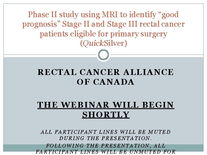
Phase II study using MRI to identify “good prognosis” Stage II and Stage III rectal cancer patients eligible for primary surgery (Quick. Silver) RECTAL CANCER ALLIANCE OF CANADA THE WEBINAR WILL BEGIN SHORTLY ALL PARTICIPANT LINES WILL BE MUTED DURING THE PRESENTATION. FOLLOWING THE PRESENTATION, ALL PARTICIPANT LINES WILL BE UNMUTED FOR
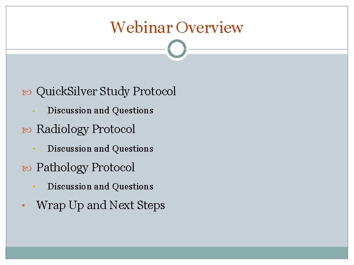
Webinar Overview Quick. Silver Study Protocol • Discussion and Questions Radiology Protocol • Discussion and Questions Pathology Protocol • • Discussion and Questions Wrap Up and Next Steps
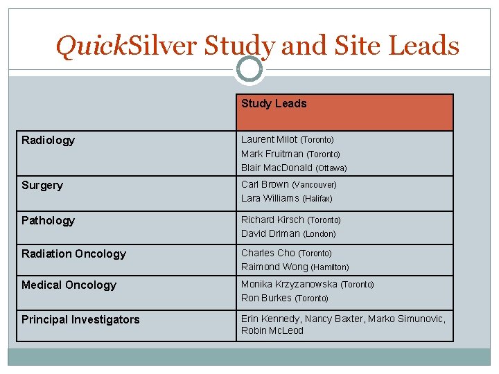
Quick. Silver Study and Site Leads Study Leads Radiology Laurent Milot (Toronto) Mark Fruitman (Toronto) Blair Mac. Donald (Ottawa) Surgery Carl Brown (Vancouver) Lara Williams (Halifax) Pathology Richard Kirsch (Toronto) David Driman (London) Radiation Oncology Charles Cho (Toronto) Raimond Wong (Hamilton) Medical Oncology Monika Krzyzanowska (Toronto) Ron Burkes (Toronto) Principal Investigators Erin Kennedy, Nancy Baxter, Marko Simunovic, Robin Mc. Leod
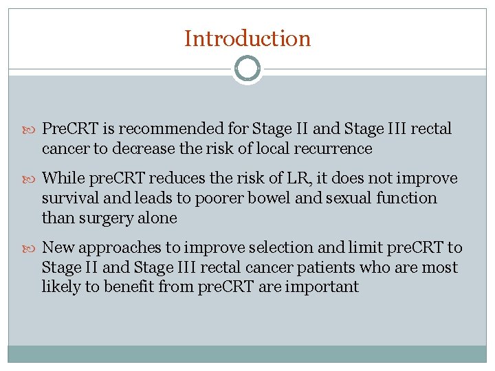
Introduction Pre. CRT is recommended for Stage II and Stage III rectal cancer to decrease the risk of local recurrence While pre. CRT reduces the risk of LR, it does not improve survival and leads to poorer bowel and sexual function than surgery alone New approaches to improve selection and limit pre. CRT to Stage II and Stage III rectal cancer patients who are most likely to benefit from pre. CRT are important
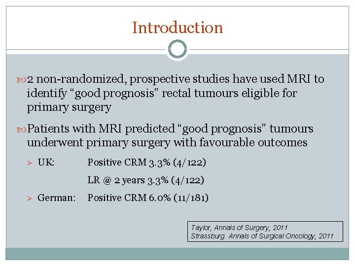
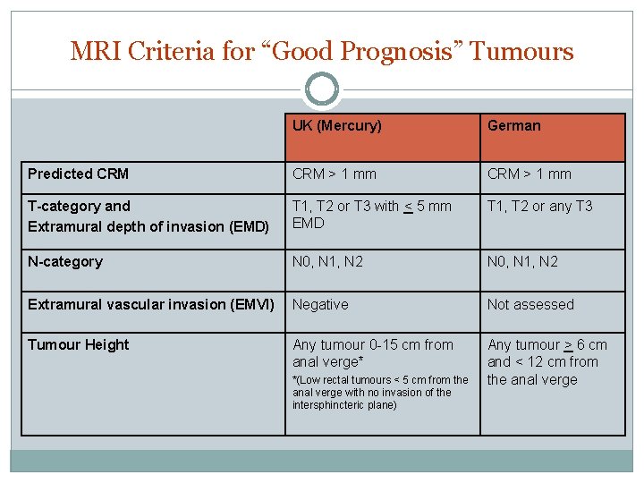
Introduction 2 non-randomized, prospective studies have used MRI to identify “good prognosis” rectal tumours eligible for primary surgery Patients with MRI predicted “good prognosis” tumours underwent primary surgery with favourable outcomes Ø UK: Positive CRM 3. 3% (4/122) LR @ 2 years 3. 3% (4/122) Ø German: Positive CRM 6. 0% (11/181) Taylor, Annals of Surgery, 2011 Strassburg. Annals of Surgical Oncology, 2011
MRI Criteria for “Good Prognosis” Tumours UK (Mercury) German Predicted CRM > 1 mm T-category and Extramural depth of invasion (EMD) T 1, T 2 or T 3 with < 5 mm EMD T 1, T 2 or any T 3 N-category N 0, N 1, N 2 Extramural vascular invasion (EMVI) Negative Not assessed Tumour Height Any tumour 0 -15 cm from anal verge* Any tumour > 6 cm and < 12 cm from the anal verge *(Low rectal tumours < 5 cm from the anal verge with no invasion of the intersphincteric plane)
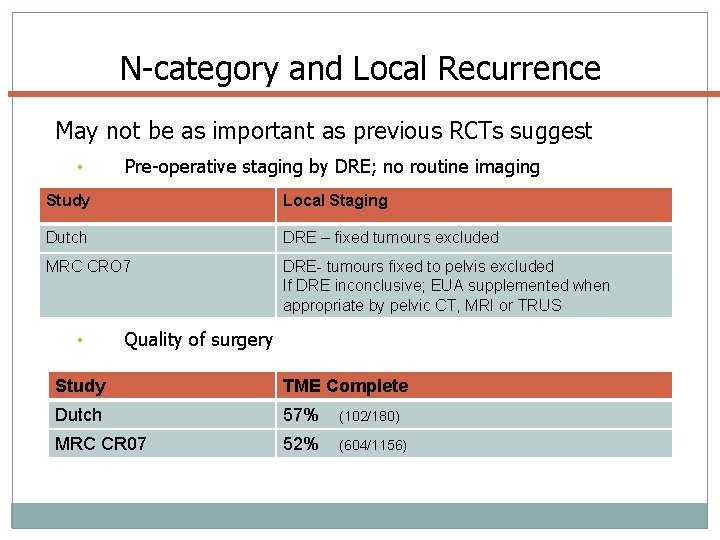
N-category and Local Recurrence May not be as important as previous RCTs suggest • Pre-operative staging by DRE; no routine imaging Study Local Staging Dutch DRE – fixed tumours excluded MRC CRO 7 DRE- tumours fixed to pelvis excluded If DRE inconclusive; EUA supplemented when appropriate by pelvic CT, MRI or TRUS • Quality of surgery Study TME Complete Dutch 57% (102/180) MRC CR 07 52% (604/1156)
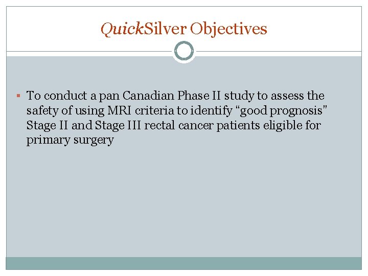
Quick. Silver Objectives § To conduct a pan Canadian Phase II study to assess the safety of using MRI criteria to identify “good prognosis” Stage II and Stage III rectal cancer patients eligible for primary surgery
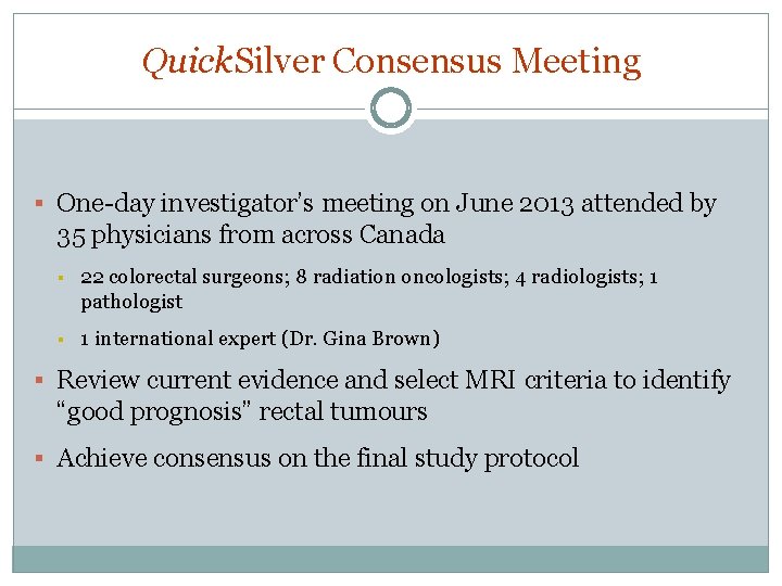
Quick. Silver Consensus Meeting § One-day investigator’s meeting on June 2013 attended by 35 physicians from across Canada § 22 colorectal surgeons; 8 radiation oncologists; 4 radiologists; 1 pathologist § 1 international expert (Dr. Gina Brown) § Review current evidence and select MRI criteria to identify “good prognosis” rectal tumours § Achieve consensus on the final study protocol
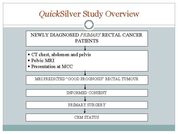
Quick. Silver Study Overview NEWLY DIAGNOSED PRIMARY RECTAL CANCER PATIENTS · CT chest, abdomen and pelvis · Pelvic MRI · Presentation at MCC MRI PREDICTED “GOOD PROGNOSIS” RECTAL TUMOUR INFORMED CONSENT PRIMARY SURGERY CRM STATUS
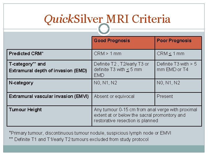
Quick. Silver MRI Criteria Good Prognosis Poor Prognosis Predicted CRM* CRM > 1 mm CRM < 1 mm T-category** and Extramural depth of invasion (EMD) Definite T 2 , T 2/early T 3 or definite T 3 with < 5 mm EMD Definite T 3 with > 5 mm EMD or T 4 N-category N 0, N 1, N 2 Extramural vascular invasion (EMVI) Absent or equivocal Present Tumour Height Any tumour 0 -15 cm from anal verge with proximal extent at or below the sacral promontory and restorative resection is planned *Primary tumour, discontinuous tumour nodule, suspicious lymph node or EMVI ** Definite T 1 and T 1/early T 2 tumours excluded from study protocol
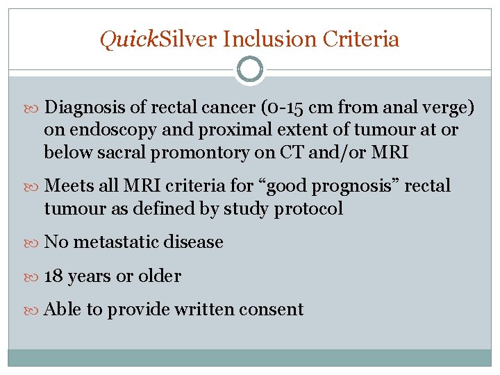
Quick. Silver Inclusion Criteria Diagnosis of rectal cancer (0 -15 cm from anal verge) on endoscopy and proximal extent of tumour at or below sacral promontory on CT and/or MRI Meets all MRI criteria for “good prognosis” rectal tumour as defined by study protocol No metastatic disease 18 years or older Able to provide written consent
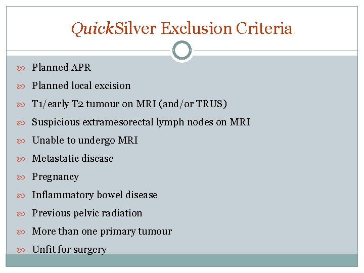
Quick. Silver Exclusion Criteria Planned APR Planned local excision T 1/early T 2 tumour on MRI (and/or TRUS) Suspicious extramesorectal lymph nodes on MRI Unable to undergo MRI Metastatic disease Pregnancy Inflammatory bowel disease Previous pelvic radiation More than one primary tumour Unfit for surgery
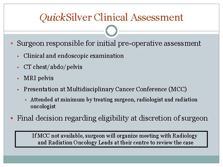
Quick. Silver Clinical Assessment § Surgeon responsible for initial pre-operative assessment § Clinical and endoscopic examination § CT chest/abdo/pelvis § MRI pelvis § Presentation at Multidisciplinary Cancer Conference (MCC) § Attended at minimum by treating surgeon, radiologist and radiation oncologist § Final decision regarding eligibility at discretion of surgeon If MCC not available, surgeon will organize meeting with Radiology and Radiation Oncology Leads at their centre to review the case
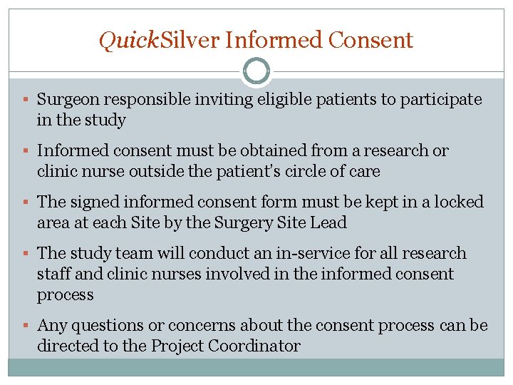
Quick. Silver Informed Consent § Surgeon responsible inviting eligible patients to participate in the study § Informed consent must be obtained from a research or clinic nurse outside the patient’s circle of care § The signed informed consent form must be kept in a locked area at each Site by the Surgery Site Lead § The study team will conduct an in-service for all research staff and clinic nurses involved in the informed consent process § Any questions or concerns about the consent process can be directed to the Project Coordinator
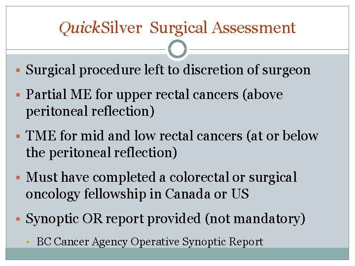
Quick. Silver Surgical Assessment § Surgical procedure left to discretion of surgeon § Partial ME for upper rectal cancers (above peritoneal reflection) § TME for mid and low rectal cancers (at or below the peritoneal reflection) § Must have completed a colorectal or surgical oncology fellowship in Canada or US § Synoptic OR report provided (not mandatory) § BC Cancer Agency Operative Synoptic Report
Quick. Silver MRI and Pathology Assessment § Specific MRI and pathology protocols for study § Specific field requirements on MRI and pathology reports § Synoptic reports recommended but not mandatory § Synoptic MRI report § CAP checklist
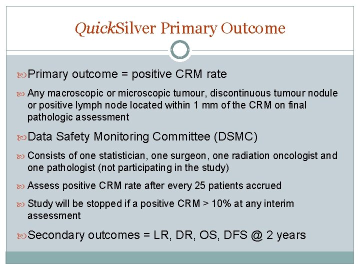
Quick. Silver Primary Outcome Primary outcome = positive CRM rate Any macroscopic or microscopic tumour, discontinuous tumour nodule or positive lymph node located within 1 mm of the CRM on final pathologic assessment Data Safety Monitoring Committee (DSMC) Consists of one statistician, one surgeon, one radiation oncologist and one pathologist (not participating in the study) Assess positive CRM rate after every 25 patients accrued Study will be stopped if a positive CRM > 10% at any interim assessment Secondary outcomes = LR, DR, OS, DFS @ 2 years
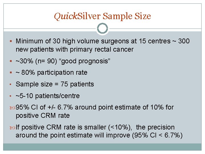
Quick. Silver Sample Size § Minimum of 30 high volume surgeons at 15 centres ~ 300 new patients with primary rectal cancer § ~30% (n= 90) “good prognosis” § ~ 80% participation rate • Sample size = 75 patients • ~5 -10 patients/centre 95% CI of +/- 6. 7% around point estimate of 10% for positive CRM rate If positive CRM rate is smaller (<10%), the precision around the point estimate will improve (95% CI < 6. 7%)
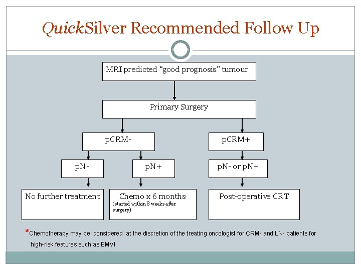
Quick. Silver Recommended Follow Up MRI predicted “good prognosis” tumour Primary Surgery p. CRMp. N- p. CRM+ p. N+ No further treatment Chemo x 6 months p. N- or p. N+ Post-operative CRT (started within 8 weeks after surgery) *Chemotherapy may be considered at the discretion of the treating oncologist for CRM- and LN- patients for high-risk features such as EMVI
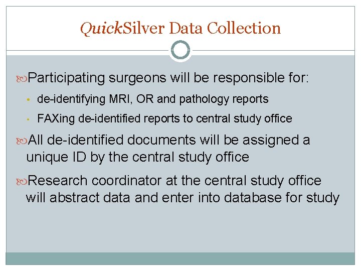
Quick. Silver Data Collection Participating surgeons will be responsible for: • de-identifying MRI, OR and pathology reports • FAXing de-identified reports to central study office All de-identified documents will be assigned a unique ID by the central study office Research coordinator at the central study office will abstract data and enter into database for study
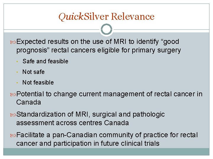
Quick. Silver Relevance Expected results on the use of MRI to identify “good prognosis” rectal cancers eligible for primary surgery • Safe and feasible • Not safe • Not feasible Potential to change current management of rectal cancer in Canada Standardization of MRI, surgical and pathologic assessment across centres Canada Facilitate a pan-Canadian community of practice for rectal cancer and participation in future clinical trials
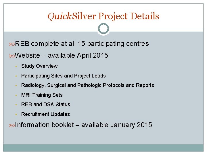
Quick. Silver Project Details REB complete at all 15 participating centres Website - available April 2015 • Study Overview • Participating Sites and Project Leads • Radiology, Surgical and Pathologic Protocols and Reports • MRI Training Sets • REB and DSA Status • Recruitment Updates Information booklet – available January 2015
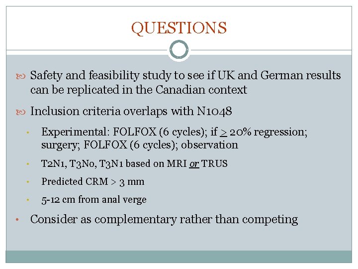
QUESTIONS Safety and feasibility study to see if UK and German results can be replicated in the Canadian context Inclusion criteria overlaps with N 1048 • • Experimental: FOLFOX (6 cycles); if > 20% regression; surgery; FOLFOX (6 cycles); observation • T 2 N 1, T 3 No, T 3 N 1 based on MRI or TRUS • Predicted CRM > 3 mm • 5 -12 cm from anal verge Consider as complementary rather than competing
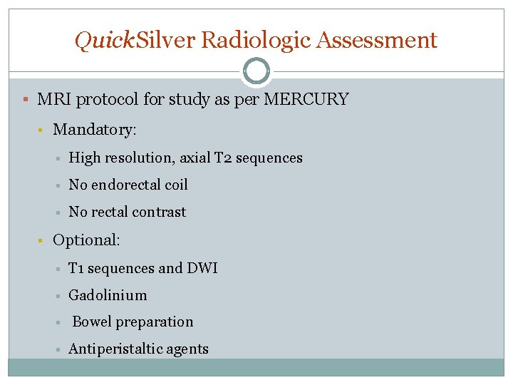
Quick. Silver Radiologic Assessment § MRI protocol for study as per MERCURY § § Mandatory: § High resolution, axial T 2 sequences § No endorectal coil § No rectal contrast Optional: § T 1 sequences and DWI § Gadolinium § § Bowel preparation Antiperistaltic agents
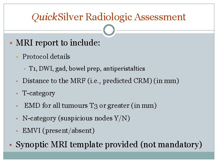
Quick. Silver Radiologic Assessment § MRI report to include: § Protocol details § T 1, DWI, gad, bowel prep, antiperistaltics • Distance to the MRF (i. e. , predicted CRM) (in mm) • T-category • EMD for all tumours T 3 or greater (in mm) • N-category (suspicious nodes Y/N) • EMVI (present/absent) • Synoptic MRI template provided (not mandatory)
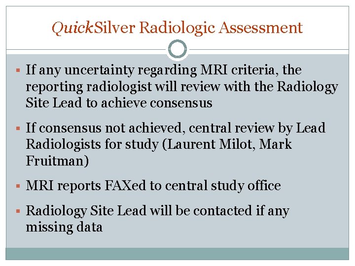
Quick. Silver Radiologic Assessment § If any uncertainty regarding MRI criteria, the reporting radiologist will review with the Radiology Site Lead to achieve consensus § If consensus not achieved, central review by Lead Radiologists for study (Laurent Milot, Mark Fruitman) § MRI reports FAXed to central study office § Radiology Site Lead will be contacted if any missing data
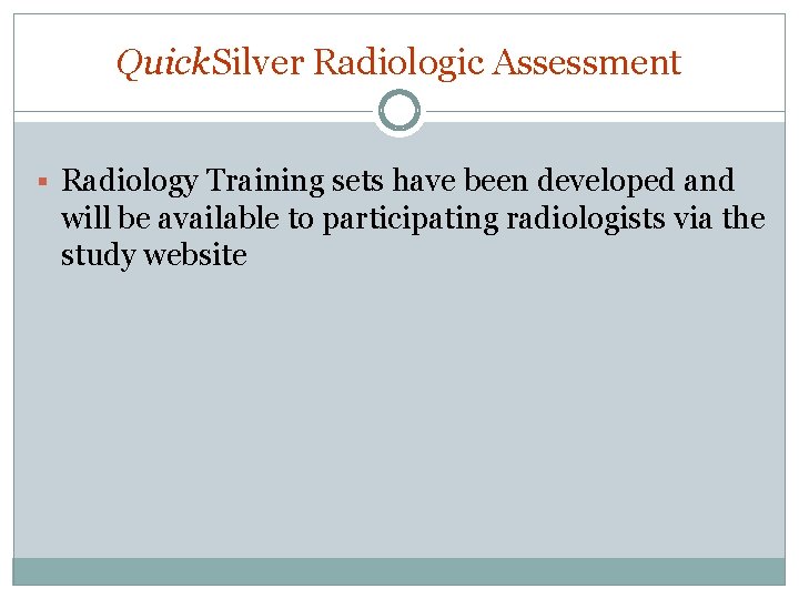
Quick. Silver Radiologic Assessment § Radiology Training sets have been developed and will be available to participating radiologists via the study website
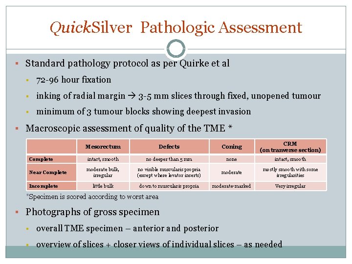
Quick. Silver Pathologic Assessment § Standard pathology protocol as per Quirke et al § 72 -96 hour fixation § inking of radial margin 3 -5 mm slices through fixed, unopened tumour § minimum of 3 tumour blocks showing deepest invasion § Macroscopic assessment of quality of the TME * Mesorectum Defects Coning CRM (on tranverse section) Complete intact, smooth no deeper than 5 mm none intact, smooth Near Complete moderate bulk, irregular no visible muscularis propria (except where levator inserts) moderate mostly smooth with some irregularities little bulk down to muscularis propria moderate-marked Very irregular Incomplete *Specimen is scored according to worst area § Photographs of gross specimen § overall TME specimen – anterior and posterior § overview of slices + closer views of individual slices – as needed
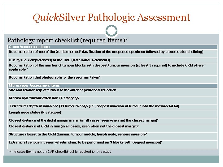
Quick. Silver Pathologic Assessment Pathology report checklist (required items)* Gross Assessment Items Documentation of use of the Quirke method* (i. e. fixation of the unopened specimen followed by cross sectional slicing) Quality (i. e. completeness) of the TME (state various elements) Documentation of the number of tumour blocks with deepest tumour invasion (at least 3 required) to include CRM where applicable * Documentation that photographs of the specimen taken* Microscopic Assessment Items Site and relationship of tumour to the anterior peritoneal reflection* Microscopic tumour extension (T-category) Extramural depth of invasion* (T 3 tumours only) (i. e. , deepest invasion of tumour into the mesorectal fat) Lymph node status (N-category) Closest distance of the distal margin in mm (in all cases, even when not the closest margin)* Closest distance of CRM in mm (in all cases, even when not the closest margin)* Structure closest to the CRM (tumour, tumour nodule, lymph node, venous invasion)* Extramural venous invasion (elastin stain: to be performed on 3 blocks with deepest invasion)* * Indicates item is not on CAP checklist but is required for this study
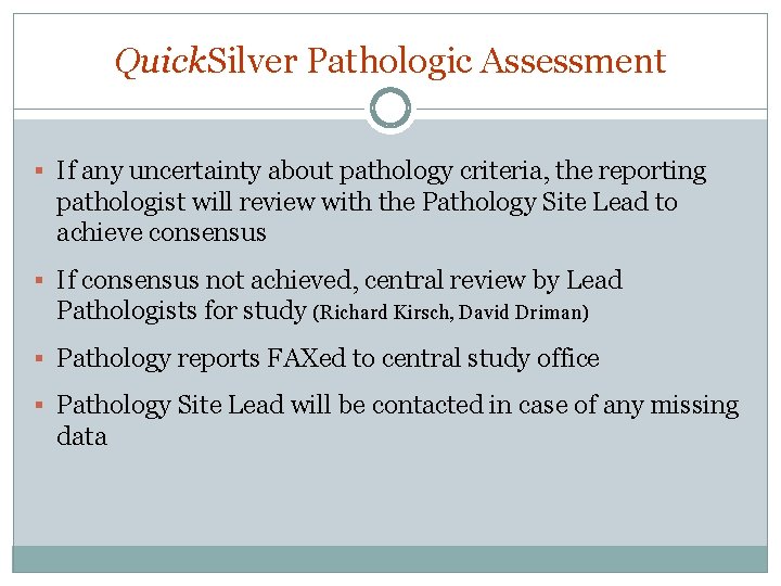
Quick. Silver Pathologic Assessment § If any uncertainty about pathology criteria, the reporting pathologist will review with the Pathology Site Lead to achieve consensus § If consensus not achieved, central review by Lead Pathologists for study (Richard Kirsch, David Driman) § Pathology reports FAXed to central study office § Pathology Site Lead will be contacted in case of any missing data
NEXT STEPS § Thank you to everyone! § Welcome any further comments about the study up until Sept 29, 2014 § Site Leads to review and finalize website and information booklet by Oct 1, 2014 § Informed consent in-service with centres as REB is approved § Plan to start study on Oct 1, 2014 to March 2016