Periferik Lenfoid Doku veya Organlar Dr mit lmez


 • T")
 in the dark")


 ---Ig. A")


- Slides: 19
Periferik Lenfoid Doku veya Organlar Dr. Ümit Ölmez
Figure 1 -7
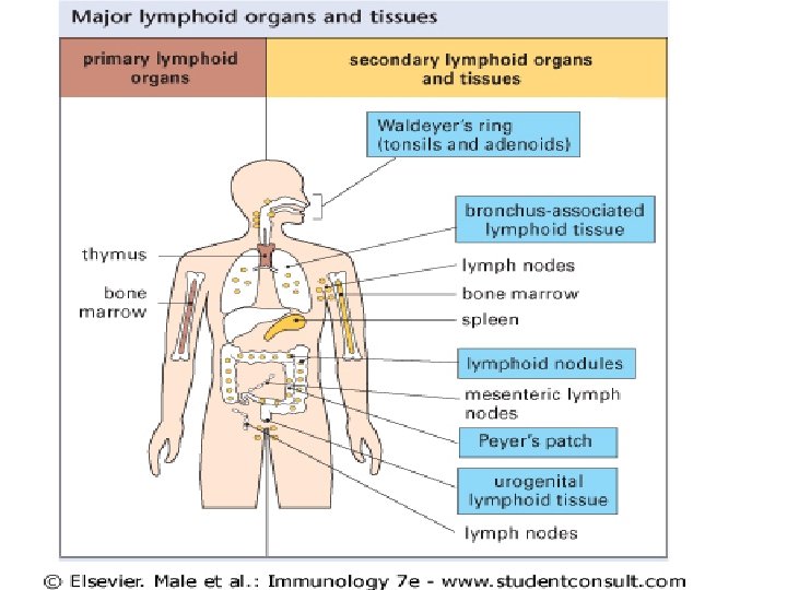
• Dalak • Lenf Bezleri • MALT: Mucosa associated lenfoid tissue – GALT: Tonsiller, adenoidler, apendiks, peyer plakları – BALT – Genitoüriner sistem – ve diğer MALT
Figure 1 -8 part 1 of 2
Figure 1 -8 part 2 of 2
Lenf Bezi • Afferent lenfatikler----- • B lenfositler : folliküllerde—ag---proliferasyon(T hüc. Yardımıyla) • T lenfositler: parakortikal alanlarda • APC---T hüc------aktive ag spesifik T hücre---- B hücre
Figure 2. 48 One or a few B cells (founder cells) in the dark zone proliferate actively. This proliferation leads to clonal expansion and is accompanied by somatic hypermutation of the immunoglobulin V region genes. B cells with the same specificity, but different affinity, are therefore generated. In the light zone, B cells with disadvantageous mutations or with low affinity undergo apoptosis and are phagocytosed by macrophages. Cells with appropriate affinity encounter the antigen on the surface of the follicular dendritic cells (FDCs) and, with the help of CD 4+ T cells, undergo class switching, leaving the follicle as memory B cells or plasma cells precursors.
Figure 1 -9 part 1 of 3
Figure 1 -9 part 2 of 3
Figure 1 -9 part 3 of 3
• Dalak ve lenf bezinden giren Ag. le karşılaşıldıktan sonra antikor yapılır, kana verilir ve lokal hücresel cevap ortaya çıkar
• MALT: Mucosa associated lenfoid tissue – GALT: Tonsiller, adenoidler, apendiks, peyer plakları – BALT – Genitoüriner sistem – ve diğer MALT
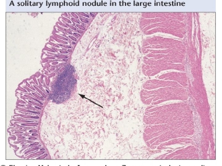
Figure 1 -10 Barsakla ilişkili lenfoid dokunun organizasyonu
Peyer's patches, as well as tonsils and other lymphoid areas of MALT, are sites of lymphocyte priming by antigens, which are internalized by M cells in the follicle-associated epithelium (FAE). The subepithelial region, the dome, is rich in APCs and also contains a subset of B cells similar to those found in the splenic marginal zone. Lymphoid follicles and intervening T-dependent zones are localized under the dome region. , Lymphocytes primed by antigens in these initiation sites of the gut mucosa migrate to the mesenteric lymph nodes and then to the effector sites (the intestinal villi), where they are found both in the lamina propria (LPLs) and within the surface epithelium (IELs)
MALT • Lamina propria lenf: LPL(aktive. T hüc, B hüc, plazma hüc) ---Ig. A • İntraepitelyal lenfositler : IEL: En çok T (%10 -40 γδ T hüc ve CD 8(%70)
The lymphocytes move through the circulation and enter the lymph nodes and MALT via the specialized endothelial cells of the postcapillary venules (i. e. high endothelial venules [HEVs]). They leave the lymph nodes and MALT through the efferent lymphatic vessels and pass through other nodes, finally entering the thoracic duct, which empties into the circulation at the left subclavian vein (in humans). Lymphocytes enter the white pulp areas of the spleen in the marginal zones, pass into the sinusoids of the red pulp, and leave via the splenic vein.
Figure 1 -11