Muscle Physiology I Muscle cells are highly specialized


- Slides: 19
Muscle Physiology I
¥ Muscle cells are highly specialized cells for the conversion of chemical energy to mechanical energy. Specifically, muscle cells use the energy in ATP to generate force or do work
Types of Muscle Tissue ¥ Skeletal muscle tissue is primarily attached to bones. It is striated and voluntary. ¥ Cardiac muscle tissue forms the wall of the heart. It is striated and involuntary. ¥ Smooth (visceral) muscle tissue is located in viscera. It is non-straited (smooth) and involuntary. ¥ In all three muscle types, force is generated by the interaction of actin and myosin molecules, a process that requires transient elevation of intracellular [Ca++].
Functions of Muscle Tissue ¥ ¥ ¥ Producing body movements Stabilizing body positions Regulating organ volumes Movement of substances within the body Producing heat
Properties of Muscle Tissue ¥ Excitability: They can react to nervous stimulation ¥ Conductivity: They can transmit the electrical impulses ¥ Contractility: They can shorten forcibly when stimulated by nervous system and hormones ¥ Extensibility: They can lengthen when stretched ¥ Elasticity: ability of muscle tissue to return to its normal resting length
Skeletal Muscle structural arrangement ¥ Skeletal muscle is composed of bundles of muscle fibers called a fasciculus. A muscle fiber represents an individual muscle cell and contains bundles of myofibrils. ¥ In addition to muscle fiber, skeletal muscle contains § § § Connective Tissue Blood vessels nerves
Filaments and the Sarcomere
The Proteins of Muscle ¥ Myofibrils are built of 3 kinds of protein § contractile proteins myosin and actin § regulatory proteins which turn contraction on & off troponin and tropomyosin § structural proteins which provide proper alignment, elasticity and extensibility titin, myomesin, nebulin and dystrophin
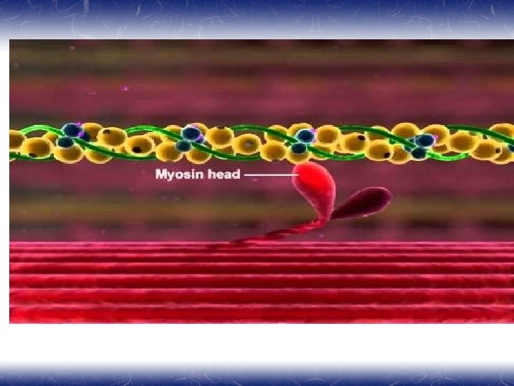
¥ ¥ ¥ Myosin is a large protein that consists of six different polypeptides with one pair of large heavy chains and two pairs of light chains. The heavy chains are wound together in an α-helical configuration to form a long rod-like segment, with the N-terminal portion of each heavy chain forming a large globular head. The head region extends away from the thick filament toward the actin thin filament and is the portion of the molecule that can bind to actin. Myosin is also able to hydrolyze ATP, and ATPase activity is located in the globular head. Two pairs of light chains are associated with the globular head. One of these pairs of light chains, termed essential light chains, is critical for the ATPase activity of myosin. The other pair of light chains, sometimes called regulatory light chains, may influence the kinetics of myosin and actin binding under certain conditions. Thus, myosin ATPase activity resides in the globular head of myosin and requires the presence of light chains (viz. , the "essential" light chains).
¥ ¥ ¥ The thick myosin filaments are tethered to the Z lines by a cytoskeletal protein called titin. Titin is a very large elastic protein that extends from the Z line to the center of the sarcomere. Titin is important for organization and alignment of the thick filaments in the sarcomere. Titin may also serve as a mechanosensor and influence gene expression and protein degradation in a mechanical activity-dependent manner. Some forms of muscular dystrophy have been attributed to defects in titin. The M line (myomesin) connects to titin and connects adjacent thick filaments together.
¥ ¥ ¥ Polymerization of monomeric actin into filamentous actin forms the backbone of the thin filament. The elongated cytoskeletal protein nebulin extends along the length of the thin filament and may participate in regulation of the length of the thin filament. Dimers of the protein tropomyosin extend over the entire actin filament and cover myosin binding sites on the actin molecules. A troponin complex consisting of three subunits (troponin T, troponin I, and troponin C) is present on each tropomyosin dimer and influences the position of the tropomyosin molecule on the actin filament and hence the ability of tropomyosin to inhibit binding of myosin to the actin filament. Troponin T binds tropomyosin, troponin I facilitates the inhibition of myosin binding to actin by tropomyosin, and troponin C binds Ca++. Binding of Ca++ to troponin C promotes the movement of tropomyosin on the actin filament, thereby exposing myosin binding sites and facilitating the interaction of myosin and actin filaments and sarcomere contraction.
¥ Additional proteins associated with the thin filament : ¥ tropomodulin, α-actinin, and cap. Z protein. Tropomodulin is located at the end of the thin filament, toward the center of the sarcomere, and may participate in setting the length of the thin filament. α-Actinin and cap. Z protein serve to anchor the thin filament to the Z line.
¥ Dystrophin links thin filaments to sarcolemma and transmits the tension generated to the tendon. ¥ The dystrophin-glycoprotein complex provides a structural link between the cytoskeleton of the muscle cell and the extracellular matrix, which appears to stabilize the sarcolemma and hence prevents contraction-induced injury (rupture). Duchenne's muscular dystrophy is associated with loss of dystrophin.
¥ ¥ ¥ Each myofibril in a muscle fiber is surrounded by sarcoplasmic reticulum (SR). The SR is an intracellular membrane network that plays a critical role in the regulation of intracellular [Ca++]. T tubules are invaginations of the plasma membrane, which are present exclusively in striated muscle. Their role is to maintain the SR calcium store under the tight control of membrane depolarization via the voltage sensor channel DHPR The SR is an intracellular network, whereas the T tubules are in contact with the extracellular space The portion of the SR nearest the T tubules is called the terminal cisternae, and it is the site of Ca++ release, which is critical for contraction of skeletal muscle.
Skeletal Muscle Structure
Mechanism of Contraction Sliding Filament Theory of Contraction ¥ Myosin cross bridges pull on thin filaments ¥ Thin filaments slide inward ¥ Z Discs come toward each other ¥ Sarcomeres shorten. The muscle fiber shortens. The muscle shortens ¥ Notice : Thick & thin filaments do not change in length
State A, In the relaxed state, ATP is partially hydrolyzed. ¥ State B, In the presence of elevated myoplasmic Ca++, myosin binds to actin. ¥ State C, Hydrolysis of ATP is completed and causes a conformational change in the myosin molecule that pulls the actin filament toward the center of the sarcomere (Pivoting). ¥ State D, A new ATP binds to myosin and causes release of the cross-bridge. Partial hydrolysis of the newly bound ATP recocks the myosin head, which is now ready to bind again and again. If myoplasmic [Ca++] is still elevated, the cycle repeats. If myoplasmic [Ca++] is low, relaxation results. ¥
Contraction Cycle