TISSUES Tissues are collections of specialized cells and


- Slides: 33

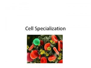
TISSUES • Tissues are collections of specialized cells and cell products organized to perform one or more select functions Histology = study of tissues • All cells in the body are classified into one of the four tissue types: Epithelial (Epithelium and Glands) Connective Nervous Muscular 119

Epithelial Tissue: our first tissue type Types of Epithelial Tissue 1. Epithelial sheets (“epithelium”) 2. Glands 120

Epithelial Tissue Lecture Outline 1. 2. 3. 4. Characteristics of Epithelial Sheets Cell-Cell Junctions : Gap Junctions, Tight Junctions, Desmosomes, and Lateral Interdigitations Naming Epithelial Sheets by Thickness and Cell shape Glands : Endocrine and Exocrine 121

Epithelium: a layer of tightly adjoining related by their embryology and function Locations: covers body surface (outermost part of skin) lines all hollow structures in body Functions: Physical protection Control permeability Produce specialized secretions 122

Characteristics of Epithelial sheets: • • • Cells are polarized Little space between cells Cells tightly held together by junctions Avascular (no blood vessels between cells) Specialized for absorption or protection Continuous rate of cell division 123

Epithelial Cells have Polarity (sided-ness) Apical (free or exposed surface) Lateral (neighboring epithelial cells) Basal (surface anchored to connective tissue) 124

Epithelial cells are attached at the basal surface to a basement membrane The epithelial cells are attached to connective tissue through the glue-like proteins of the basement membrane. We sometimes use the phrases “basal lamina” and “basement membrane” interchangeably 125

The avascular concept Epithelium one layer in thickness Epithelium multiple layers in thickness Little space between adjacent cells for blood vessels. Epithelial cells are usually kept alive by diffusion of oxygen and nutrients from the blood vessels in the adjacent connective tissue below the basal lamina Epithelial layers are limited in thickness – diffusion is poor over long distances or dense areas 126

Some Epithelial Cells may have specializations on their apical surfaces MICROVILLI Small fingerlike projections of the apical or basal surface of an epithelial cell (may be hundreds per cell) Microvilli increase surface area for the cell 127

Some Epithelial Cells have specializations on their apical surfaces CILIA Long slender extensions of the apical cell surface Cilia have a core of microtubules Cilia exhibit rhythmical movement 128

cilia 129

Epithelial Cells are tightly held to their neighboring epithelial cells Cells attach via cell adhesion molecules Cells attach at specialized cell junctions: Gap junctions, Desmosomes, Tight junctions, Lateral Interdigitations 130

Connections between Epithelial Cells : Gap Junctions Cells are connected by membrane proteins that form pores or channels between cells Cytoplasm of one cell is continuous with cytoplasm of adjacent cell allowing ions to move between cells Important in electrical signaling and organs where cells work in close synchrony 131

Connections between Epithelial Cells : Tight Junctions The plasma membranes of adjacent cells are “fused” together by membrane proteins 132

Connections between Epithelial Cells : Tight Junctions Apical surface Tight junctions form an occluded zone just under the apical surface where the neighboring cells have fused their membranes together Extends all the way around the cell, with different neighbors Water and solutes cannot pass between cells 133

Connections between Epithelial Cells : Desmosomes Adjacent cells joined by membrane proteins and a glue-like material 134

Review: Tight Junctions, Gap Junctions, and Desmosomes and their relative locations 135

Tight Junction Lateral interdigitations Adjacent cells have intertwined membranes Desmosome Lateral Interdigitation 136

Other features of typical epithelium Basement membrane joins epithelial layer to underlying connective tissue layer Epithelial stem cells replace short-lived epithelial cells – Because epithelial sheets are in vulnerable body positions, they have a high rate of cell division to constantly replace damaged cells – Remember that carcinomas, tumors of epithelial cells, account for 90% of all cancers. 137

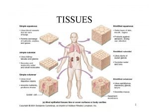
Epithelial sheets are described with 2 adjectives • Number of cell layers – Simple One cell thick – Stratified More than one cell thick • Shape of apical surface cells – Squamous – Cuboidal – Columnar Flat, broad cells Cube-shaped Tall, narrow cells 138

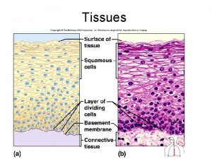
Simple Squamous Epithelium • Single layer of flat cells; little protection, allows for fast diffusion • Locations: lines all the cardiovascular organs (heart, vessels), air spaces in lungs, etc. 139

Simple Cuboidal Epithelium One layer thick; cells block–like in shape (as tall as they are wide) Function: allows for regulated exchange; cytoplasm typically holds many mitochondria to drive active transport processes; may have microvilli Locations: kidneys, ducts 140

Simple Columnar Epithelium • Cells taller than they are wide, one layer thick; cells often have microvilli or cilia on apical surface • Functions: Some protection; active transport; often have mucous cells interspersed • Locations: intestines 141

Stratified Squamous Epithelium • Many layers of cells thick • apical surface is flattened • grows from basal surface Functions: Mainly as a barrier - denies access to connective tissue below; does not allow for exchange through epithelium Locations: surface of skin, lining of mouth, anus, vagina, etc. 142

Transitional Epithelium Unique to urinary system 143

Ciliated Pseudostratified Columnar Epithelium the special epithelium of the respiratory system The ciliated surface of the respiratory tract 144

Glandular epithelia cells of epithelial line which are specialized to produce and secrete materials outside the cell Endocrine glands Release hormones into surrounding fluid to be picked up and carried away by the blood Exocrine glands Secrete through ducts onto the surface of the organ Merocrine (product released through exocytosis) Apocrine (involves the loss of both product and cytoplasm) Holocrine (destroys the cell) 145

The Structure of Endocrine Glands Product is released from cell into extracellular fluid Product is picked up and carried away by bloodstream where it may affect distant tissues 146

Mechanisms of Exocrine Gland Secretion: Merocrine Product is packaged into secretory granules Released from cell into duct by exocytosis Example: salivary glands, pancreas 147

Mechanisms of Exocrine Gland Secretion Apocrine Apical portion of cell is shed, taking product with it into duct Example: mammary glands 148

Mechanisms of Exocrine Gland Secretion: Holocrine The entire cell bursts, releasing product into duct. Cell dies and must be replaced Example: oil glands associated with hair follicles 149

Structure of Unicellular Glands Individual secretory cells are embedded within an epithelium (example: Goblet cells) 150

Structural Classification of Multicellular Glands Don’t memorize these terms, just appreciate the different levels of complexity 151
 Collection of specialized cells and cell products
 Body tissue
 Chapter 3 cells and tissues answer key
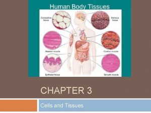
 Body tissues chapter 3 cells and tissues
 Stained cheek cell
 Eisonophil
 Insidan region jh
 What are specialized connective tissue
 Chapter 3 cells and tissues figure 3-7
 Chapter 3 cells and tissues figure 3-1
 Chapter 3 cells and tissues figure 3-1
 Specialized cells in animals
 Specialized cells
 Tissues are groups of similar cells working together to
 Tissues are groups of similar cells working together to
 Where are loins on a human
 A group of cells similar in structure and function
 Development of paranasal sinuses
 Chlorocruorin
 Plant and animal cells venn diagram
 Masses of cells form and steal nutrients from healthy cells
 Loop of henle
 Pineal gland
 Gametes vs somatic cells
 Somatic vs germ cells
 What organisms are made of eukaryotic cells
 Prokaryotes vs eukaryotes venn diagram
 Why did robert hooke name cells “cells”?
 Younger cells cuboidal older cells flattened
 4 types of eukaryotic cells
 Prokaryotic cells vs eukaryotic cells
 Chapter 8 cellular reproduction cells from cells
 Cell substance
 Pseg credit and collections
