Muscle Contraction Connective Tissue Epimysium Perimysium Endomysium surrounds

 Muscle")


 • myogram - graphic")


- Slides: 29
Muscle Contraction
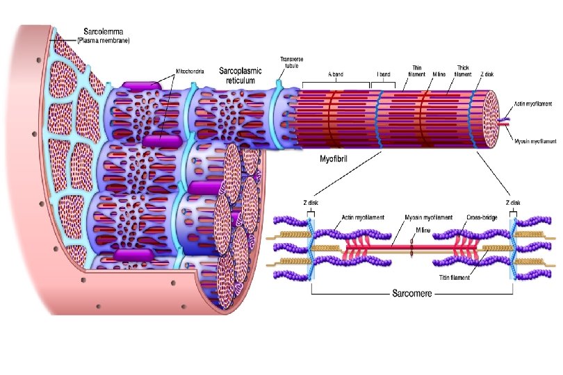
Connective Tissue Epimysium Perimysium Endomysium surrounds Structure Gross muscle Fascicle (bundles of cells) Muscle fiber (muscle cell) organ tissues cells Myofibril organelles Myofilaments molecules (contracting organelle) Actin – thin filament Myosin – thick filament
Molecular Structure of Myofilaments
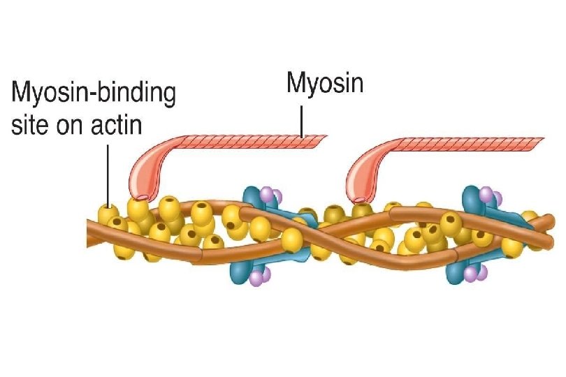
Myosin tail • rod like tail or axis ending in 2 globular heads - “crossbridges” - heads contain ATPase (enzyme) heads
Actin • globular subunits - G actin are found in long strands, like a double strand of twisted beads
Actin • tropomyosin spirals around beads, to add strength and stiffen, thin ribbon • troponin molecules bond actin to tropomyosin, has binding sites that will join with myosin crossbridge Binding sites
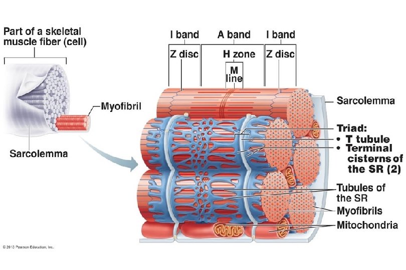
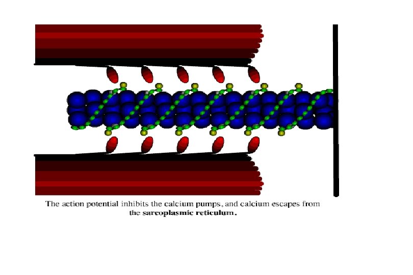
Sarcoplasmic Reticulum
Sarcoplasmic Reticulum • smooth endoplasmic reticulum of muscle cells • storage area for Ca ions • At H zones and A - I junctions tubes fuse and form lateral channels - terminal cisternae which feed into transverse tubules (T tubules) tubules at each Z line. • T tubules receive nerve stimuli and provide a pathway for oxygen, glucose, and Ca ions. (because continuous with membrane, nerve impulse allowed deep into muscle)
Mechanism of Contraction when muscle cells contract their sarcomeres shorten (Z to Z distance decreases)
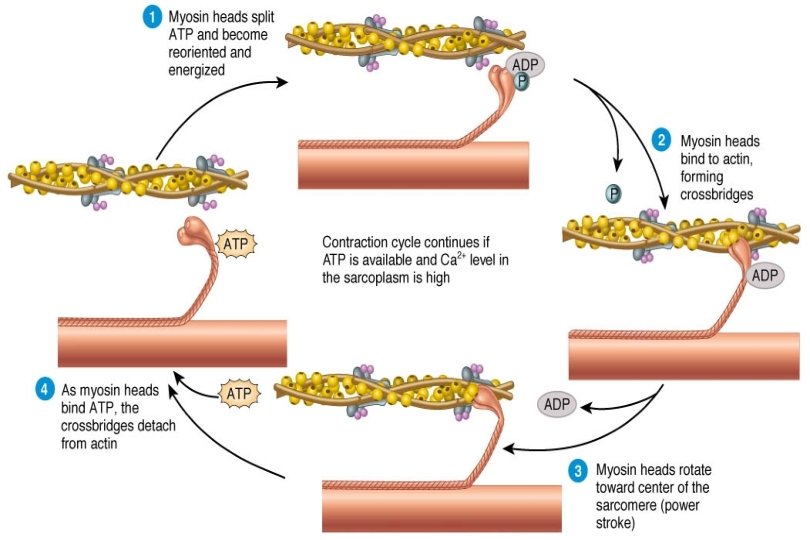
Sliding Filament Theory of Muscle Contraction 1954 Hugh Huxley • In absence of Ca, myosin - binding sites on actin are physically blocked by tropomyosin • When Ca ions are released through T tubules, tubules they bond with troponin • The resulting change in molecular conformation physically moves away tropomyosin from binding sites.
Exposure of binding sites leads to the following events in rapid succession: 1. crossbridges on myosin heads bond to troponin binding sites 2. as heads bind, they change shape to a bent shape, which causes heads to pull on actin sliding it to the center at the same time, ADP released from head (energy supplied the movement)
Exposure of binding sites leads to the following events in rapid succession: 3. As a new ATP molecule binds to myosin head, the bridge is released from actin 4. Hydrolysis of ATP to ADP + P (ATPase) provides energy needed to cock myosin heads to high energy position (potential energy).
• Now, myosin head is ready to repeat action further down thin filament. The cross bridges walk along the actin. • The contraction ends when Ca ions are withdrawn from sarcoplasm (lack of nerve stimulus).
Relaxed Sarcomere Contracted Sarcomere
Rigor Mortis • Injured and dying cells are unable to exclude Ca, so crossbridge binding is promoted. • • BUT in absence of ATP synthesis (shortly after breathing stops) detachment is impossible causing rigor mortis • • Muscles begin to stiffen 3 - 4 hr. after death. • • Peak about 12 hr. and dissipates over next 48 - 60 hr. due to breakdown of tissue.
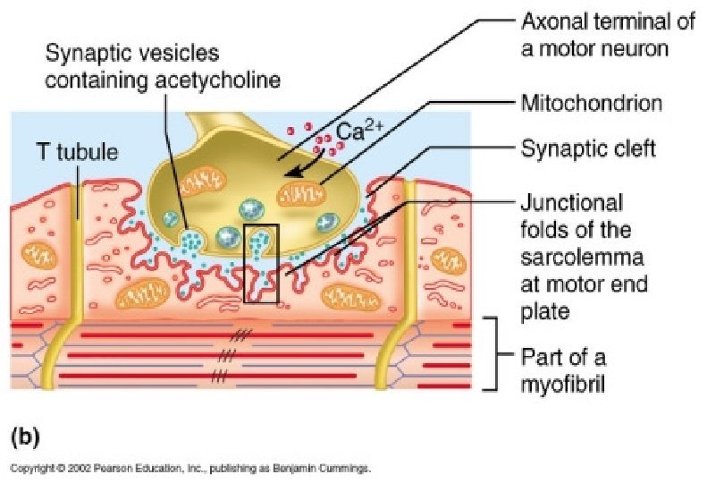
Regulation of Contraction • In order for a nerve impulse to trigger a muscle response, the impulse must travel over the gap (neuromuscular junction) junction where two cells meet. (neuron and muscle) • In the gap (extracellular space = synapse) synapse are sacs containing the neurotransmitter - acetylcholine (Ach).
Muscle Twitch • studies done in vitro (outside the body) • myogram - graphic recording of contractile activity - traces twitch • muscle twitch - muscle responds to a single brief stimulus (contracts and relaxes) - may be strong or weak 1. latent period - time between stimulus and response 2. contraction 3. relaxation - no more contractile force, tension decreases (in vivo (inside body) - longer and smoother)
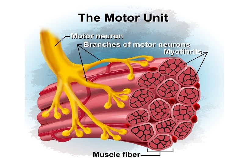
• The strength of the muscle twitch depends on the number of motor units involved. • motor unit - a motor neuron and all the muscle fibers it supplies • Muscles with fine control have small units. ex: fingers, eyes • Large weight bearing muscles have less precise movement and larger motor units. ex: hip Motor Units
• As impulses continue at faster rate all evidence of relaxation disappears and muscle has a smooth sustained contraction. - tetanus • Prolonged tetanus will lead to fatigue
Types of Muscle Contractions • Muscles do not always shorten during a contraction - active process of generating force within a muscle by cross bridge activity muscle tension - force exerted by a contracting muscle on some object load - wt/ resistance to movement exerted by the object on the muscle • muscle tension and load are opposing forces • to move a load, the muscle tension must be greater than the load
isotonic contractions - “equal tension” • muscle shortens to move the load • most common • best for developing strength ex: muscles moving the leg
isometric contractions - “equal distance” • result in an increase if tension but no shortening because the load is too great ex: trying to lift a piano single handedly, muscles maintaining posture • In the body most movement is a combination because muscles work together.