MECHANISM OF MUSCLE CONTRACTION AND RELAXATION Muscle contraction


- Slides: 25
MECHANISM OF MUSCLE CONTRACTION AND RELAXATION
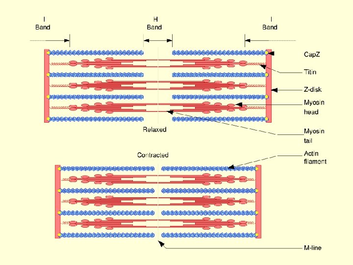
Muscle contraction • Muscle contraction is accomplished by a sliding together or telescoping of the inter digitating thick and thin filament array without and detectable shortening of the filaments themselves. • The force causing the thick and thin filaments to slide past one another is generated by the cross-bridges or myosin heads that project outward from the surface of the thick filament.
• Although it is still unclear how the chemical energy in the ATP molecule is converted to the mechanical energy of muscle contraction, a great deal is known about the anatomical and biochemical events that accompany this energy transduction
• The events that occur during muscle contraction can be divided into two general categories • (1) those events that occur in the thin filament and • (2) those events that occur in the thick filament.
Muscle contraction flow chart Contraction Phase Resting state ↓ Motor nerve action potential arrives at motor end plate ↓ Acetylcholine released, sarcolemma and membranes depolarized (Na+ flux into fiber) ↓ Action potential transmitted via T-tubules to SR ↓ Ca++ released from SR terminal cisternae into sarcoplasm ↓ Ca++ bound by troponin
Myosin ATPase activated and ATP hydrolyzed ↓ Tropomyosin shift from actin binding site ↓ Actin-myosin crossbridge formation (Contraction) ↓ Repeated formation & breaking of crossbridges resulting in sliding of filaments and sarcomere shortening
Relaxation Phase Cholinesterase released and acetylcholine breakdown ↓ Sarcolemma & T-tubules repolarized ↓ SR Ca++ pump activated & Ca++ returned to SR terminal cisternae ↓ ↓Actin-myosin crossbridge formation terminated ↓ Return of tropomyosin to actin binding site ↓ Mg++ complex formed with ATP ↓ Passive sliding of filaments ↓ Sarcomeres return to resting state
1. Changes in Thin Filaments during Muscle Contraction • A motor nerve impulse reaching the muscle cell passes along the sarcolemma and into the T-tubules. • Passage of an electrical signal along the extra cellular Ttubule causes the adjoining, intracellular sarcoplasmic reticular membranes to momentarily lose their ability to retain the calcium that they have accumulated against a concentration gradient. • This Ca 2+ therefore floods into the interior of the cell, and intracellular free Ca 2+ concentration rises from approximately 10 -8 M in resting muscle to 10 -5 to-10 -6 M.
• Because troponin-C has a binding constant near 5 x 10 -6 M for Ca 2+, an intracellular Ca 2+ concentration of 10 -5 to 10 -6 M is sufficiently high to permit binding of Ca 2+ to troponin-C. • In the absence of Ca 2+, troponin C binds to troponin – T and troponin-I, and troponin-I binds loosely to troponin-T, but firmly to actin. • In the presence of Ca 2+ troponin-T binds strongly to tropomyosin, troponin-C binds strongly to Troponin-T and troponin-I, and troponin-I binds to troponin-T but loses its affinity for actin
• In resting muscle, the tropomyosin stand is located out of the groove of the double-stranded actin filament in a positon where it physically block or at least sterically interferes with the myosin-binding site of actin. • Troponin is necessary to hold tropomyosin in this out position, and a firm interaction between troponin-I and actin is evidently necessary to maintain this out position of tropomyosin.
• When troponin-C binds Ca 2+, however, the firm linkage between troponin-I and actin is weakened, and troponin can no longer hold the tropomyosin strand in the out or “off” position. • The tropomyosin strand rolls back into the groove of the double-stranded actin filament, and the myosinbinding site on actin is expose. •
• Myosin binds to actin, contraction ensues and continues until the Ca 2+ is removed from the troponin-C and troponin-I binds to actin and forces the tropomyosin strand back out of the groove of the actin filament to block the myosin-binding site of actin. • Turning muscle contraction on and off in vertebrate striated muscle is accomplished by moving the tropomyosin strand in (on) and (off) of the groove in the double-stranded actin filament.
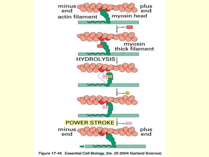
2. Change in Thick Filaments During Muscle Contraction • During muscle contraction, myosin cross-bridges extend outward, attach to the thin or actin filament and then swivel or rotate so that the tip of the cross-bridge undergoes approximately a 10 nm translocation, and pushes the actin filament toward the center of the sarcomeres. • At the end of this stroke, the spent cross-bridge detaches from the thin filament, is reoriented, and is ready to repeat the cycle.
• During a single twitch of a muscle fiber, each myosin crossbridge may perform many cycles of attaching to actin, swiveling and then dissociating from the actin filament, but only 10 -20% of the total cross-bridges in a single sarcomeres are ever attached to thin filaments at any given instant during contraction. • The remaining cross-bridges are in various other stages of the cross-bridge cycle.
• Because cross-bridges interact asynchronously with actin, the thick and thin filaments slide past each other at a smooth, uniform rate during muscle contraction rather than with a “jerky”, ratchet-like motion that would be produced by synchronous cross-bridge actin. • ATP hydrolysis is clearly involved in providing energy for muscle contraction in the presence of Mg 2+, myosin binds and splits ATP very rapidly
• Myosin molecule passes through at least five conformation states during hydrolysis of ATP in the presence of Mg 2+. • One of these conformational states is free myosin. • Binding of ATP very quickly conformational state of myosin. produces a second • Immediately after the conformational changes resulting from binding of ATP to myosin, the bound ATP induces an additional conformation state designated as M. ATP. • Hydrolysis of the bound ATP in this third conformational state occurs at an intermediate rate and produces a fourth conformation form.
• In living resting muscle, almost every myosin crossbridge is “energized” and contains one molecule each of ADP and inorganic phosphate, the hydrolysis products of ATP. • “Energized” here refers to some unknown conformational state of myosin in which the energy of ATP is stored; this state is represented by Pi. M. ADP.
• In resting muscle, the energy in energized myosin is dissipated slowly through step 4 of the transient state mechanisms. • The energized cross-bridge is unable to bind to actin because thin filament is “turned off”, i. e. , tropomyosin is blocking the myosin-binding site on actin.
• Only after ADP has been released from the spent crossbridge the myosin head can bind a new molecule of ATP. • ATP is a potent dissociator of the actin-myosin complex, binding of a new molecule of ATP immediately dissociates the myosin cross-bridge from the actin filament.
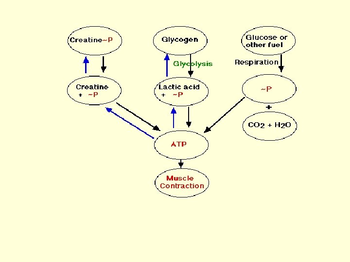
Fueling Muscle Contraction • ATP is the immediate source of energy for muscle contraction. Although a muscle fiber contains only enough ATP to power a few twitches, its ATP "pool" is replenished as needed. There are three sources of highenergy phosphate to keep the ATP pool filled. • creatine phosphate • glycogen • cellular respiration in the mitochondria of the fibers.
• The phosphate group in creatine phosphate is attached by a "high-energy" bond like that in ATP. Creatine phosphate derives its high-energy phosphate from ATP and can donate it back to ADP to form ATP. • Creatine phosphate + ADP ↔ creatine + ATP • The pool of creatine phosphate in the fiber is about 10 times larger than that of ATP and thus serves as a modest reservoir of ATP.