
1 To understand the cellular processes for synthesis






- Slides: 44
1. To understand the cellular processes for synthesis of ATP. 2. To compare and contrast aerobic and anaerobic processes in the muscle cell. 3. To examine the differences in ATP synthesis among different types of muscle cells
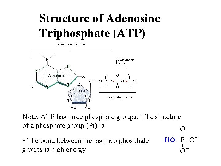
Structure of Adenosine Triphosphate (ATP) Note: ATP has three phosphate groups. The structure of a phosphate group (Pi) is: • The bond between the last two phosphate groups is high energy
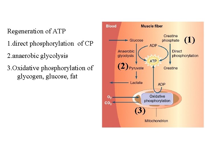
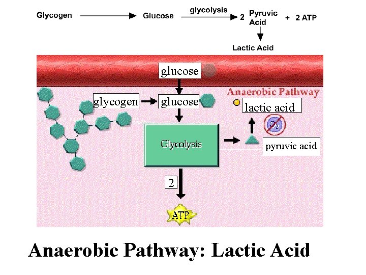
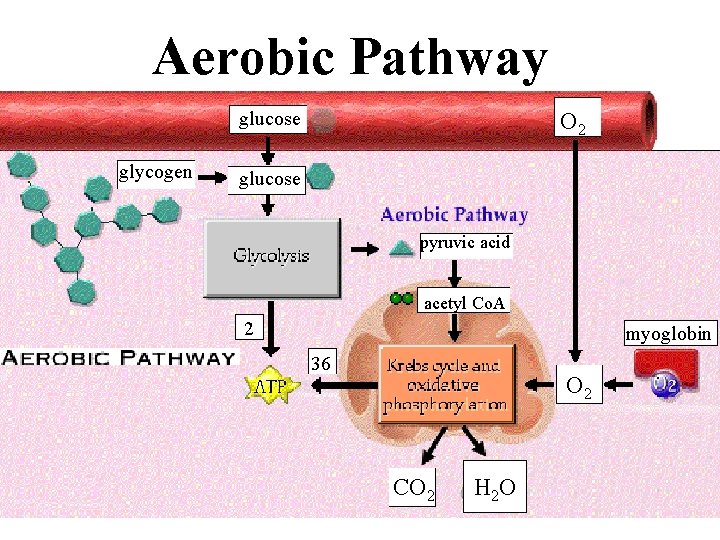
Regeneration of ATP (1) 1. direct phosphorylation of CP 2. anaerobic glycolysis 3. Oxidative phosphorylation of glycogen, glucose, fat (2) (3)
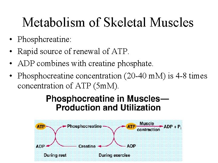
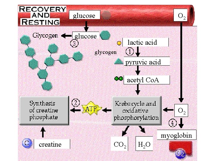
Metabolism of Skeletal Muscles • • Phosphcreatine: Rapid source of renewal of ATP. ADP combines with creatine phosphate. Phosphocreatine concentration (20 -40 m. M) is 4 -8 times concentration of ATP (5 m. M).
Sources of Glucose • Two sources of glucose to muscles: 1. Blood glucose. 2. Breakdown of glycogen into glucose within the muscle cell
glucose glycogen glucose lactic acid pyruvic acid 2 Anaerobic Pathway: Lactic Acid
Aerobic Pathway glucose glycogen O 2 glucose pyruvic acid acetyl Co. A 2 myoglobin 36 O 2 CO 2 H 2 O
Regeneration of ATP 1. direct phosphorylation of CP: 1 ATP produced per CP 2. anaerobic glycolysis: 2 ATP per glucose 3. Oxidative phosphorylation of glycogen, glucose, fat; ? ? ATP per glucose
glucose O 2 glucose lactic acid glycogen pyruvic acid acetyl Co. A O 2 myoglobin creatine CO 2 H 2 O
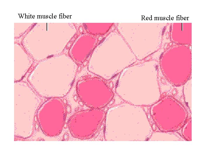
White muscle fiber Red muscle fiber
White muscle fiber Features of White Muscle Fibers Characteristics of white muscle fibers: 1. Large in diameter 2. Light in color due to reduced or absent myoglobin 3. Surrounded by only a few capillaries 4. Have relatively few mitochondria 5. Have a high glycogen content 6. Synthesize ATP mainly by glycolysis
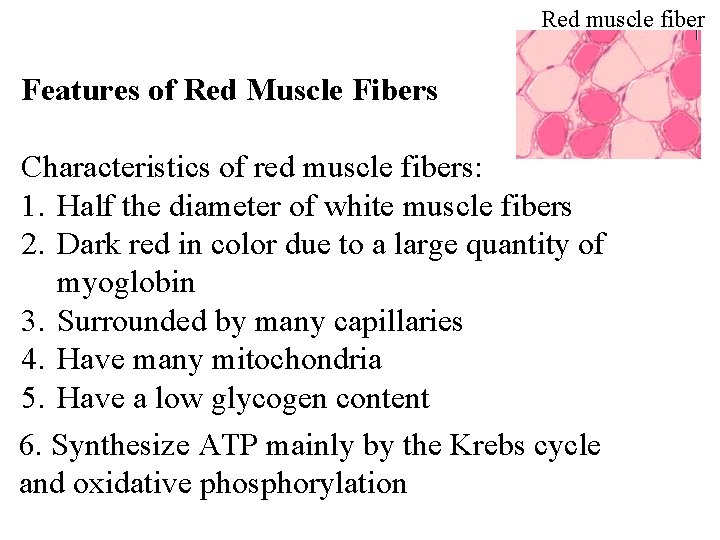
Red muscle fiber Features of Red Muscle Fibers Characteristics of red muscle fibers: 1. Half the diameter of white muscle fibers 2. Dark red in color due to a large quantity of myoglobin 3. Surrounded by many capillaries 4. Have many mitochondria 5. Have a low glycogen content 6. Synthesize ATP mainly by the Krebs cycle and oxidative phosphorylation
Factors Affecting Muscle Tension Three factors which affect muscle tension in a whole muscle: 1. Frequency of stimulation 2. Number of motor units recruited 3. Degree of muscle stretch
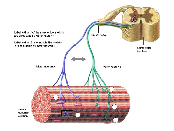
Contraction of Motor Units 1. To examine the components of a motor unit. 2. To understand the relationship between motor unit size and precision of muscle movement. 3. To explore the relationship of motor units to muscle tone.
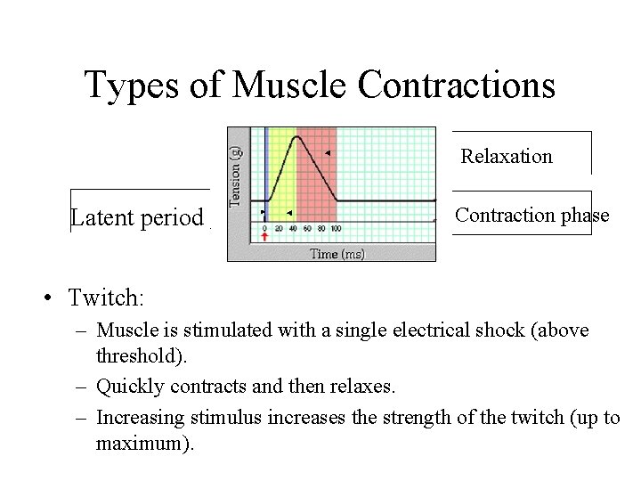
Types of Muscle Contractions Relaxation Latent period Contraction phase • Twitch: – Muscle is stimulated with a single electrical shock (above threshold). – Quickly contracts and then relaxes. – Increasing stimulus increases the strength of the twitch (up to maximum).
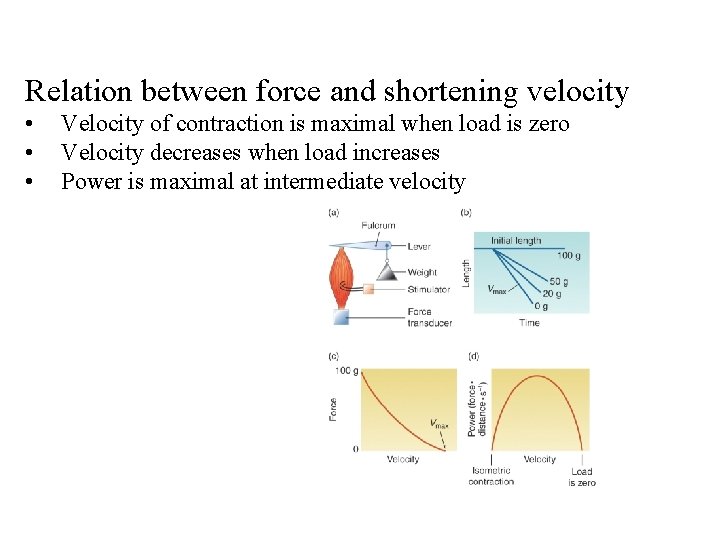
Relation between force and shortening velocity • • • Velocity of contraction is maximal when load is zero Velocity decreases when load increases Power is maximal at intermediate velocity
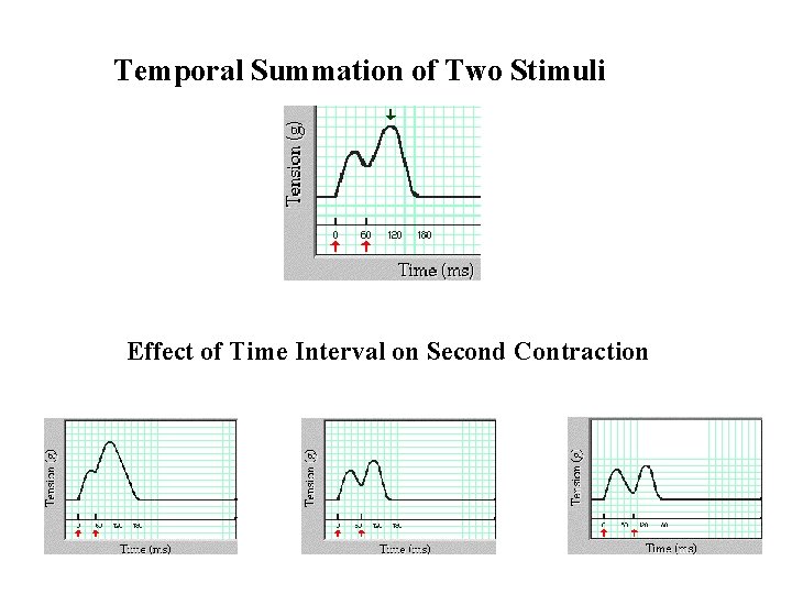
Temporal Summation of Two Stimuli Effect of Time Interval on Second Contraction
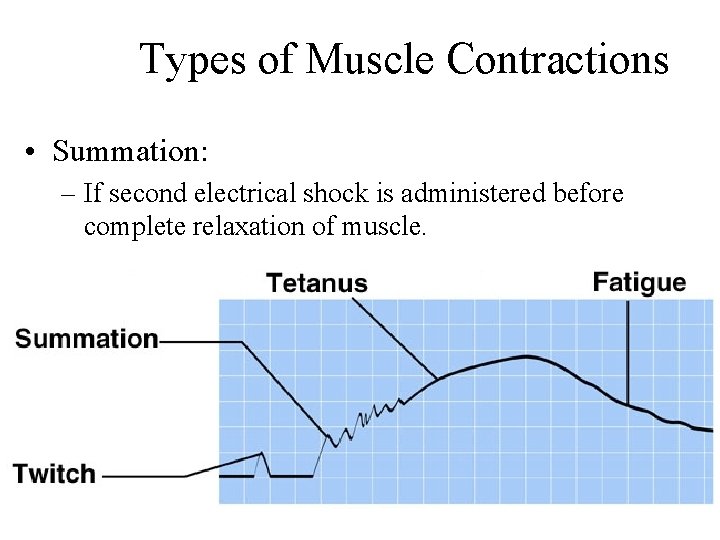
Types of Muscle Contractions • Summation: – If second electrical shock is administered before complete relaxation of muscle.
Types of Muscle Contractions • Incomplete tetanus: – Stimulator delivers an increasing frequency of electrical shocks. – Relaxation period shortens between twitches. – Strength of contraction increases. • Complete tetanus: – Fusion frequency of stimulation. – No visible relaxation between twitches. – Smooth sustained contraction.
Second & Third Factor Affecting Muscle Tension Three factors which affect the development of muscle tension: 1. Frequency of Stimulation 2. Number of Motor Units Recruited 3. Degree of Muscle Stretch
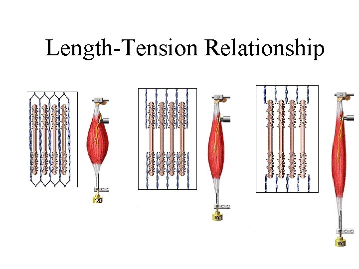
Length-Tension Relationship
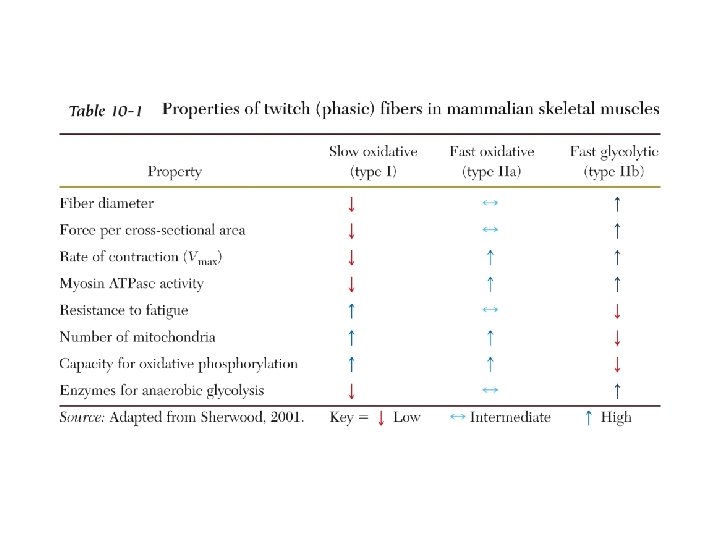
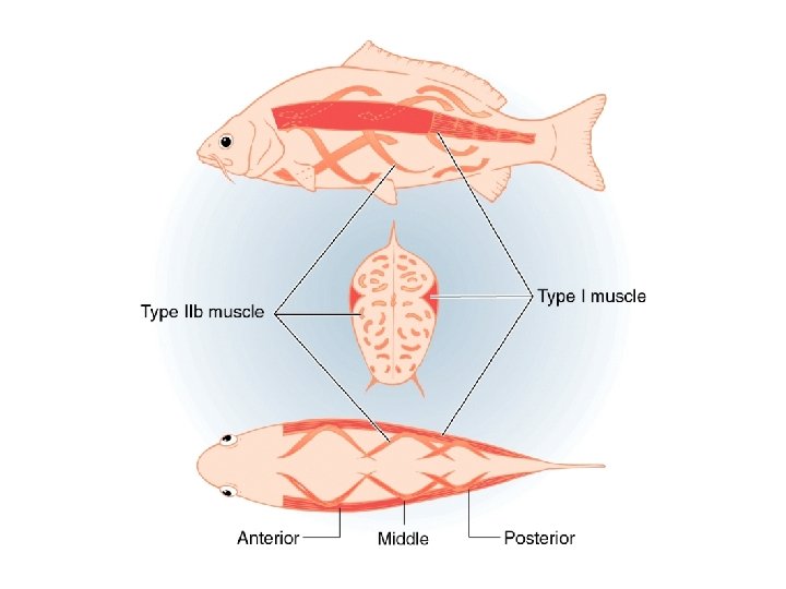
Slow- and Fast-Twitch Fibers • Slow-twitch (type I fibers): – High oxidative capacity: – Resistant to fatigue. – Have rich capillary supply. – Numerous mitochondria and aerobic enzymes. – Use ATP slowly – Red muscle (dark-colored meat of fish and fowl)
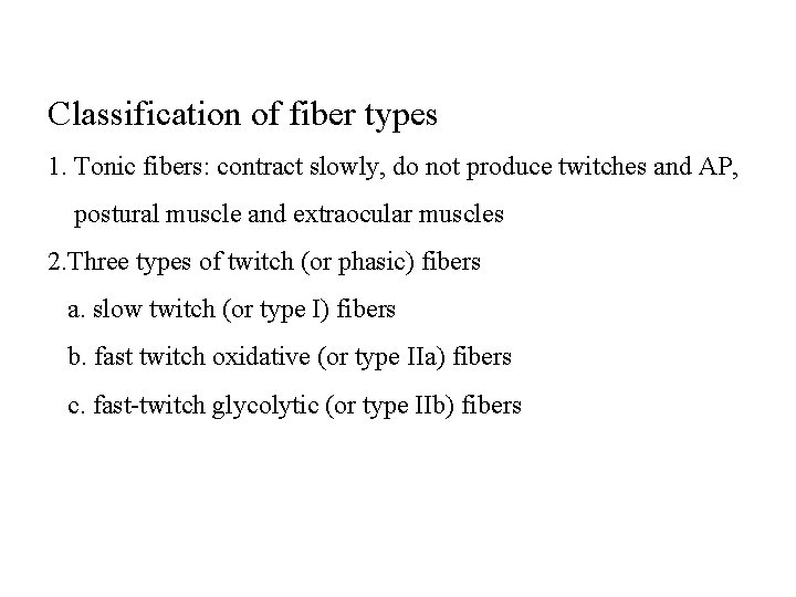
Classification of fiber types 1. Tonic fibers: contract slowly, do not produce twitches and AP, postural muscle and extraocular muscles 2. Three types of twitch (or phasic) fibers a. slow twitch (or type I) fibers b. fast twitch oxidative (or type IIa) fibers c. fast-twitch glycolytic (or type IIb) fibers
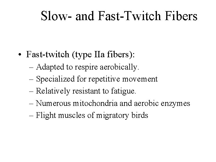
Slow- and Fast-Twitch Fibers • Fast-twitch (type IIa fibers): – Adapted to respire aerobically. – Specialized for repetitive movement – Relatively resistant to fatigue. – Numerous mitochondria and aerobic enzymes – Flight muscles of migratory birds
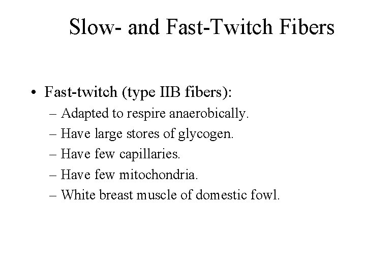
Slow- and Fast-Twitch Fibers • Fast-twitch (type IIB fibers): – Adapted to respire anaerobically. – Have large stores of glycogen. – Have few capillaries. – Have few mitochondria. – White breast muscle of domestic fowl.
Ration for different fiber types Fast-twitch fibers: rapid movement of its limb or fins, use large amount of ATP. Slow muscles: move slowly, use less amount of energy
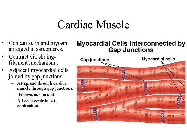
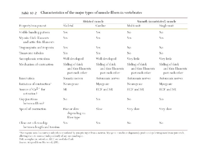
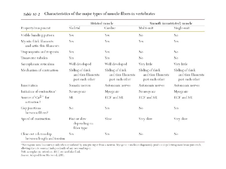
Cardiac Muscle • Contain actin and myosin arranged in sarcomeres. • Contract via slidingfilament mechanism. . • Adjacent myocardial cells joined by gap junctions. – AP spread through cardiac muscle through gap junctions. – Behaves as one unit. – All cells contribute to contraction.
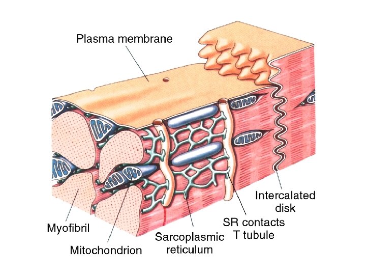
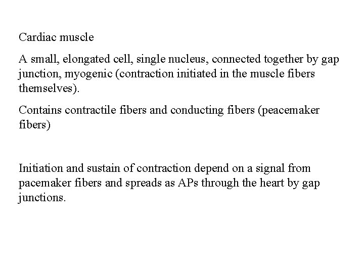
Cardiac muscle A small, elongated cell, single nucleus, connected together by gap junction, myogenic (contraction initiated in the muscle fibers themselves). Contains contractile fibers and conducting fibers (peacemaker fibers) Initiation and sustain of contraction depend on a signal from pacemaker fibers and spreads as APs through the heart by gap junctions.
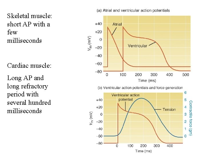
Skeletal muscle: short AP with a few milliseconds Cardiac muscle: Long AP and long refractory period with several hundred milliseconds
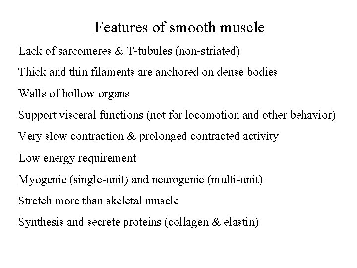
Features of smooth muscle Lack of sarcomeres & T-tubules (non-striated) Thick and thin filaments are anchored on dense bodies Walls of hollow organs Support visceral functions (not for locomotion and other behavior) Very slow contraction & prolonged contracted activity Low energy requirement Myogenic (single-unit) and neurogenic (multi-unit) Stretch more than skeletal muscle Synthesis and secrete proteins (collagen & elastin)
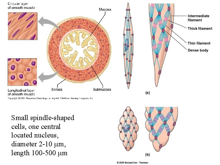
Small spindle-shaped cells, one central located nucleus, diameter 2 -10 mm, length 100 -500 mm
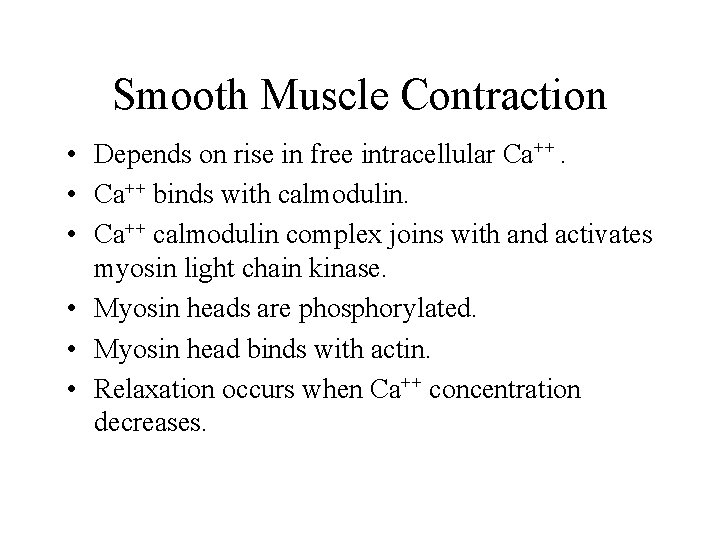
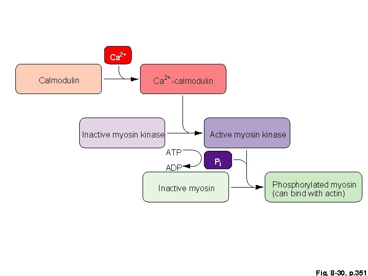
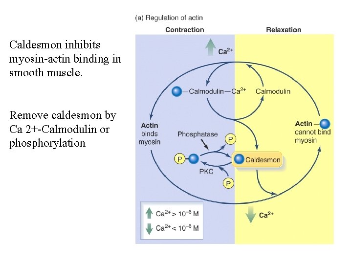
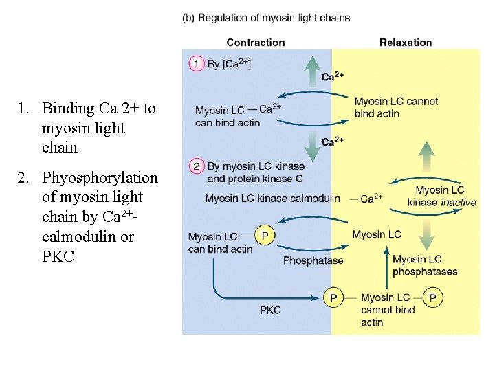
Smooth Muscle Contraction • Depends on rise in free intracellular Ca++. • Ca++ binds with calmodulin. • Ca++ calmodulin complex joins with and activates myosin light chain kinase. • Myosin heads are phosphorylated. • Myosin head binds with actin. • Relaxation occurs when Ca++ concentration decreases.
Ca 2+ Calmodulin Ca 2+ -calmodulin Inactive myosin kinase ATP ADP Pi Inactive myosin Phosphorylated myosin (can bind with actin) Fig. 8 -30, p. 351
Caldesmon inhibits myosin-actin binding in smooth muscle. Remove caldesmon by Ca 2+-Calmodulin or phosphorylation
1. Binding Ca 2+ to myosin light chain 2. Phyosphorylation of myosin light chain by Ca 2+calmodulin or PKC