Excitation contraction coupling Excitationdepolarization of muscle fiber Development
 of muscle fiber Development of AP (electrical phenomenon) ca ++")
Excitation –contraction coupling Excitation(depolarization) of muscle fiber Development of AP (electrical phenomenon) ca ++ Muscular contraction (mechanical phenomenon)

Steps in excitation contraction coupling propagation of AP in the motor axon release of Ach into synaptic cleft Ach binds with Ach receptors development of EPP depolarization of muscle membrane generation of MAP inward spread of MAP along t-tubules

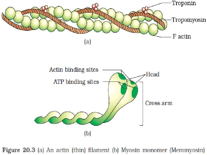
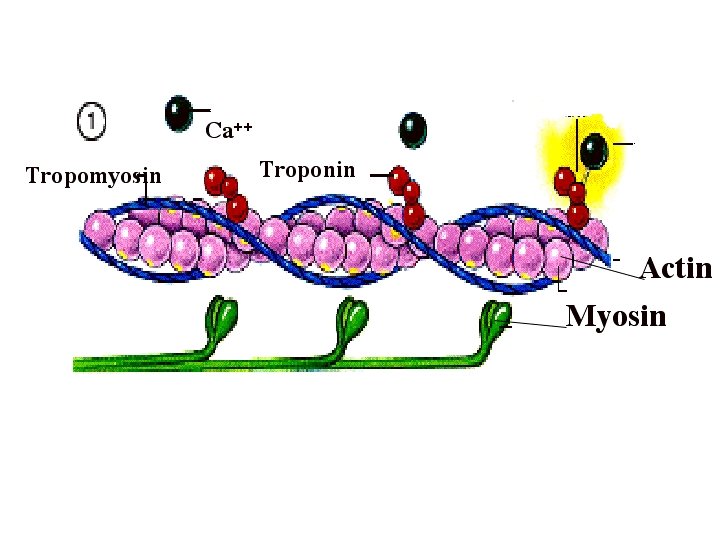
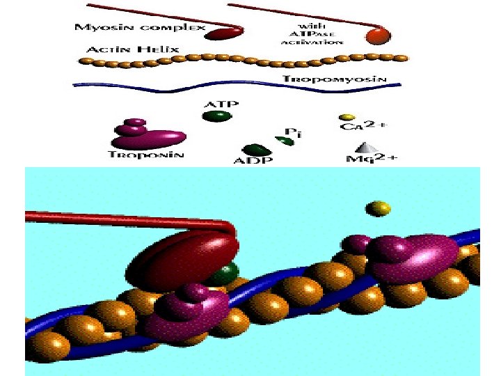
Spread of depolarization to terminal cisterns release of Ca ++ binds to troponin c tropomysosin move laterally uncovers the binding site for myosin head on activates ATPase provides the energy for cross bridge actomysosin complex and sliding filament mechanism.

Steps in E-C Coupling: Sarcolemma Voltage-sensitive tubule protein T tubule 1 Action potential is propagated along the sarcolemma and down the T tubules. Ca 2+ release channel 2 Calcium ions are released. Terminal cisterna of SR Ca 2+ Actin Troponin Ca 2+ Tropomyosin blocking active sites Myosin 3 Calcium binds to troponin and removes the blocking action of tropomyosin. Active sites exposed and ready for myosin binding 4 Contraction begins Myosin cross bridge The aftermath 5 Figure 9. 11, step 2

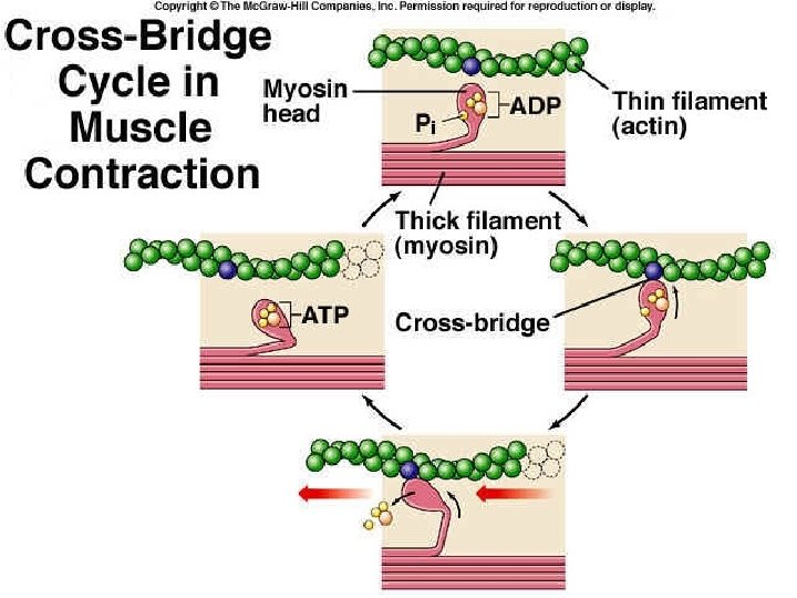
Molecular basis of muscle contraction • Cross bridge cycle v ATP molecule binds to myosin head and ATPase get activated causing the split of ATP to ADP+Pi v myosin head is positioned at an angle of 90 ◦ pointing towards actin but not attached to it. v Thus the myosin head is energized as the hydrolyze product of ATP remain attached to it. v. Thus most of the cross bridges are energized state keeping the muscle in relaxed state.

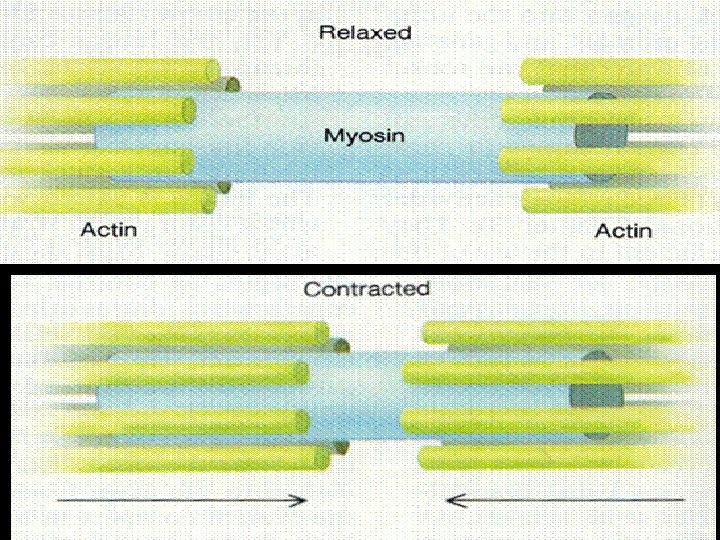
Sliding filament mechanism • Due to release of ca ++ from terminal cistern , the myosin binding site on actin is exposed. • Actomyosin complex is formed • The hydrolyze product of ATP is released which causes conformational change in myosin head which is now bend at an angle of 45 degrees. . • This causes power stroke movement that leads to the movement of thin filament towards centre of sarcomere. • This causes shortening of muscle by 1%. • Complete contraction of muscle by repeating the cycle.
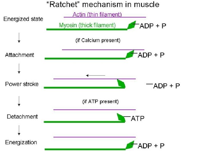

- cross-bridges attach to thin filament pull towards center detach attach")
• Huxley (1969)- cross-bridges attach to thin filament pull towards center detach attach further down ratchet theory or walk-along theory
 ADP+Pi Cross bridge dissociated low actin afffinity relaxation ADP+P Cross bridge")
High actin affinity(actin+myosin) ADP+Pi Cross bridge dissociated low actin afffinity relaxation ADP+P Cross bridge Actomyosin ADP pi contraction Actomyosin Absence of ATP causes Rigor mortis

Muscle relaxation sarcoplasmic Ca++ level Activation of Ca++ Mg 2+ Atpase Ca 2+ pumped into SR sarcoplasmic Ca 2+ level Detachment of Ca 2+ from troponin inhibit actin-myosin interaction muscle relaxation

• Rigor mortis • • Absence of ATP causes of binding of myosin with actin Cross bridge is not broken Leads to stiffening of muscle called rigor mortis. Observed after death
- Slides: 18