Mid brain cerebellum By Dr Amjed Hassan midbrain

Mid brain & cerebellum By Dr. Amjed Hassan

midbrain

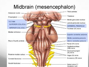
Gross A ppearance of M i dbrai n: • connectsthe pons and cerebell um wi th the f orebrain. • Its long axis ascends through theopening in the tentorium cerebell i. • The midbrain is traversed by a narrow channel, the cerebral aqueduct, whi ch is filled wi th cerebrospinal fluid

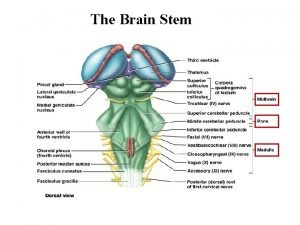
posterior surface 1. Four colliculi These are rounded eminences that are divi ded by a vertical and a transverse groove into : • Superi or colliculi : are centers for vi sual refl exes • I nferi or colliculi : are lower auditory centers.

2. Trochlear nerves : emerge In the midline below the i nferi or colliculi, (These are slender cranial nerves that wi nd around the lateral aspect of the midbrain to enter the l ateral wall of the cavernous sinus).

• On the lateral aspect of the midbrain, 3. Superior brachium passes f rom the superior colliculus to the lateral geniculate body and the opti c tract. 4. I nferi or brachium connects the inferior colliculus to the medial geniculate body.

A nterior aspect 1. there is a deep depression in the midline, call ed : I nterpeduncular f ossa, 2. Thi s depression is bounded on either side by the : Crus cerebri. M any small blood vessels perforate the floor of the i nterpeduncular f ossa, and this region is termed the : Posterior perforated substance

3. The occulomotor nerve emerges f rom a groove on themedial side of the crus cerebri and passes f orward in the l ateral wall of the cavernous sinus.

A rteri al supply: is supplied by: 1. Posterior cerebral 2. Superior cerebell ar 3. Basil ar arteries. Venous drainage : i nto the basal or great cerebral veins

I nternal Structure Of M i dbrain

Level I nferior colliculi Cavity Cerebral aqueduct Nuclei Inferi or coll i culus, Substantia nigra, Trochlear nucleus, M esencephali c nuclei of cranial nerve V Motor Tracts Sensory Tracts Corti cospinal and corti conuclear tracts, Temporopontine, Frontopontine, M edial l ongitudinal f asciculus, L ateral, tri geminal, spinal, and medial l emnisci; decussati on of superior cerebell ar peduncles

Nucl i e: 4. M esencephali c nuclei of cranial nerve V 1. I nferi or coll i culus, 2. Trochlear nucleus, 3. Substantia ni gra,

M otor Tracts: 1. Temporopontine 2. Corti cospinal & corti conuclear 3. Frontopontine, 4. M edial l ongitudi nal f asciculus

Se nsory tracts 1. Lemnisci ( Lmn. ) L ateral L mn. Tri geminal Lmn Spinal L mn. M edial L mn. Cerebral aqueduct 2. Decussati on of superior cerebell ar peduncles

Level Superior colliculi Cavity Cerebral aqueduct Nuclei Superior coll i culus, substantia nigra, Oculomotor nucleus, Edinger-Westphal nucleus, red nucleus, M esencephali c nucleus of cranial nerve V Motor Tracts Corti cospinal and corti conuclear tracts, temporoponti ne, f rontopontine, medial l ongitudi nal f asciculus, decussati on of rubrospinal tract Sensory Tracts Tri geminal, spinal, and medial l emnisci

Cerebral aqueduct

Nucl i e: 1. Superior coll i culus, 2. M esencephali c nucleus of cranial nerve V 2. Oculomotor nucleus, 3. Edinger-Westphal nucleus, 4. Red nucleus 5. Substantia nigra,

M otor Tracts: 1. Temporopontine 2. Corti cospinal & corti conuclear 3. Frontopontine, 5. M edial l ongitudi nal f asciculus 4. Decussati on of rubrospinal tract

Se nsory tracts Lemnisci ( Lmn. ) Tri geminal Lmn Spinal L mn. M edial L mn.

cerebellum

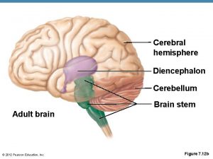
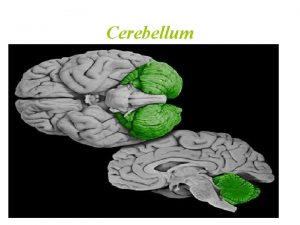
Defi niti on: • The trilobed structure of the brain, lying posterior to the pons and medulla oblongata and i nferior to occipital lobes of the cerebral hemispheres, thus it lies in the posterior cranial f ossa. • Responsible f or theregulation and coordination of complex vol untary muscular movements and the maintainence of postureand balance

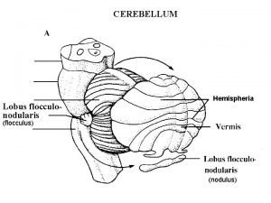
Gross A ppearance of the Cerebell um • situated in theposteri or crani al f ossa • covered superiorl y by thetentori um cerebell i • lies posterior to the fourth ventricle, the pons, and the medulla oblongata • i s somewhat ovoi d in shape and constricted in its median part.

It consists of: 1. two cerebell ar hemispheres 2. Vermis : joining both hemispheres.

Connected to posterior aspect of the brainstem by three symmetrical bundles of nerve fibers called the: 1. Superior cerebell ar peduncle 2. Middle cerebellar peduncle 3. inferior cerebell ar peduncle

The cerebell um is divi ded i nto three main l obes: 1. Anterior lobe : may be seen on the superior surface of the cerebell um and is separated f rom the middle lobe by a wide V -shaped f i ssure call ed the primary f i ssure.

2. Middle lobe : Superior veiw (sometimes call ed the posterior l obe), whi ch is the l argest part of the cerebell um, is situated between the primary and posterolateral f i ssures. • • • Fl occul onodul ar l obe: is situated posterior to the posterolateral f i ssure. Formed by two f l occuli and the nodule I nferi or veiw

Tonsils • Are roughly spherical lobules on the inferior aspect of posterior l obe. • The tonsil may be displaced down through thef oramen magnum in conditi ons of severeraised i ntracranial pressureor in congenital malf ormations

• hori zontal fissure : that is found along the margin of the cerebell um separates the superior f rom the inferior surf aces.

The vermis • consists of ; A. Superior part B. I nferi or part • Superior Vermis lies between superior medull ary velum & pri mary f i ssure • I s composed of: 1. Li ngula 2. Central l obule 3. Culmuen

• Inferior Vermis lies between pri mary fissure and posterol ateral f i ssure, and consists of : 1. Decli ve 2. Foli um 3. Tuber 4. Pyramid 5. Uvul a 6. Nodule

l ongi tudi nal di vi sion: 1. Vermis (medial zone) 2. Paravermal Region ( I ntermediate zone) 3. Cerebell ar Hemisphere: (L ateral zone)

Arterial s upply of The cerebellum is by: 1. Superior cerebell ar 2. A nterior i nferi or cerebell ar, 3. Posterior i nferi or cerebellar Ve nous drainage by veins that empty i nto the • Great cerebral vein • V enous sinuses.

Intracerebe llar Nuclei • Four masses of gray matter are embedded in the white matter of the cerebell um on each side of the midline. From l ateral to medial, these nuclei are: 1. Dentate nucleus, 2. Emboli f orm nucleus, 3. Globose nucleus, 4. Fastigial nucleus.

Fi stugial nucleus Globose nucleus Emboli f orm nucleus Dentate nucleus 4 th Ventri cle Pons

A fferent Cerebell ar Pathways

1. Cerebellar Afferent Fibers From the Cerebral Cortex I nformation regarding the initiation of movement in the cerebral cortex is probably transmitted to the cerebell um so that the movement can be monitored and appropriate adjustments in the voluntary muscle activity can be made.

Pathway Function Ori gin Destination 1. corticoponto cerebellar Conveys control Frontal, parietal, Via pontine signals from temporal, and nuclei to cerebral cortex occipital l obes cerebell ar cortex 2. cerebroolivocerebellar Conveys control Frontal, parietal, Via inferior signals from temporal, and ol i vary nuclei to cerebral cortex occipital l obes cerebell ar cortex 3. cerebroreticulocerebellar Conveys control signals from cerebral cortex Sensori motor areas Via reticular f ormation to cerebell ar cortex

Corti copontocerebell ar pathway Corti coreticulocerebell ar pathway Corti co-oli vocerebell ar pathway Reticular f ormation I nferi or oli vary nucleus Pontine nucli e

2. Cerebellar Afferent Fibers From Spinal Cord • The spinal cord sends i nformation to the cerebell um f rom somatosensory receptors by three pathways: (1) the anterior spinocerebell ar tract: is found at all segments of the spinal cord, and its fibers convey muscle joint i nformation f rom theupper and l ower limbs (2) the posterior spinocerebell ar tract: receives muscle joint i nformation f rom thetrunk and l ower l i mbs. (3) the cuneocerebell ar tract: receives muscle joint i nformation f rom theupper limb and upper part of the thorax

3. Cerebellar Afferent Fibers From The Vestibular Nerv e • The vestibular nerve receives i nformation f rom the inner ear concerning: A. M oti on f rom the semicircular canals B. positi on relative to gravity f rom: Utri cle Saccule.

4. Other Afferent Fibers • In additi on, the cerebell um receives small bundles of aff erent fibers from: 1. the red nucleus 2. the tectum.

Superior cerebell ar peduncle I nf erior cerebell ar peduncle Cerebell um I nf erior cerebell ar peduncle Medulla A nterior spinocerebell ar tract (mi nority of f i bers) V estibular nucli e Dentate nucleus Nucleus cunatus V estibular nucleus Posterior spinocerebell ar tract A nterior spinocerebell ar tract (majori ty of f i bers)

The Efferent Cerebell ar Pathways

Pathway Function I nfl uences Globosei psil ateral emboliform motor -rubral activi ty Origin Destination contralateral red nucleus, then via crossed Globose and rubrospinal emboli f orm tract to nuclei i psil ateral motor neurons in spinal cord

Pathway Function Origin Destination dentothalamic contralateral ventrol ateral nucleus of thalamus, I nfl uences contralateral motor i psil ateral Dentate cerebral cortex; motor nucleus corti cospinal tract activi ty crosses midline and controls i psil ateral motor neurons in spinal cord

Pathway Function Origin Destination Mainly to i psil ateral and to contralateral I nfl uences l ateral vestibular i psil ateral fasti. Gial Fastigial nuclei; vestibuloextensor vestibular nucleus spinal tract to muscle i psil ateral motor tone neurons in spinal cord

Pathway Function Origin Destination neurons of reticular I nfl uences f ormation; reticulofasti. Gial i psil ateral Fastigial spinal tract to ipsireticular muscle nucleus l ateral motor neurons tone to spinal cord

Thanks
- Slides: 48
