Cerebellum Dr K Shankar Bhat Sr Prof and

Cerebellum Dr K Shankar Bhat. Sr. Prof and HOD Dept of Physiology Yenepoya Medical College

Cerebellum Controls Subconscious Skeletal Muscle Contractions Required For: 1. Co Ordination 2. Posture 3. Equilibrium/ Balance

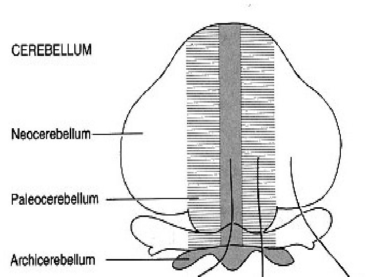
DIVISIONS OF CEREBELLUM

1. ARCHICEREBELLUM: flocculonodular lobe connections with vestibular apparatus and vestibular nuclei. Function : Maintenance of Equilibrium & Eye Movement. 2. PALEOCEREBELLUM: Anterior lobe Function: Concerned with the regulation of Toner & Posture. 3. NEOCEREBELLUM: It is of recent origin best developed in man, connected with cerebral cortex. Function: Co-ordinates every type of motor activities.

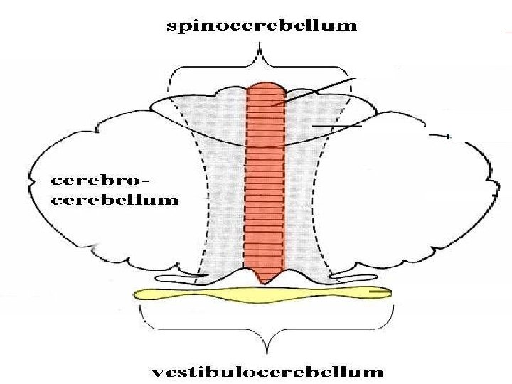
Functional divisions of cerebellum

1. Vestibulocerebellum: Concerned with balance and eye movements. Connected is vestibular apparatus – vestibulonuclei and reticular nuclei. 2. Spinocerebellum: Concerned with motor executions. Connected to spinal cord and descending system. 3. Cerebrocerebellum: Concerned with motor planning and initiation of movements. Connected with the cerebral cortex.

Vestibulo-cerebellum Superior cerebellar peduncle 1. Vestibulo cerebellum pons flocculonodular cortex Inferior cerebellar peduncle Vestibular system Functions: Regulation of gait and posture and equlibrium Coordination of head movements with eye movements (vestibular nuclei & vestibular nerve)

Spino-cerebellum red nucleus 2. Spinocerebellum Superior cerebellar peduncle pons reticular formation nuclei Function: Speech Coord’n Muscle Tone Adjustment Limb movement Coord’n Nuclei Fastigial Interposed Inferior cerebellar peduncle Dorsal & Ventral Spinocerebellar tracts (Somatosensory inputs Reticulo-spinal and Rubrofrom proprioceptors and spinal tracts exteroceptors) (to spinal cord-motor neurons) Somatotopic organization in the cerebellum cortex spinal cord
")
Cerebro-cerebellum 3. Cerebrocerebellum Cerebral cortex (MI, SMA, S 1, PP & primary visual cortex) thalamus Superior cerebellar peduncle Function: Coordination of skilled movements Tactics of immediate movements Cognitive function Attention, processing of language Emotional control Reticular formation & pontine nuclei pons Dentate nucleus Middle cerebellar Peduncle inferior olive nuclei Inferior cerebellar peduncle
 Control of limbs and trunk Cerebrocerebellum (Lateral")
Cerebellar divisions Spinocerebellum (Vermis + Intermed. Hem) Control of limbs and trunk Cerebrocerebellum (Lateral hemisphere) Planning of movement Vestibulo-cerebellum (Floculo-nodular lobe) Spinocerebellum: Vermis Intermediate hem. Cerebrocerebellum: Lateral hem. IVth vent Vermis Intermediate hem. Lateral hem. Control of eye & head movements Balance Floculo-nodular lobe
 Input Vestibular sys")
Vestibulocerebellum Spino-cerebellum Cerebro-cerebellum Age/ Region Oldest Incl vermis Newest (Lateral lobe) Input Vestibular sys (INF) Dorsal & Ventral Spinocerebellar tracts (INF) Reticular form’n & pontine N. (MED) Inf olive N. (INF) Functions n Gait, posture & eqbm n Coordinate head w/ eye movements n Coordinate speech n Adjust muscle tone n Coordinate limb movements n Output Vestibular sys (INF) (i) Vermis Fastigial N. (ii) Intermediate Interposed N. Dentate N. & Cortex Skilled motion n Immediate motion n. Cognitive fxn n. Attn, Lang n. Emotional control

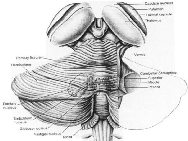
The white matter of cerebellum consists: 1. Tracts. 2. Four pairs of nuclei (Deep nuclei) § Fastigial § Globosus – globose § Emboliformis – inter positus § Dentatus

Internal structures Cerebellar cortex Fastigial nucleus Globose nucleus Dentate nucleus medullary center Emboliform nucleus

Connections of cerebellum

Afferents: Inputs to cerebellum 1. 2. 3. 4. 5. 6. 7. Dorsal spino cerebellar tract Ventral spinocerebellar tract Olivo cerebellar tract Tectocerebellar tract Vestibulocerebellar tract Cuneocerebellar tract Pontocerebellar tract

Above afferents bring NONCONSCIOUS Sensory inputs from Muscle, Joint Proprioceptors and the skin. Thus cerebellum gains information about: 1. Degree and distribution of tone 2. Position of limbs in space 3. Body posture
 2) 3) 4) Dentato – Rubro- Thalamo cortical pathway")
Efferents: Outputs from cerebellum 1) 2) 3) 4) Dentato – Rubro- Thalamo cortical pathway Dentato- Vestibulo- spinal tract Dentato – reticulospinal tract Cerebello- pontine tract

The efferent fibers arise from the Deep cerebellar nuclei. 1. 2. 3. 4. Nucleus Globosus Nucleus Emboliformis Nucleus Fastigius Nucleus Dentatus Deep Nuclei receive signals from 2 sources: 1. Cerebellar cortex 2. Sensory tracts (Afferents) of cerebellum

Cytoarchitecture of cerebellar cortex and cerebellar circuitry

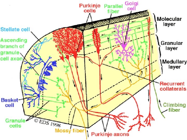
Cerebellar cortex, shows 3 layers: 1. Molecular layer 2. Purkinje layer 3. Granule cell layer

There are 5 types of neurons: 1. Basket cell 2. Stellate cell 3. Golgi cell These neurons are inhibitory interneuron's which release GABA. Their activity regulates the functions of 4. Purkinje and 5. Granule cells The neurotransmitter for excitatory output from cerebellar nucleus is GLUTAMATE.

LAYERS AND CELLS OF THE CEREBELLAR CORTEX

Cerebellar Cortex Inputs Climbing fibers • from Inferior olive Mossy fibers Output Purkinje neurons Interneurons Granule neurons Stellate neurons Basket neurons Molecular Purkinje Granular NTA Fig. 13 -11 Golgi neurons

CEREBELLAR CIRCUITRY:

Neuronal circuit formed by: • • Basket cell. satellite cell. Golgi cell. purkinje cell. granule cell. climbing fibers. mossy fibers. parallel fibers in cerebellar cortex.
")
The function of cerebellar circuitry is MODULATING OR TIMING the excitatory output (Efferent impulses) of the deep cerebellar nuclei to the brain stem and Thalamus.

Cerebellum is connected to brainstem through 1. Superior peduncle 2. Middle peduncle 3. Inferior peduncle Afferents & efferent's pass through these.

Main Connections of the Vestibulocerebellum Vestibular Organ Floculonodular Lobe VESTIBULAR NUCLEUS Vermis vestibulospinal tract MLF lower motor neuron LMN FASTIGIAL NUCLEUS ARCHICEREBELLUM

Main Connections of Spinocerebellum RED NUCLEUS rubrospinal tract lower motor neuron SPINAL CORD NUCLEUS INTERPOSITUS Inferior Olivry Nucleus ANTERIOR LOBE PARAVERMAL ZONE SPINOCEREBELLUM spinocerebellar tract

Main Connections of the Neocerebellum CEREBRAL CORTEX pyramidal tract lower motor neuron LMN THALAMUS Pontine Nucleus DENTATE NUCLEUS POSTERIOR LOBE CEREBELLAR HEMISPHERE NEOCEREBELLUM

Functions of cerebellum

Cerebellum controls subconscious skeletal muscle contractions required for: 1. Co ordination 2. Posture 3. Equilibrium/ balancing. Controls Modulates Coordinates All movements

1. Maintenance of Normal Muscle tone-: by Ant. Lobe. α -γ linkage of stretch reflexes. 2. Maintenance of normal erect posture of body. Posture is defined as the active muscular resistance to displacement of the body by gravity or acceleration. This function is by the flocculonodular lobe.

3. Equilibrium or Balancing: During standing, running and jumping etc cerebellum helps maintain equilibrium by working in conjunction with the vestibular apparatus i. e. adjusting tone among various groups of muscles engaged in the particular act.

4. Synchronization of Movements: During any voluntary activity cerebellum synchronizes the firing of Protagonists, Antagonists And Synergists simultaneously. Thus it makes the voluntary movements Smooth, non oscillating and regular smooth, graceful voluntary movements.

5. Control of rate, range, force, speed and direction of movement for particular purpose. 6. Predictive function: Cerebellum predicts the future position of the body part during a particular movement. Eg. Walking.

7. Detection of errors in movements and its correction through cerebrum: Lesion produces pendular jerks. Cerebellum acts as a “ Comparator of servo mechanism”. A control system for maintaining the operation of another system. Thus cerebellum acts as a control system for the operation of the cerebrum.

8. Role in expression of emotions is exteriorization of emotion. 9. Helps in learning of new movements Eg. Driving with training.

Signs of cerebellar disease

Damage to Neocerebellum causes most of the dysfunctions. Characteristic disturbances of 1. Posture 2. Voluntary movements are seen. In Unilateral lesions signs are predominantly seen in on the same side of the body

Disturbances of posture 1. 2. 3. 4. 5. Atonia Attitude Spontaneous deviation Nystagmus Pendular deep reflexes

1. Atonia: Epsilateral Atonia muscles are flabby and the limbs swing about like a flail. 2. Attitude: Face rotated to the opposite side lateral shoulder is lower than its fellow. Leg is abducted and rotated outwards. Body weight is thrown on sound leg so that the trunk is bent with the concavity towards the affected side. 3. Spontaneous deviation: When the eyes are closed and the arms are held straight out in front of the body the Epsilateral arm sways laterally.

4. Nystagmus: Rhythmic oscillations of the eye balls pendular or Jerky. 5. Pendular deep reflexes: Knee jerk is characteristically pendular

Disturbances of voluntary movement: 1. 2. 3. 4. 5. Asthenia Ataxia Intention tremor Drunken gait Slow lalling speech.

1. Asthenia: Feeble movements, muscles tire readily, voluntary movements are carried out- very slowly. 2. Ataxia: Inco- ordination of movements, severe. unaffected by closing the eyes. a. Decomposition of movement occurs in stages. b. Asynergia: Lack of coordination between protagonists, Antagonists & synergists. c. Adiadokinesis: Difficulty in carrying out alternate movements quickly.
 during")
3. Intention tremor: Very conspicuous of Neocerebellar lesions. Coarse tremor (4 -6/5 ) during voluntary movement. Its cause is damage to Dentato- Thalamic pathway. ATAXIA can be demonstrated by: 1. Finger – Nose test 2. Adiadokokinesis: Rapid pronation & Supination of hands not possible.

4. Gait: Patient tends to deviate to the affected side and tries to correct- zig zag path. Drunken gait.

Posture Gait – Ataxia Tremor

a d b c Cerebellar Ataxia Ataxic gait and position: Left cerebellar tumor a. Sways to the right in standing position b. Steady on the right leg c. Unsteady on the left leg d. ataxic gait

Cerebellar Medulloblastoma Cerebellar tumors on vermis - Truncal Ataxia - Frequent Falling The child in this picture: - would not try to stand unsupported - would not let go of the bed rail if she was stood on the floor.

5. Speech: Slow and lalling. imperfect execution of the movements of laryngeal muscles and tongue. Nystagmus Intention tremor Lalling speech Charcot’s triad


Further details:

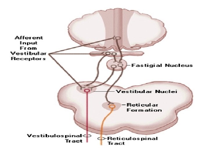
The Vestibulocerebellum Ø It receives input mainly from the vestibular system. Ø It is richly interconnected with UMNs of the medial brainstem motor pathway, especially those in the vestibular nuclei. Ø Some efferent fibers from the Vestibulocerebellum also end on reticulospinal neurons. Ø The pathways to the vestibular nuclei are via direct projections of Purkinje cell axons from the flocculonodular lobe and via efferent fibers from the Fastigial nucleus. Ø The Vestibulocerebellum provides for coordination of movements involving whole body equilibrium and posture.

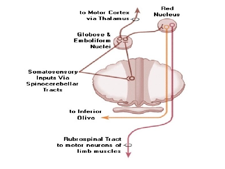
The Spinocerebellum Ø Its inputs are mainly from Proprioceptors and exteroceptors of the limbs via spinocerebellar pathways. Ø It is richly interconnected, via efferent fibers from the globose and emboli form nuclei (together called the interposed nucleus), with UMNs of the lateral brainstem motor pathway (the red nucleus). Ø Ø It also connects with reticulospinal neurons and sends information rostrally to the motor cortex via the ventrolateral (VL) nucleus of the thalamus. Ø The Spinocerebellum provides for coordination of limb movements for posture, progression, and other purposes. It also is important for "updating evolving movements, " a role it shares to some extent with the Cerebrocerebellum.

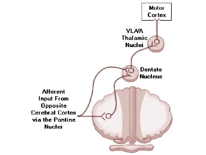
The Cerebrocerebellum ØIts inputs arrive mainly from cerebral cortex via the pontine nuclei. ØBecause of this fact it is also sometimes called the Pontocerebellar. ØIt is richly interconnected, mostly via the ventrolateral (VL) nucleus of the thalamus, with UMNs of the corticospinal motor pathway. ØThe Cerebrocerebellum provides for coordination of independent limb movement and skilled movement, especially in terms of preprogramming movements.

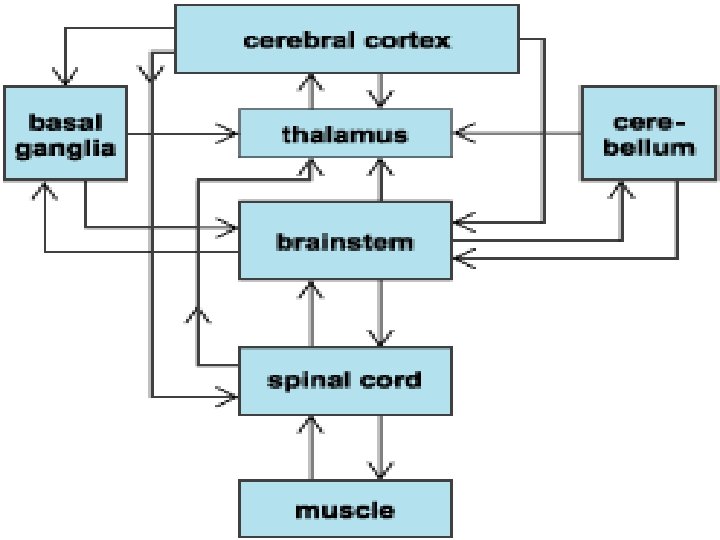
Cerebellum and Automatic Motor Control Motor Cortex CEREBELLUM Red Nucleus Reticular Formation Lower Motor Neuron (LMN) Vestibular Nucleus Proprioceptors

Pyramidal Tract and Associated Circuits upper motor neuron UMN BASAL GANGLIA Cerebellum pyramidal tract lower motor neuron UMN
- Slides: 66