Nervous system brain spinal cord Dr L Kiss

Nervous system: brain, spinal cord Dr. L. Kiss Anna Department of Anatomy, Histology and Developmental Biology, Semmelweis University 2018

Nervous system Central nervous system Spinal cord brain Peripheral nervous system Spinal nerves, cranial nerves, ganglions Functionally: somatic (axial muscles) vegetatíve: (internal organs)
 - voluntary - unvoluntary -")
Functional division of the nervous system Somatic Autonomic (vegetative) - voluntary - unvoluntary - starts and stops functions - regulates the functions (slow down-speed up) - segmental - network sympathetic thora-columbar parasympathetic cranio-sacral

Central nervous system: Spinal cord

Spinal cord: meningeal layers Dura mater: outer layer: endorachis Epidural spce inner layer Subdural space Arachnoid layer Subarachnoid space Pia mater

MENINGES Epidural space Subarachnoid space Denticulate ligament

Meningeal layers of the spinal cord ganglion spinale fila radicularia dorsalia arachnoidea dura mater ligamentum denticulatum

MENINGES Dura mater Arachnoid Pia mater: Denticulate ligament
 Lumbosacral enlargement (L 2 -S 3)")
MACROSCOPY Enlargements: Cervical enlargement (C 4 -T 1) Lumbosacral enlargement (L 2 -S 3) Conus terminalis Cauda equina Filum terminale Central nervous system Brain Spinal cord Peripheral nervous system Nerves Ganglia

cauda equina

Lumbal punction

Blood supply of the spinal cord

Blodd supply of the spinal cord radicular arteries vertebral artery subclavian artery Aortic arch radicular arteries pathway of the vertebral artery
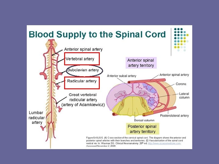
 • a spinalis posterior (2)")
Vasocorona medullaris Vertebral artery: • a. spinalis anterior (united) • a spinalis posterior (2) Radicular arteries: • cervical: vertebral a. ascending. cerv. a. deep cerv. a. • thoracal: intercostal aa. • lumbal: lumbal aa. • sacral: lateral sacr. art.

CROSS SECTION Dorsal median sulcus Gray matter White matter Ventral median fissure Central canal
 median sulcus post. (dorsalis)horn intermediate")
Cross section of the spinal cord post. (dors. ) median sulcus post. (dorsalis)horn intermediate zone (Th and L 2 -L 3): lat. horn central canal ant. (ventral) horn ant. (ventr. ) median fissure

SPINAL CORD SEGMENT

SPINAL NERVES Dorsal branch Ventral branch

SPINAL CORD SEGMENTS Spinal cord ends at the level of the L 1 vertebra in adults Lumbar and sacral roots travel several vertebrae down: cauda equina
 12 thoracic 5 lumbar 5")
SPINAL CORD SEGMENTS Number of segments: 8 cervical (!) 12 thoracic 5 lumbar 5 sacral 1 coccygeal

SEGMENTAL INNERVATION Dermatomes: segmental symptomes
: Phrenic nerve Branchial plexus")
CERVICAL PLEXUS AND BRACHIAL PLEXUS Cervical plexus (C 1 -4): Phrenic nerve Branchial plexus (C 5 -T 1): Superior trunk (C 5 -6) Middle trunk (C 7) Inferior trunk (C 8 -T 1)
: Lateral cord (C")
CERVICAL PLEXUS AND BRACHIAL PLEXUS Branchial plexus (C 5 -T 1): Lateral cord (C 5 -7) Medial cord (C 8 -T 1) Posterior cord (C 5 -T 1)
: Sacral plexus (L 4 -S 3):")
LUMBOSACRAL PLEXUS Lumbar plexus (T 12 -L 4): Sacral plexus (L 4 -S 3):

Brain
 Cerebellum Diencephalon (thalamus, hypophysis) Cerebrum Brain stem")
Brain Medulla oblongata Pons Midbrain (mesencephalon) Cerebellum Diencephalon (thalamus, hypophysis) Cerebrum Brain stem

MEDIANSAGITTAL SECTION OF THE BRAIN STEM M M Mo P Mo

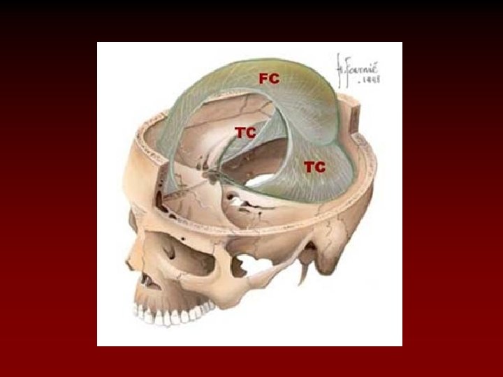
Meningeal layers of the brain Dura mater: falx cerebri, tentorium cerebelli, falx cerebelli sinuses: venous blood Aracnhoir layer: cisterns CSF Pia mater: blood vessels

VENTRICLES Lateral ventricle 3 rd ventricle 4 th ventricle

CEREBROSPINAL FLUID Production: choroid plexus: into ventricles Absorption: arachnoid villi: from subarachnoid space into superior sagittal sinus

BRAIN: Cerebrum SUPERIOR VIEW Frontal lobe Parietal lobe Occipital lobe Longitudinal cerebral fissure

BRAIN: LATERAL VIEW Parietal lobe Central sulcus Frontal lobe Occipital lobe Transverse cerebral fissure Lateral cerebral sulcus Temporal lobe Cerebellum Pons Medulla oblongata

BRAIN: MEDIANSAGITTAL CUT Longitudinal cerebral fissure Parietal lobe Corpus callosum Occipital lobe Frontal lobe Transverse cerebral fissure Temporal lobe Cerebellum

central sulcus praecentral gyrus postcentral gyrus
 Optic nerve (II)")
BRAIN: INFERIOR VIEW Longitudinal cerebral fissure Frontal lobe Olfactory nerve (I) Optic nerve (II) Optic chiasm Temporal lobe Optic tract Diencephalon Pons Medulla oblongata Cerebellum Occipital lobe

BLOOD SUPPLY: ARTERIES Arterial supply: Vertebral artery Internal carotid artery internal carotid artery vertebral artery

CIRCLE OF WILLIS Anterior communicating a. Anterior cerebral a. Internal carotid a. Middle cerebral a. Posterior communicating a. Posterior cerebral a. Basilar a. Vertebral a.

BLOOD SUPPLY: VEINS Venous drainage: Internal jugular vein: Sinuses Cerebral veins
 Olive Pyramid Vagus n. (X) Accessory n. (XI)")
BRAIN STEM: MEDULLA Glossopharyngeal n. (IX) Olive Pyramid Vagus n. (X) Accessory n. (XI) Hypoglossal n. (XII)

BRAIN STEM Cerebral aqueduct Cerebellum Midbrain Pons Fourth ventricle Medulla Central canal

CEREBELLUM Vermis Tonsil Cerebellar hemisphere

CEREBELLUM Fastigial nucleus Cerebellar cortex Globose nucleus Emboliform nucleus White matter Dentate nucleus

CEREBELLUM superior cerebellar peduncle midbrain middle cerebellar peduncle pons inferior cerebellar peduncle medulla

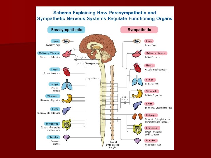
Functional division of the nervous system Somatic Autonomic - voluntary - unvoluntary - starts and stops functions - regulates the functions (slow down-speed up) - segmental - network sympathetic thora-columbar parasympathetic cranio-sacral

Autonomic nervous system: efferent part Sympathetic Parasympathetic - thoracolumbar segments - craniosacral region - speeds up the functions (except for digestive system) - slows down the functions (except for the digestive system) - empties the stores - filles in the stores - adrenalin, noradrenalin (sympatho-adrenal system) - acetylcholine - last longer - shorter effect -general effect - local effect -short praeganglionic fibers -longer postganglionic fibers shorter postganglionic fibers

Dr. Nándor Nagy picture

References Lecture and pictures of Dr. Nandor Nagy Sobotta atlas Wikipaedia
- Slides: 51