Microscopical structure of the spinal cord Rexed zones


 Pacinian corpuscle Concentrically arranged layers of modified Schwann cells around nerve endings. Are")


 C-fibres unmyelinated, Aδ-fibres thin myelin sheath Aα, Aβ-fibres thick myelin sheath, sensory ganglion")


 CNS directly controls skeletal and visceral striated muscles through the")


")


- Slides: 34
Microscopical structure of the spinal cord, Rexed zones. Receptors and effectors, spinal reflexes, proprioceptive reflex arc. http: //siennaartstudios. com/shop/thermal-dols-spinal-cord-nociceptors Mark Kozsurek, M. D. , Ph. D. mark@kozsurek. hu EM II 9 -17, 24 Sept,
INPUT: sensory fibres OUTPUT: motor fibres Terms: white and gray matter, spinal nerve, dorsal and ventral roots, spinal nerves, spinal ganglion (dorsal root ganglion -
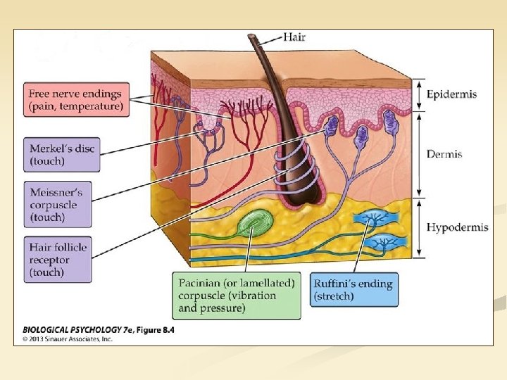
1. Where does the information come from? RECEPTORS ancient - free nerve endings • invade the epithelium of the skin and mucosa, occure in the walls of viscera • no highly specialized structures around the peripheral processes of sensory neurons • pain and temperature, with relatively high threshold relatively new - specialized structures • • • rather in the dermis and subcutis, in joints, muscles and tendons complex structures around the peripheral nerve endings composed of different cell types including modified Schwann-cells low threshold for touch, pressure, vibration, stretch
Meissner’s corpuscle Lamellar structure formed by modified Schwann cells around nerve endings. Are found in the conn. tissue papillae of the skin.
(Vater-) Pacinian corpuscle Concentrically arranged layers of modified Schwann cells around nerve endings. Are found in dermis or even deeper in the subcutis. (Can not only occur in the skin but also in
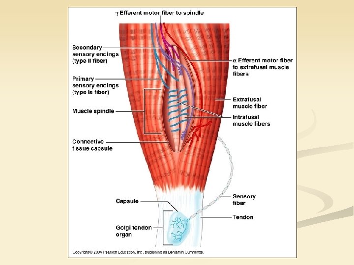
Muscle spindle
2. How does the information reach the spinal cord? NERVE FIBRES Please note, that even the unmyelinated axons are supported by Schwann cells but in a different
) C-fibres unmyelinated, Aδ-fibres thin myelin sheath Aα, Aβ-fibres thick myelin sheath, sensory ganglion with pseudounipolar neurons
3. What happens in the spinal cord? Information is processed, decision is made and commands are sent to the effectors. brain viscerosensory visceromotor spinal cord somatosensory skin, muscles, joints, tendons internal organs somatomotor skeletal muscles smooth muscles, glands
3. What happens in the spinal cord? Information is processed, decision is made and commands are sent to the effectors. brain viscerosensory visceromotor spinal cord SENSORY GGL. skin, muscles, joints, tendons internal organs somatomotor AUTONOMIC GGL. skeletal muscles smooth muscles, glands v somatosensory
EFFECTORS (muscles and glands) CNS directly controls skeletal and visceral striated muscles through the neuromuscular junctions: there is no need for synapses on the periphery.
Axons of autonomic neurons of the CNS terminate in autonomic ganglia and only the axons of those neurons resting in them will reach the target organs. Without at least one synapse on the periphery CNS has no direct influence on smooth muscles and glands.
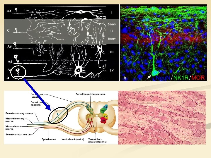
4. How do the neuronal networks of the spinal cord contribute to decision making? Laminar organization of the spinal cord (Rexed, 1952) : especially in the dorsal spinal horn, lamina I -VI
Dorsal horn lamina II: substantia gelatinosa Rolandi
n High-threshold, slow-conducting, thin-myelinated Aδ and unmyelinated C fibres convey predominantly nociceptive information (pain, heat) to the lamina I-III. Low-threshold, high-conducting, thick-myelinated Aβ fibres carry nonpainfull stimuli and terminate in deeper laminae of the spinal dorsal horn. n Dorsal spinal horn plays a crutial role in sensory information processing. Local circuit neurons are more numerous, projection neurons forwarding stimuli to higher levels of the CNS constitute only 5 -10%. n Lamina II is densly packed with mu-opioid receptors binding morphine and other opioid analgesics.
Intermediate substance IML CLARK-STILLING IMM: intermediomedial nucleus, IML: intermediolateral nucleus
n Lateral horn exists only in the thoracic and upper lumbar segments. At these levels appearance of the lateral horn can be explained by the lateral transposition of the IML nucleus. n IML nucleus contains preganglionic neurons of the sympathetic (thoraco-lumbar levels) and parasympathetic (sacral segments) nervous system, with other words visceromotor fibres arise from this nucleus. IMM nucleus is considered as an autonomic spinal reflex centre. n n Dorsal or thoracic nucleus (of Clarke-Stilling) found in the Th 9 -L 3 segments is the source of the dorsal spinocerebellar tract (of Flechsig). Downward continuation of this nucleus in L 4 -S 3 segments is the approximate site of origin of the ventral spinocerebellar tract (of Gowers). Both of these tracts convay proprioceptiv information to the cerebellum.
Ventral horn
Recurrent inhibition: alpha-motoneurons have an axon collateral that terminates on the Renshaw-neuron inhibiting the alphamotoneuron from which the recurrent collateral arised.
A typical neuron in the microscope: prominent nucleolus in the large, oval or rounded, poorly stained nucleus, surrounded by dark, basofilic, granulated cytoplasm, which is always darker then the nucleus.
Spinal reflexes n Reflex: involuntary and automatic response to a stimulus n A (1) receptor is responsible for detecting a stimulus and converting it into a neuronal signal. Evoked action potentials reach the central nervous system through the (2) afferent limb. Decision is made and response is generated by the (3) central processing neuronal networks. (4) Efferent limb exits the central nervous system and terminates on the (5) effector, which might be a muscle or a gland. (1) to (5) components altogether constitute the reflex arc! n In the case of spinal reflex arcs the stimulus is received and processed by the spinal cord and the response is organized and performed by the spinal neuronal circuits. Under normal circumstances stimuli evoking spinal reflexes also reach the brain and descending fibres from higher centres may modulate spinal reflexes but experimentally spinal reflexes
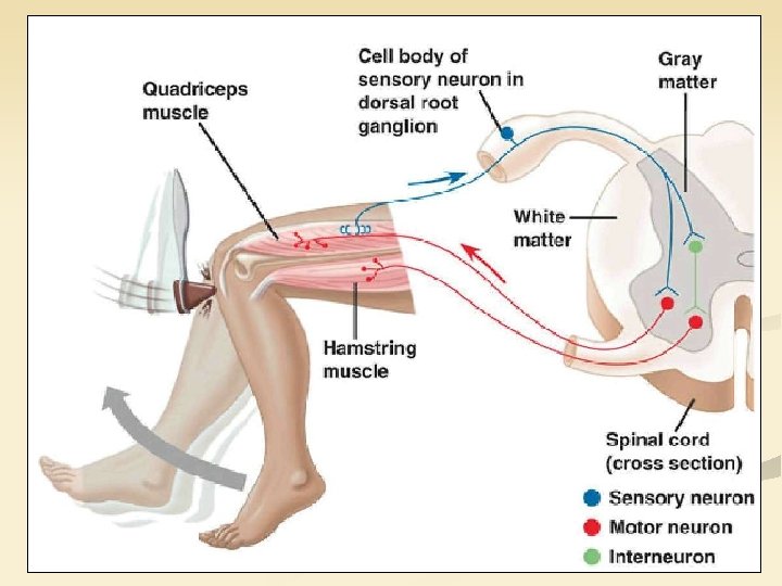
Proprioceptive reflex arc n Synonyms: stretch reflex, monosynaptic spinal reflex, myotatic reflex n Stretching a muscle results in its contraction providing automatic regulation of skeletal muscle length and the posture of the body. The „stretch receptors” are the muscle spindles.
MUSCLE SPINDLE flower-spray anulospiral terminals alpha-motoneuron gamma-motoneuron
n Muscle spindles are special stretch receptors enclosed by a connective tissue capsule. Within the capsule the intrafusal fibres are present, while outside the extrinsic muscle fibres of the host muscle can be found. Intrafusal fibres are innervated by gamma-motoneurons and extrafusal fibres receive stimuli from alphamotoneurons. n Within the muscle spindle two types of sensory terminals, the anulospiral endings and the flower-spray terminals can be distinguished. These inform the CNS about the actual length and movement of the muscles.
Muscle spindle → spinal ganglion → dorsal root → alpha-motoneuron in the ventral horn → peripheral nerve → skeletal muscle fibres surrounding the muscle spindle. Collaterals through inhibitory interneurons will avoid simultaneous contraction of the antagonist muscle groups.
Object held in hands stretches the flexors of the arm and due to the proprioceptive reflex arc the flexors will contract and hold the weight. Proprioceptive spinal reflex developed in response to gravity. Knee joints tend to flex under the weight of the body, but stretching of the quadriceps femoris muscle activates the proprioceptive reflex which increases the tone of the extensors avoiding the collapse of the human body.
Gamma-loop descending pathways adjusting the threshold of muscle spindles gamma motoneuron alpha motoneuron
Golgi tendon organ
Both the gamma-loop and the GTOs contribute to the regulation of motor functions at spinal level. n Gamma-loop sets the threshold of muscle spindles: the shorter the spindle the more sensitive to stretching. n In GTOs axons among collagen bundles can be activated by transformations of the musculotendinous junction. GTO is connected in series to the muscle fibres. Inhibits overstimulation of muscles and prevevents bone, tendon or joint injuries. n n
Thank you for your attention!