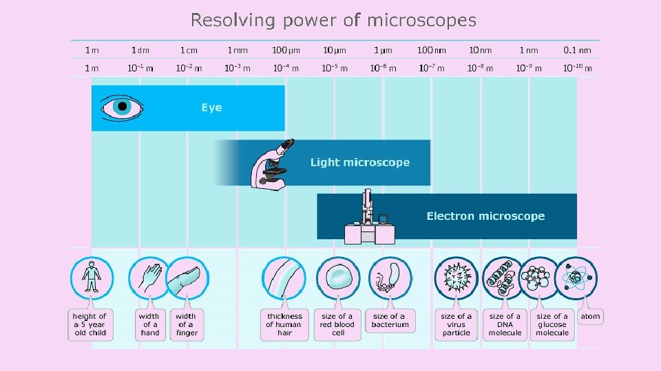
Microscopes THEIR TYPES AND USES Light microscope Key


")
 Real size (scale bar) Scale bar")


 Once you have calibrated the graticule, determine")
- Slides: 17
Microscopes THEIR TYPES AND USES
Light microscope Key: A=ocular lens area B=objective lens area C and D - slide and stage opening E=mechanical stage F=light source
Dark Field Darkfield microscopy is a specialized illumination technique that capitalizes on oblique illumination to enhance contrast in specimens that are not imaged well under normal brightfield illumination conditions. Basic concepts of microscopy
Phase Contrast Basic concepts of microscopy
Electron microscope
Types of electron microscopes Transmission electron microscopes pass a beam of electrons through the specimen. Electron gun Thin sections of specimen are needed for transmission electron microscopy as the electrons have to pass through the specimen for the image to be produced. Dense areas stop the electrons and appear dark. Specimen Monitor Sensor
Types of electron microscopes Scanning electron microscopes pass a beam of electrons over the surface of the specimen in the form of a ‘scanning’ beam. Larger, thicker structures can thus be seen under the scanning electron microscope as the electrons do not have to pass through the sample in order to form the image. However the resolution of the scanning electron microscope is lower than that of the transmission electron microscope. Monitor Electron gun r o s n e S Specimen Electrons are reflected creating a 3 D image of troughs and ridges
Scanning vs transmission
Electron microscopes Better resolution: The ability to distinguish between two points on an image. Like pixels in a digital camera. Greater magnification: How much bigger a sample appears to be under the microscope than it is in real life.
Same magnification, different resolution Light microscope Scanning electronmicroscope
What microscope to use ? Light Electron Cheap to purchase (200 – 1000 $) Expensive (over 2, 000$) Cheap to operate Expensive to produce electron beams Small and portable Large and requires special rooms Simple and easy preparations Lengthy and complex preparations Material rarely distorted by preparation Preparation distorts material Vacuum is not required Vacuum is required Natural color maintained All images in black and white Magnifies objects only up to 2000 times Magnifies over 500, 000 times
Magnification and scale bars Magnification = Magnified size(ruler) Real size (scale bar) Scale bar = 10 µm
DETERMINING THE SIZE OF CELLS Using a graticule
Calibration How much is each division on the graticule worth? This depends on the magnification! Use the lowest Graticule magnification. We do not have a stage micrometer so we will need to use a transparent ruler where the smallest unit is in mm. At 20 x magnification: 5 mm = 5000µm ∴ each division on the graticule is equal to: 5000/100 = 50µm It is easier to turn the eyepiece to align the graticule with the ruler.
The values for the graticule will decrease as we increase the magnification. In the case given on the previous page: Magnification Calculation Graticule value per smallest division x 20 x 100 x 400 See previous slide 50µm (20/100) x 50µm 10µm (20/400) x 50µm 2. 5µm Each microscope needs to be calibrated separately.
Determining the size of cells (onion epidermis) Once you have calibrated the graticule, determine the length and width of an onion epidermis cell. Measuring the length of many cells and calculating the mean will give a more reliable estimate!